. 2024 Dec;47(6):100713.
doi: 10.1016/j.bj.2024.100713. Epub 2024 Mar 3.

Altered purinergic P2X7 and A2B receptors signaling limits macrophage-mediated host defense in schistosomiasis

Affiliations

Altered purinergic P2X7 and A2B receptors signaling limits macrophage-mediated host defense in schistosomiasis

Maria Luiza Thorstenberg et al. Biomed J. 2024 Dec.

Abstract

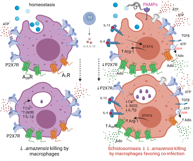
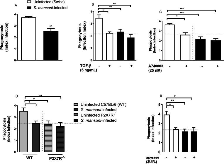
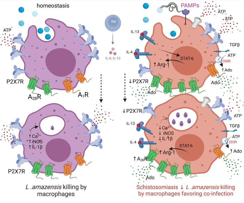
Background: The occurrence of co-infections during schistosomiasis, a neglected tropical disease, with other parasites have been reported suggesting an impaired host immune defense. Macrophage purinergic P2X7 receptor (P2X7R) plays an important role against intracellular pathogens. Therefore, we investigated the P2X7R-mediated phagocytosis and killing capacity of Leishmania amazonensis by macrophages during schistosomiasis in vitro and in vivo.

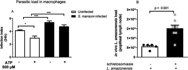
Methods: Swiss and C57BL/6 (Wild type) and P2X7R-/- were randomized in two groups: control (uninfected) and Schistosoma mansoni-infected. Alternatively, control Swiss and S. mansoni-infected mice were also infected with L. amazonensis.

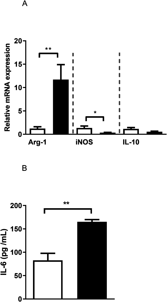
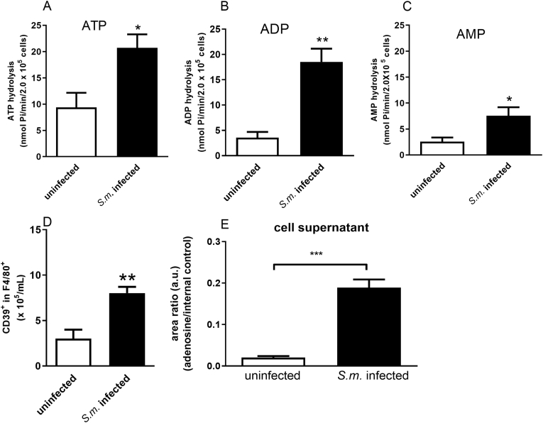
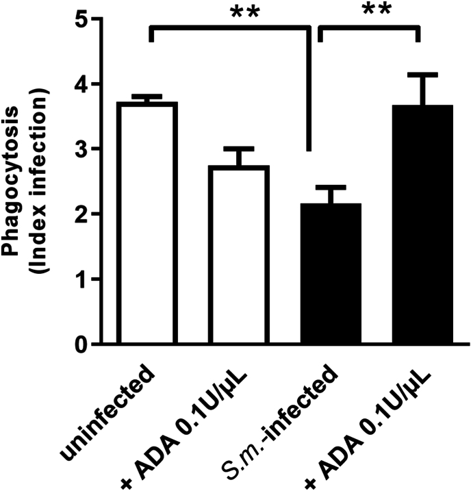
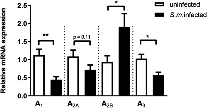
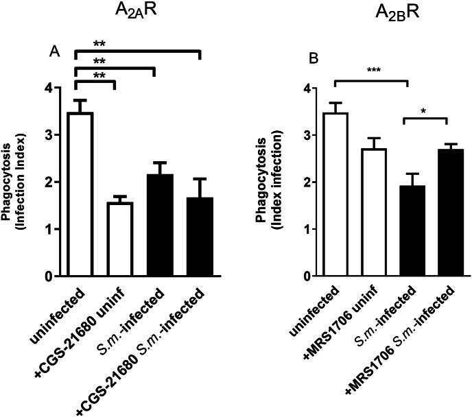
Results: The pre-treatment of control macrophages with the P2X7R antagonist (A74003) or TGF-β reduced the phagocytosis index, mimicking the phenotype of cells from S. mansoni-infected mice and P2X7R-/- mice. Apyrase also reduced the phagocytosis index in the control group corroborating the role of ATP to macrophage activation. Moreover, l-arginine-nitric oxide pathway was compromised during schistosomiasis, which could explain the reduced killing capacity in response to ATP in vitro and in vivo. We found an increased extracellular nucleotide (ATP, ADP and AMP) hydrolysis along with an increased frequency of F4/80+ CD39+ macrophages from the S. mansoni-infected group. Moreover, the content of adenosine in the cell supernatant was higher in the S. mansoni-infected group in relation to controls. Schistosomiasis also increased the expression of macrophage adenosine A2BR. In good accordance, both ADA and the selective A2BR antagonist restored the phagocytosis index of macrophages from S. mansoni-infected group.

Conclusions: Altogether, the altered P2X7R and A2BR signaling limits the role of macrophages to host defense against L. amazonensis during schistosomiasis, potentially contributing to the pathophysiology and clinically relevant co-infections.

Keywords: Adenosine receptor; Host defense; Leishmaniasis; Macrophages; P2X7 receptor; Schistosomiasis.


Conflict of interest statement

Declaration of competing interest none.

Figures

Image 1
Graphical abstract
Fig. 1
Fig. 1
Macrophages with reduced P2X7 receptor function due to schistosomiasis showed a reduced phagocytosis index of L. amazonensis in vitro. Macrophages from Swiss control and S. mansoni-infected mice were incubated with TGF-β (5 ng/mL, 12h), with A740003 (25 nM, 30 min) or apyrase (2UI/L, 30 min) before exposition to L. amazonensis. White and light gray bars = uninfected (control) Swiss (A, B, C, E) or WT C57BL/6 (D) mice, respectively. Black bars = S. mansoni-infected mice. Light and dark gray hatched bars = P2X7R −/− uninfected or S. mansoni-infected mice, respectively (D). Data expressed as mean and SEM. ∗p < 0.05, ∗∗p < 0.01, ∗∗∗p < 0.001 (n = 4–5 individual animals for each group, Student's t-test (A) or one way ANOVA followed by Tukey's multiple comparisons test (B–D). E) ∗p < 0.05, ∗∗p < 0.01, n = 5–7 individual animals for each group, one way ANOVA followed by Tukey's multiple comparisons test.
Fig. 2
Fig. 2
Schistosomiasis alters macrophage expression of enzymes regulating the l-arginine-NO pathway and immunomodulatory cytokines. A) Relative mRNA expression of Arg-1, iNOS and IL-10. B) IL-6 concentration (ELISA). White and black bars = uninfected or S. mansoni-infected mice, respectively. Data expressed as mean and SEM. ∗p < 0.05, ∗∗p < 0.01, n = 5 individual animals for each group, Student's t-test.
Fig. 3
Fig. 3
The reduced macrophage P2X7 receptor function during schistosomiasis impairs L. amazonensis killing in response to ATP in vitro and in vivo. A) Macrophages from control and S. mansoni-infected mice were exposed to L. amazonensis for 4 h (MOI 10:1) and washed to remove non-internalized protozoan. After 24 h, cells were treated with 500 μM ATP for 30 min, and 24 h later, cells were washed, fixed, and imaged to calculate the infection index (see Methods section). Data expressed as mean and SEM. ∗p < 0.05, ∗∗∗p < 0.001, n = 4–5 individual animals for each group, one way ANOVA followed by Tukey's multiple comparisons test. B) Reduced control of L. amazonensis proliferation during schistosomiasis in vivo. Control and S. mansoni-infected mice were infected through an intradermal injection of 106 L. amazonensis promatigotes. The popliteal lymph nodes were analyzed 35 days later. n = 5–8 individual animals for each group, p = 0.001, Student's t-test.
Fig. 4
Fig. 4
Schistosomiasis alters nucleotide hydrolysis and increases extracellular adenosine. Macrophages from control and S. mansoni-infected mice (2 × 105 cells/well) were plated for enzymatic activity (A–C), CD39 expression (D) or adenosine quantification (E). Cells were incubated with 1 mM ATP, ADP or AMP for 30 min. Data expressed as nmol of Pi released/min/2 × 105 cells (mean and SEM). A-C) n = 4–6 individual animals for each group, ∗p < 0.05, ∗∗p < 0.01, Student's t-test. D) CD39+ cells in the gate of F4/80+ cells, n = 3–4 individual animals for each group, ∗∗p < 0.01, Student's t-test. E) n = 12 samples, ∗∗∗p < 0.001, Student's t-test. S.m. = S. mansoni.
Fig. 5
Fig. 5
Adenosine deaminase (ADA) restores the phagocytic index of macrophages from S. mansoni-infected mice. Macrophages from control and S. mansoni-infected groups were incubated with vehicle or ADA (0.1U/μL) for 30 min before exposition to L. amazonensis. Data expressed as mean and SEM. n = 4–7 individual animals for each group, ∗∗p < 0.01, one way ANOVA followed by Tukey's multiple comparisons test. S.m. = S. mansoni.
Fig. 6
Fig. 6
Schistosomiasis alters the macrophage expression of purinergic A1, A2B and A3 receptors. Data expressed as mean and SEM. n = 5–6 individual animals for each group, ∗p < 0.05, ∗∗p < 0.01, Student's t-test. S.m. = S. mansoni
Fig. 7
Fig. 7
The pharmacological blockage of purinergic A2B receptor restores the phagocytic index of macrophages from S. mansoni-infected mice. Macrophages from both groups were incubated with the selective A2A receptor agonist CGS21680 (1 μM, A) or with the selective A2B receptor antagonist MRS1706 (1 μM, B) for 30 min before exposition to L. amazonensis. Data expressed as mean and SEM. n = 5–8 individual animals for each group, ∗p < 0.05, ∗∗p < 0.01, ∗∗∗p < 0.001, one way ANOVA followed by Tukey's multiple comparisons test (A, B) or Student's t-test (B, S.m.-infected + MRS1706). S.m. = S. mansoni
Fig. 8
Fig. 8
Schistosomiasis negatively impacts macrophage purinergic P2X7 receptors and elevates A2B receptor signaling, leading to a decreased ability to eliminate L. amazonensis. Extracellular ATP is an important DAMP (damage-associated molecular pattern) and the main agonist of purinergic signaling. Under normal conditions (homeostasis), macrophage P2X7 receptors trigger Ca2+ influx and inflammasome activation, resulting in IL-1β release, NO production, and efficient phagocytosis of promastigotes (blue circles) and killing of L. amazonensis. However, in the context of chronic schistosomiasis, the recognition by host innate immune cells of pathogen-associated molecular patterns (PAMPs; pink) released by S. mansoni and eggs triggers an adaptative immune response that reshapes the host's purinergic signaling. Elevated TGF-β levels diminish macrophage P2X7 receptor signaling. We also observed an increase in extracellular ATP hydrolysis by CD39 favoring adenosine (Ado) formation. Additionally, increased IL-6 levels, A2B receptor (green) and Arg-1 expressions, and decreased iNOS expression compromised microbicidal mechanisms, fostering a favorable environment for L. amazonensis co-infection in vitro and in vivo. The blockage of A2B receptors restored macrophage phagocytosis during schistosomiasis. These alterations in purinergic signaling may offer insights into the molecular basis of co-infections observed in individuals enduring chronic schistosomiasis. Understanding these mechanisms not only enhances our comprehension of host-pathogen interactions but also opens avenues for developing targeted therapeutic strategies to address co-infections in patients with chronic schistosomiasis. Created with Biorender.

Similar articles

References

    1. King CH. Health Metrics for helminth infections. Acta Trop. 2015;141:150–160. - PMC - PubMed
    1. Dunne DW, Cooke A. A worm’s eye view of the immune system: consequences for evolution of human autoimmune disease. Nature Rev Immunol. 2005;5(5):420–426. - PubMed
    1. Silva CLM., Morel N, Lenzi HL, Noël F. Increased reactivity to 5-hydroxytryptamine of portal veins from mice infected with Schistosoma mansoni. Comp Biochem Physiol Mol Integr Physiol. 1998;120(3):417–423. - PubMed
    1. Costain AH, MacDonald AS, Smits H. Schistosoma egg migration: mechanisms, pathogenesis and host immune responses. Frontiers Immunol. 2018;9:3042. - PMC - PubMed
    1. Brunet RL, Beall M, Dunne DW, Pearce EJ. Nitric oxide and the Th2 response combine to prevent severe hepatic damage during Schistosoma mansoni infection. J Immunol. 1999;163(9):4976–4984. - PubMed

Substances

LinkOut - more resources