. 2024 Mar 5;15(1):2004.
doi: 10.1038/s41467-024-46391-9.

DJ-1 protects proteins from acylation by catalyzing the hydrolysis of highly reactive cyclic 3-phosphoglyceric anhydride

Affiliations

DJ-1 protects proteins from acylation by catalyzing the hydrolysis of highly reactive cyclic 3-phosphoglyceric anhydride

Aizhan Akhmadi et al. Nat Commun. .

Abstract

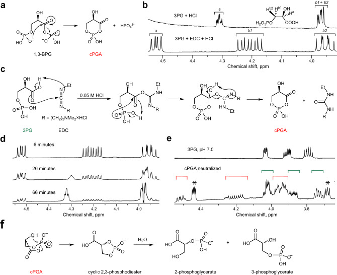
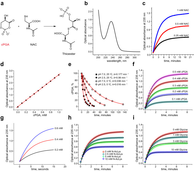
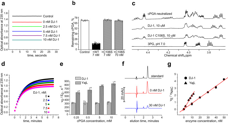
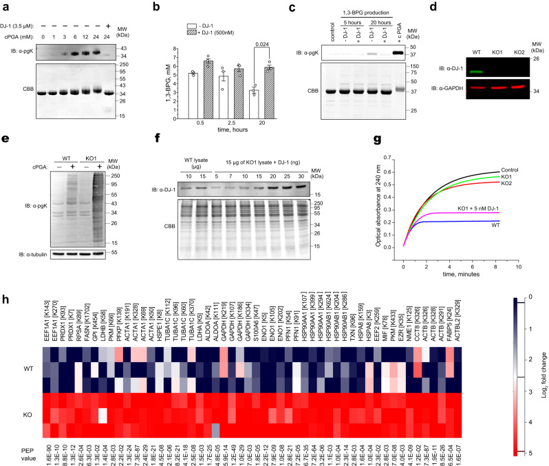
Mutations in the human PARK7 gene that encodes protein DJ-1 lead to familial Parkinsonism due to loss of dopaminergic neurons. However, the molecular function of DJ-1 underpinning its cytoprotective effects are unclear. Recently, DJ-1 has been shown to prevent acylation of amino groups of proteins and metabolites by 1,3-bisphosphoglycerate. This acylation is indirect and thought to proceed via the formation of an unstable intermediate, presumably a cyclic 3-phosphoglyceric anhydride (cPGA). Several lines of evidence indicate that DJ-1 destroys cPGA, however this enzymatic activity has not been directly demonstrated. Here, we report simple and effective procedures for synthesis and quantitation of cPGA and present a comprehensive characterization of this highly reactive acylating electrophile. We demonstrate that DJ-1 is an efficient cPGA hydrolase with kcat/Km = 5.9 × 10⁶ M-1s-1. Experiments with DJ-1-null cells reveal that DJ-1 protects against accumulation of 3-phosphoglyceroyl-lysine residues in proteins. Our results establish a definitive cytoprotective function for DJ-1 that uses catalytic hydrolysis of cPGA to mitigate the damage from this glycolytic byproduct.


Conflict of interest statement

The authors declare no competing interests.

Figures

Fig. 1
Fig. 1. Synthesis and decomposition of cPGA.
a Proposed mechanism of cPGA formation from 1,3-BPG. b In the presence of equimolar amounts of EDC and HCl, 3PG is immediately and completely converted to another molecule. 1H-NMR spectra of 50 mM 3PG, or 3PG + EDC (50 mM each) in D2O supplemented with 50 mM HCl were recorded immediately after mixing of all reagents. Signals from the Ha, Hb1, and Hb2 protons are labeled as a, b1 and b2. c Proposed mechanism of formation of cPGA in reaction of 3PG with EDC. d A series of 1H-NMR spectra of a mixture of 3PG, EDC and HCl (50 mM each) were recorded at indicated times after mixing of all reagents to illustrate the gradual spontaneous decomposition of cPGA back to 3PG. e 3PG, EDC, and HCl (50 mM each) were mixed in D2O, incubated for 1 min, neutralized with 50 mM NaOH, and analyzed by 1H-NMR. Signals of 3PG and cPGA are labeled with green and red square brackets, respectively. Signals that likely correspond to 2,3-phosphodiester of glyceric acid are labeled with asterisk. The 1H-NMR spectrum of 3PG at pH 7.0 is shown for reference: a complex splitting pattern and peak broadening suggest that at neutral conditions 3PG is present in at least two different forms (e.g., monomer-dimer) that exist in quick equilibrium with each other. f Proposed mechanism of cPGA conversion into cyclic 2,3-phosphodiester at neutral pH. All NMR spectra are representative of at least 5 independent experiments.
Fig. 2
Fig. 2. cPGA is a highly reactive electrophile.
Unless otherwise indicated, all experiments were conducted in 50 mM sodium phosphate buffer (pH 7.0) at 25 °C. a Scheme for thioester formation in the reaction of cPGA with NAC. b UV absorbance spectrum of 0.5 mM of pure thioester. c Kinetics of thioester formation in the reaction of 0.75 mM cPGA with indicated concentrations of NAC was monitored by optical absorbance at 235 nm. d A standard curve for cPGA was obtained by derivatization with NAC. The linear fit to the data is shown as a red line. e Kinetics of spontaneous decomposition of cPGA at 0 and 25 °C in neutral and acidic conditions. cPGA concentrations were measured at indicated time points using the thioester-based end-point assay. Exponential decay fits are shown as red lines. f cPGA at indicated concentrations was allowed to react with 1.5 mM of NAC. The data were fit globally (solid lines) to derive the rate constant. g Kinetics of formation of thioester in the reaction of cPGA (0.2, 0.4, and 0.6 mM) with 10 mM NAC at pH 7.4 and 37 °C to mimic physiological conditions. Note that the reaction is essentially over within 10 s. h and i Reaction kinetics of NAC (5 mM) and cPGA (0.5 mM) in the presence of indicated concentrations of Nα-acetyllysine (h) and glycine (i) as competitors. Global fits (solid black lines) were performed to derive rate constants of reaction of cPGA with Nα-acetyllysine and glycine. Results shown in (ce and g) are representative of 3–5 independent experiments. Results shown in (f, h, and i) represent one of the two independent series of experiments that showed the same results. Source data are provided as a Source Data file.
Fig. 3
Fig. 3. Characterization of DJ−1 as cPGA hydrolase.
Unless otherwise indicated, all experiments were conducted in 50 mM sodium phosphate buffer (pH 7.0) at 25 °C. a cPGA (500 µM) was incubated with indicated concentrations of DJ−1 for 1 min at room temperature followed by an immediate conversion into NAC-thioester. Optical absorbance at 235 nm was recorded for 20 s to ensure the stability of signal after derivatization. b cPGA (500 µM) was incubated with DJ-1 or its C106S mutant at the indicated concentrations for 2 min followed by quantitation of the remaining cPGA by NAC assay (mean ± s.d. for n = 4 independent experiments). c 1H-NMR spectra of 50 mM cPGA neutralized with 50 mM Na2HPO4. DJ-1 or the C106S mutant were added immediately after neutralization. 1H-NMR spectrum of 3PG at pH 7.0 is shown for reference. d 0.5 mM of cPGA was allowed to react with 1.5 mM of NAC in the presence of DJ-1 at indicated concentrations. Thioester formation was monitored by optical absorbance at 235 nm. e Apparent catalytic constants (v0/E0) for DJ-1 and YajL were determined for the indicated concentrations of cPGA using end-point NAC assay (mean ± s.d. for n = 3 independent experiments for each concentration of cPGA). f HPLC quantitation of NAC-thioester of phosphoglyceric acid. Synthetic thioester standard (50 µM) was used to quantify the amount of thioester formed in the reaction of 10 mM NAC with 100 µM cPGA (10 µM of cPGA were added 10 times every 3 min) in the presence or absence of 30 nM DJ-1. g To determine kcat/Km of DJ-1 and YajL, enzymes at the indicated concentrations were allowed to compete with 10 mM of NAC for cPGA. The concentration of cPGA was kept in the low micromolar range to ensure a linear dependence of the rate of enzymatic conversion on the concentration of cPGA. The ratio of the enzymatic conversion rate of cPGA (vE) to the reaction rate of cPGA with NAC (vNAC) was determined by HPLC from amounts of accumulated thioester at the end of the experiment (mean ± s.e.m. for n = 3 independent experiments for each concentration of enzymes). Representative kinetic curves (a, d), NMR spectra (c) or HPLC traces (f) from three independent experiments are shown. Source data are provided as a Source Data file.
Fig. 4
Fig. 4. DJ-1 protects proteins from acylation by 1,3-BPG and cPGA.
a Immunoblot and Coomassie-stained gel of GAPDH (0.2 and 2 µg of GAPDH respectively) show that acylation by cPGA can be specifically detected by anti-3PG antibodies. b 1,3-BPG was produced continuously in the presence of 2.5 mg/mL of GAPDH that was later assayed for acylation by immunoblot. Concentration of 1,3-BPG was measured at indicated times and shown as the mean ± s.e.m. P-values were calculated using two-tailed paired Student’s t-test (n = 3, independent experiments). c Acylation level of GAPDH in the presence of continuously produced 1,3-BPG was assessed by immunoblot. DJ-1 (500 nM) completely prevents acylation that is visible at 20 h of incubation. d Immunoblot analysis of DJ-1 expression in wild-type (WT) and DJ-1-null (KO1 and KO2) HCT116 cells. Bands corresponding to the molecular weight of DJ-1 are absent in both of the KO1 and KO2 clones. GAPDH was used as a loading control. e Immunoblot analysis of phosphoglyceroyl modifications in cytosol of WT and DJ-1-null cells (KO1) before and after treatment of cytosolic extracts with 0.5 mM cPGA for 20 min. Tubulin was used as a loading control. f To quantify DJ-1 in HCT116 cells, 10 and 15 µg of wild-type cytoplasmic protein extract was immunoblotted along with 15 µg of extract from KO1 cells spiked with 5–30 ng of purified recombinant DJ-1. Coomassie brilliant blue (CBB) stained gel of the same samples is shown below. g Cytoplasmic protein extracts from wild-type HCT116 (0.1 mg/mL), KO1 (0.3 mg/mL), and KO2 (0.3 mg/mL) cells were added to a reaction of cPGA (0.3 mM) with NAC (1 mM). The formation of thioester was monitored by optical absorbance at 240 nm instead of 235 nm because of the strong absorbance of cell extracts. A strong inhibitory effect of WT extract on thioester accumulation could be recapitulated with KO1 lysate by adding 5 nM of DJ-1. h Quantitation of pgK modifications of cytoplasmic proteins from wild-type and DJ-1 knockout cells treated with cPGA. Protein extracts were treated by cPGA (1 mM of cPGA was added 5 times every 3 min) followed by proteolytic digestion and quantitation by LC/MS. Three technical replicates from a representative experiment are shown. Representative gels/blots (a, cf) or kinetic curves (g) from three independent experiments are shown. Source data are provided as a Source Data file.

Similar articles

Cited by

References

    1. Bonifati V, et al. Mutations in the DJ−1 gene associated with autosomal recessive early-onset parkinsonism. Science. 2003;299:256–259. doi: 10.1126/science.1077209. - DOI - PubMed
    1. Smith N, Wilson MA. Structural biology of the DJ−1 superfamily. Adv. Exp. Med. Biol. 2017;1037:5–24. doi: 10.1007/978-981-10-6583-5_2. - DOI - PubMed
    1. Canet-Aviles RM, et al. The Parkinson’s disease protein DJ-1 is neuroprotective due to cysteine-sulfinic acid-driven mitochondrial localization. Proc. Natl Acad. Sci. USA. 2004;101:9103–9108. doi: 10.1073/pnas.0402959101. - DOI - PMC - PubMed
    1. Blackinton J, et al. Formation of a stabilized cysteine sulfinic acid is critical for the mitochondrial function of the parkinsonism protein DJ-1. J. Biol. Chem. 2009;284:6476–6485. doi: 10.1074/jbc.M806599200. - DOI - PMC - PubMed
    1. Lee JY, et al. Human DJ-1 and its homologs are novel glyoxalases. Hum. Mol. Genet. 2012;21:3215–3225. doi: 10.1093/hmg/dds155. - DOI - PubMed