. 2024 Mar 5;15(3):187.
doi: 10.1038/s41419-024-06572-2.

Synergistic antitumor activity between HER2 antibody-drug conjugate and chemotherapy for treating advanced colorectal cancer

Affiliations

Synergistic antitumor activity between HER2 antibody-drug conjugate and chemotherapy for treating advanced colorectal cancer

Hongfu Liu et al. Cell Death Dis. .

Abstract

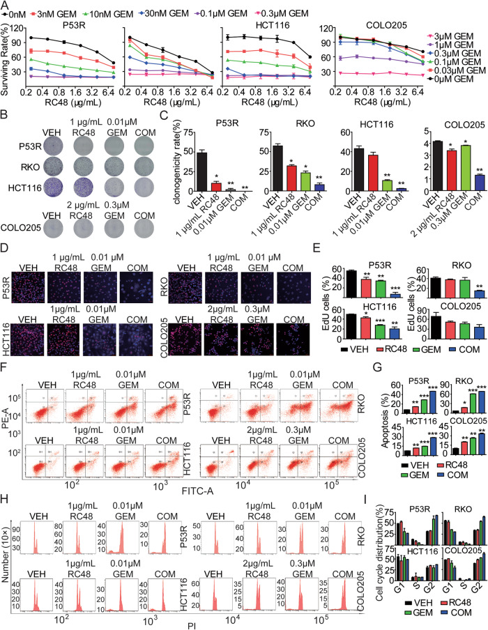
Colorectal cancer (CRC) is the third most common cancer associated with a poor prognosis. Effective targeted therapy alone or in combination for treating advanced CRC remains to be a major clinical challenge. Here, we propose the therapeutic efficacy and molecular mechanism underlying RC48, a FDA-approved anti-HER2 antibody conjugate via a cleavable linker to the microtubule inhibitor monomethyl auristatin E (MMAE), either alone or in combination with gemcitabine (GEM) in various models of HER2-positive advanced CRC. Our findings demonstrated that HER2 was widely expressed and located on the plasma membrane of CRC patient specimens, PDX xenograft tumors and cell lines. It confirmed that RC48 alone significantly targeted and eradicated HER2 positive CRC tumor in these models. Moreover, we screened a panel of FDA-approved first-line chemotherapy drugs in vitro. We found that GEM exhibited stronger antiproliferative activity compared to the other first-line anti-cancer agents. Furthermore, combination therapy of RC48 and GEM significantly showed synergetic antitumor activity in vitro and in vivo. To gain further mechanistic insights into the combination therapy, we performed RNA-seq analysis. The results revealed that combination treatment of RC48 and GEM regulated multiple signaling pathways, such as PI3K-AKT, MAPK, p53, Foxo, apoptosis, cell cycle and cell senescence, etc., to exert its antitumor activity in CRC cells. Collectively, these preclinical findings demonstrated that RC48 alone or combinational therapy exerted promising antitumor activity, and meriting the preclinical framework for combinational therapy of anti-HER2 drug conjugate drug and chemotherapy drugs for HER2-positive patients with advanced CRC.


Conflict of interest statement

The authors declare no competing interests.

Figures

Fig. 1
Fig. 1. RC48 significantly inhibited cell proliferation in HER2-positive CRC cell lines.
A DR5 mRNA overexpression in human CRC versus normal colon. Box plot has been downloaded from the GEPIA database. B Percent of 538 CRC tumors in each of the four staining intensity, including strong, moderate, low, and negative. C Representative pictures of the 4 staining intensity of HER2 IHC on human CRC tumor tissue microarray. D The expression of human HER2 in COLO205, P53R, RKO and HCT116 cells quantified by Western blotting. E Plasma membrane and intracellular subcellular localization of HER2 protein (green) by immunofluorescence in COLO205, P53R, RKO and HCT116 cells. Overlay with DAPI (blue) is depicted. F P53R, RKO, COLO205 and HCT116 cells were treated with RC48, T-DM1, T-DXd and trastuzumab for 72 h, respectively. The cell viability was detected by Cell Titer-Glo Cell Viability assay. G P53R, RKO, COLO205 and HCT116 cells were treated by 0, 1 and 5 μg/ml RC48 for 48 h, then cell proliferation was determined by the EdU assay. Fluorescence images were obtained and analyzed with a fluorescence microscope. Data are presented as means ± SEM of three independent experiments. Images captured at 400×magnification, respectively. Scale bars = 20 µm. Data represent the mean ± SEM of at least three independent experiments and statistical significance was assessed by unpaired T-test (*p < 0.05; **p < 0.01; ***p < 0.001).
Fig. 2
Fig. 2. RC48 induced apoptosis and cell cycle arrest in HER2-positive CRC cells.
P53R, RKO, COLO205 and HCT116 cells were treated with indicated concentrations of RC48 for 48 h, respectively. A, B, E, F The cells were stained with an anti-Annexin V-FITC antibody and PI for apoptosis analysis by flow cytometry. Annexin V-FITC-positive cells were defined as apoptotic (A, B). And the induction of cell cycle analysis in the CRC cells was detected by flow cytometry (E, F). The expression of critical molecules in the antiapoptotic (C, D) and cell cycle (G, H) pathway in CRC cells were examined by immunoblot analysis. Quantification was performed using Image J software. Data represent the mean ± SEM of at least three independent experiments and statistical significance was assessed by unpaired T-test (*p < 0.05; **p < 0.01; ***p < 0.001).
Fig. 3
Fig. 3. Antitumor activity of RC48 alone in mouse CRC CDX and PDX models.
A, B Antitumor activity of RC48 in mouse COLO205 and HCT116 CDX models. BALB/c nude mice were injected s. c. with 5× 10⁶ COLO205 or HCT116 cells. Mice bearing xenografts approximately averaged 100–150 mm3 were intravenously received saline and 10.0 mg/kg of RC48. Tumor sizes and body weight were measured twice a week. Tumor growth curve and transplanted tumor weights were assessed at the end of the experiment in mouse COLO205 (A) and HCT116 (B) CDX models. Immunofluorescence analysis (C) and immunohistochemical (D) of HER2 expression in the transplanted tumors derived from the CRC054, CRC082, CRC090 and CRC073 PDX mice. Antitumor activity (E, G, I, K) and survival benefit (F, H, J, L) of RC48 in the CRC054 (E, F), CRC082 (G, H), CRC090 (I, J) and CRC073 (K, L) PDX models. RC48 and vehicle groups were intravenously administered. Immunofluorescence images captured at 630× magnification, scale bars = 10 µm. Immunohistochemical images captured at 100× and 400× magnification, scale bars = 100 and 20 µm, respectively.
Fig. 4
Fig. 4. The synergistic antitumor effects of RC48 and/or GEM on HER2-positive CRC cells.
A Cytotoxicity of RC48 combined with GEM in P53R, RKO, HCT116 and COLO205 cells were determined by CellTiter-Glo® cytotoxicity assays. The dose of RC48 monotherapy was set as 0, 0.25, 0.5, 1, 2, 4 and 8 μg/mL in P53R, RKO, HCT116 and COLO205 cells, while GEM was set as 0, 3, 10, 30, 100 and 300 nM in P53R, RKO and HCT116 cells, and GEM was set as 0, 0.03, 0.1, 0.3, 1 and 3 μM in COLO205 cells. B, C P53R, RKO, HCT116 and COLO205 cells were treated with RC48, GEM and their COM for 14 days. The proliferative ability of CRC cells was investigated via colony formation assays. D, E P53R, RKO, HCT116 and COLO205 cells were treated with RC48, GEM, or their COM for 48 h, then cell proliferation was determined by EdU assay. F, G P53R, RKO, HCT116 and COLO205 cells were treated with RC48, GEM, or their COM for 48 h. The cells were stained with an anti-Annexin V-FITC antibody and PI for apoptosis analysis by flow cytometry. Annexin V-FITC-positive cells were defined as apoptotic. H, I P53R, RKO, HCT116 and COLO205 cells were treated with RC48, GEM, or their COM for 48 h.The induction of cell cycle analysis in P53R, RKO, HCT116 and COLO205 cells was detected by flow cytometry. Data represent the mean ± SEM of at least three independent experiments and statistical significance was assessed by unpaired T-test (*p < 0.05; **p < 0.01; ***p < 0.001).
Fig. 5
Fig. 5. Synergic effects of RC48 and GEM combinational treatment on global gene expression profile in CRC cells.
A All DEGs in the vehicle-treated group (VEH; N = 3), 1 μg/mL RC48 (N = 3), 0.01 μM GEM (N = 3) and RC48 plus GEM (COM; N = 3) treated groups in P53R cell. B Numbers of overlapped DEGs in the RC48, GEM, and COM treated samples as compared with the VEH group in P53R cell. KEGG analysis of DEGs in the RC48 (C), GEM (D) and COM (E) treated samples compared with the VEH-treated samples in P53R cells. F, G Analysis of the PI3K/AKT pathway, AMPK pathway, p53 pathway, FOXO pathway, cell cycle-related proteins and antiapoptotic proteins in P53R and RKO cells. Quantification was performed using Image J software. GAPDH was used as the loading control.
Fig. 6
Fig. 6. RC48 plus GEM treatment affected unique CRC-associated prognostic genes.
A Heatmap showing the mRNA levels of the top 100 genes (50 up or downregulated) after RC48 plus GEM combinational treatment (COM). The expression levels of each gene were normalized to the total mRNA abundance of each sample and compared with that of vehicle-treated group (VEH). B The top ranked positively and negatively enriched gene sets identified using GSEA in response to combinational treatment. GSEA was conducted with top common DEGs in P53R and RKO cells after combinational treatment using 50 HALLMARK gene sets database in MSigDB. C GSEA plots showing strong negative enrichment of the cell cycle, and positively enrichment of the P53 and FOXO pathway in P53R and RKO cells in response to combinational treatment. NES Normalized Enrichment Score, FDR false discovery rate. D, E Kaplan–Meier estimate of overall survival based on expression of DEGs (TCGA CRC Cohort). Relative high expression of IGF2BP3, PHF19, FUT11 and C2orf48 genes (D), and relative low expression of IL1B, GPA33, PPP1R14C and UGT2B7 (E) were associated with poor overall survival in CRC cohort. Log rank (Mantel–Cox) test was used for significance; p < 0.05 was considered significant.
Fig. 7
Fig. 7. RC48 plus GEM synergistically inhibited CRC tumor growth in mouse CDX and PDX models.
Tumor growth curve, transplanted tumor weight, and images are displayed in COLO205 (A−C) and HCT116 (D−F) CDX models. G Images of immunohistochemical staining for Ki67 in COLO205 and HCT116 CDX mouse xenograft tumors. Tumor growth curve, transplanted tumor weight, and images were evaluated in CRC054 (HM) and CRC082 (KM) PDX models. N Images of immunohistochemical staining for Ki67 in CRC054 and CRC082 PDX mice xenograft tumors. O Images of immunofluorescence TUNEL of CRC054 and CRC082 PDX mouse xenograft tumors. RC48 and vehicle groups were intravenously administered and GEM groups were intraperitoneally administered. IHC Images captured at 400×magnification, Scale bars = 20 µm. TUNEL Images captured at 200×magnification, respectively. Scale bars = 500 µm. And statistical significance was assessed by unpaired T-test (*p < 0.05; **p < 0.01; ***p < 0.001).

Similar articles

Cited by

References

    1. Siegel RL, Miller KD, Jemal A. Cancer statistics, 2020. CA Cancer J Clin. 2020;70:7–30. doi: 10.3322/caac.21590. - DOI - PubMed
    1. Sung H, Ferlay J, Siegel RL, Laversanne M, Soerjomataram I, Jemal A, et al. Global Cancer Statistics 2020: GLOBOCAN Estimates of Incidence and Mortality Worldwide for 36 Cancers in 185 Countries. CA Cancer J Clin. 2021;71:209–49. doi: 10.3322/caac.21660. - DOI - PubMed
    1. Keum N, Giovannucci E. Global burden of colorectal cancer: emerging trends, risk factors and prevention strategies. Nat Rev Gastroenterol Hepatol. 2019;16:713–32. doi: 10.1038/s41575-019-0189-8. - DOI - PubMed
    1. Dekker E, Tanis PJ, Vleugels JLA, Kasi PM, Wallace MB. Colorectal cancer. Lancet. 2019;394:1467–80. doi: 10.1016/S0140-6736(19)32319-0. - DOI - PubMed
    1. Xiao Y, Zhong J, Zhong B, Huang J, Jiang L, Jiang Y, et al. Exosomes as potential sources of biomarkers in colorectal cancer. Cancer Lett. 2020;476:13–22. doi: 10.1016/j.canlet.2020.01.033. - DOI - PubMed

Publication types