. 2024 Mar 5;15(1):1992.
doi: 10.1038/s41467-024-46221-y.

In-cell NMR suggests that DNA i-motif levels are strongly depleted in living human cells

Affiliations

In-cell NMR suggests that DNA i-motif levels are strongly depleted in living human cells

Pavlína Víšková et al. Nat Commun. .

Abstract

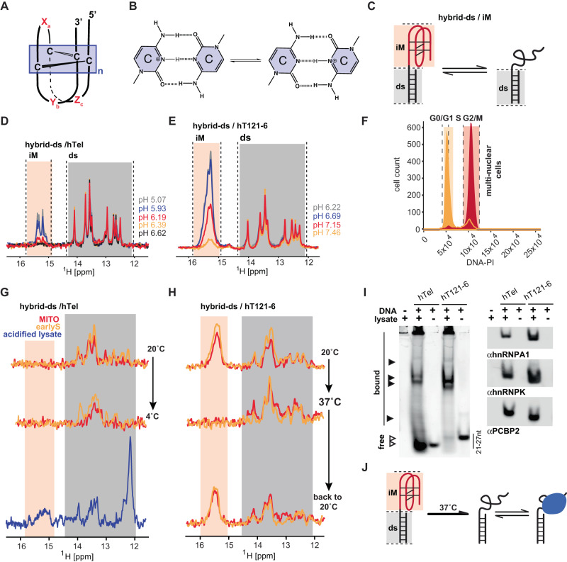
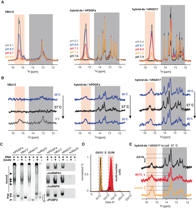
I-Motifs (iM) are non-canonical DNA structures potentially forming in the accessible, single-stranded, cytosine-rich genomic regions with regulatory roles. Chromatin, protein interactions, and intracellular properties seem to govern iM formation at sites with i-motif formation propensity (iMFPS) in human cells, yet their specific contributions remain unclear. Using in-cell NMR with oligonucleotide iMFPS models, we monitor iM-associated structural equilibria in asynchronous and cell cycle-synchronized HeLa cells at 37 °C. Our findings show that iMFPS displaying pHT < 7 under reference in vitro conditions occur predominantly in unfolded states in cells, while those with pHT > 7 appear as a mix of folded and unfolded states depending on the cell cycle phase. Comparing these results with previous data obtained using an iM-specific antibody (iMab) reveals that cell cycle-dependent iM formation has a dual origin, and iM formation concerns only a tiny fraction (possibly 1%) of genomic sites with iM formation propensity. We propose a comprehensive model aligning observations from iMab and in-cell NMR and enabling the identification of iMFPS capable of adopting iM structures under physiological conditions in living human cells. Our results suggest that many iMFPS may have biological roles linked to their unfolded states.


Conflict of interest statement

The authors declare no competing interests.

Figures

Fig. 1
Fig. 1. Hybrid-ds/hTel and hybrid-ds/hT121-6 do not form iM in cells at 37 °C.
A Schematic representations of the DNA i-motif structure with the minimal i-motif segment highlighted in the box. Xn, Yn, and Zn mark i-motif loop regions of variable length. B C:CH+ base-pair, according to ref. . C Hybrid-ds/iM reporter of iM-associated structural equilibrium; ds and iM correspond to double-stranded and i-motif segments, respectively. D, E show overlays of the imino regions of 1D 1H NMR spectra of hybrid-ds/hTel and hybrid-ds/hT121-6, respectively, acquired at 20 °C as a function of the pH in-vitro (IC buffer: 25 mM KPOi, 10.5 mM NaCl, 110 mM KCl, 1 mM MgCl2, 130 nM CaCl2). Spectral regions specific for the imino protons involved in C:CH+ (iM) and Watson-Crick (ds) base pairs are indicated in light orange and gray, respectively. F shows propidium iodide (PI) DNA content staining of cells transfected with (FAM-)hybrid-ds/hT121-6 and synchronized in M (red) and early-S (green) cell-cycle phase. G, H show imino regions of 1D 1H in-cell NMR spectra of hybrid-ds/hTel and hybrid/hT121-6, respectively, acquired as a function of the temperature (indicated) in mitotic- (red, MITO) and early-S (green, earlyS) synchronized cells. The displayed in-cell NMR spectra are representative images of two independent experiments. In blue is the NMR spectrum of the hybrid-ds/hTel acquired in the acidified (pH < 6) cell lysate prepared from the respective in-cell NMR sample at 4 °C (folding control). I LEFT: native PAGE of the Cy3-labeled hTel and hT121-6 in the absence and the presence of lysates from HeLa cells visualized via the Cy3-fluorescence. RIGHT: immuno-stained electroblots (PVDF membrane) of the native PAGE with αhnRPNP A1, αhnRNP K, and αPCBP2. The gel and blot are representative images of three independent experiments. Source data are provided as a Source Data file. J Projected representation of structural equilibria involving hybrid-ds/iMs in living cells.
Fig. 2
Fig. 2. Hybrid-ds/hRAD17 folds into iM in cells; its iM levels change depending on the cell cycle phase.
A shows overlays of the imino regions of 1D 1H NMR spectra of hBcl-2, hybrid-ds/hPDGFa, and hybrid-ds/hRAD17 acquired at 20 °C as a function of the pH in-vitro (IC buffer: 25 mM KPOi, 10.5 mM NaCl, 110 mM KCl, 1 mM MgCl2, 130 nM CaCl2). Spectral regions specific for the imino protons involved in C:CH+ (iM) and Watson-Crick (ds) base pairs are indicated in light orange and gray, respectively. Note: the Watson-Crick part of the NMR spectra of hybrid-ds/RAD17 display some extra signals compared to the other hybrid-ds/iM constructs. The extra signals come from GC base pairs in the hRAD17 iM segment. B shows imino regions of 1D 1H in-cell NMR spectra of hBcl-2, hybrid-ds/hPDGFa, and hybrid-ds/hRAD17 acquired as a function of the temperature (indicated) in asynchronous HeLa cells. C LEFT: native PAGE of the Cy3-labeled hBcl-2, hPDGFa, hRAD17, and G-quadruplex forming control (hcMycG) in the absence and the presence of lysates from HeLa cells visualized via the Cy3-fluorescence. RIGHT: immuno-stained electroblots (PVDF membrane) of the native PAGE with αhnRPNP A1, αhnRNP K, and αPCBP2. The gel and blot are representative images of three independent experiments. Source data are provided as a Source Data file. D shows PI DNA content staining of cells transfected with (FAM-)hybrid-ds/hRAD17: asynchronous (black line) and synchronized in M (red, G2/M) and early-S (green, G0/S1) cell-cycle phase. E shows imino regions of 1D 1H in-cell NMR spectra of hybrid-ds/hRAD17 acquired at 37 °C in HeLa cells: asynchronous (black) and synchronized in M (red, MITO) and early-S (green, earlyS) cell-cycle phase. The dashed-horizontal lines highlight iM level differences among early-S phase, M-phase, and asynchronous cells. The displayed in-cell NMR spectra are representative images of two independent experiments.

Similar articles

Cited by

References

    1. Gehring K, Leroy J-L, Guéron M. A tetrameric DNA structure with protonated cytosine-cytosine base pairs. Nature. 1993;363:561–565. doi: 10.1038/363561a0. - DOI - PubMed
    1. Lieblein AL, Krämer M, Dreuw A, Fürtig B, Schwalbe H. The nature of hydrogen bonds in cytidine⋅⋅⋅H+⋅⋅⋅Cytidine DNA base pairs. Angew. Chem. Int. Ed. 2012;51:4067–4070. doi: 10.1002/anie.201200549. - DOI - PubMed
    1. Abou Assi H, Garavís M, González C, Damha MJ, i-Motif DNA. structural features and significance to cell biology. Nucleic Acids Res. 2018;46:8038–8056. doi: 10.1093/nar/gky735. - DOI - PMC - PubMed
    1. Iaccarino N, et al. Assessing the influence of pH and cationic strength on i-motif DNA structure. Anal. Bioanal. Chem. 2019;411:7473–7479. doi: 10.1007/s00216-019-02120-6. - DOI - PubMed
    1. Bhavsar-Jog YP, Van Dornshuld E, Brooks TA, Tschumper GS, Wadkins RM. Epigenetic modification, dehydration, and molecular crowding effects on the thermodynamics of i-motif structure formation from C-rich DNA. Biochemistry. 2014;53:1586–94. doi: 10.1021/bi401523b. - DOI - PMC - PubMed