

Quantification of myocardial scar of different etiology using dark- and bright-blood late gadolinium enhancement cardiovascular magnetic resonance
- PMID: 38443457
- PMCID: PMC10914833
- DOI: 10.1038/s41598-024-52058-8
Quantification of myocardial scar of different etiology using dark- and bright-blood late gadolinium enhancement cardiovascular magnetic resonance
Abstract
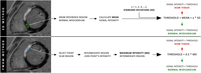
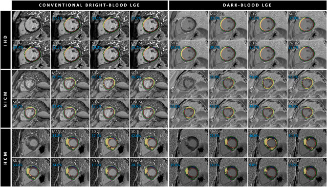
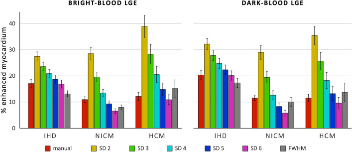
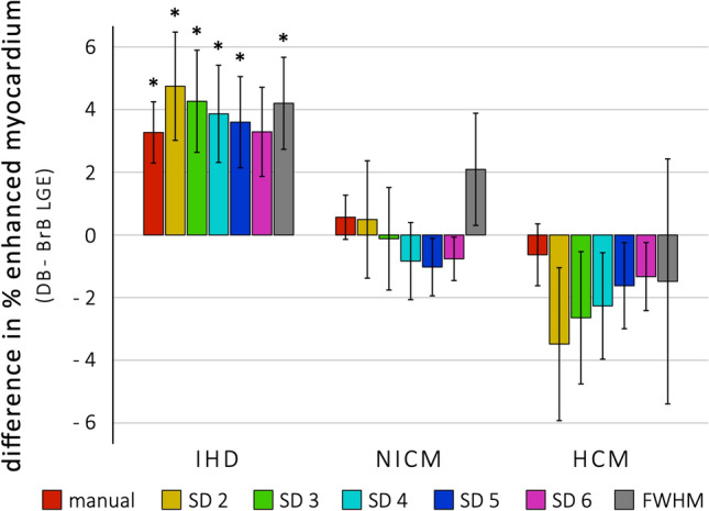
Dark-blood late gadolinium enhancement (LGE) has been shown to improve the visualization and quantification of areas of ischemic scar compared to standard bright-blood LGE. Recently, the performance of various semi-automated quantification methods has been evaluated for the assessment of infarct size using both dark-blood LGE and conventional bright-blood LGE with histopathology as a reference standard. However, the impact of this sequence on different quantification strategies in vivo remains uncertain. In this study, various semi-automated scar quantification methods were evaluated for a range of different ischemic and non-ischemic pathologies encountered in clinical practice. A total of 62 patients referred for clinical cardiovascular magnetic resonance (CMR) were retrospectively included. All patients had a confirmed diagnosis of either ischemic heart disease (IHD; n = 21), dilated/non-ischemic cardiomyopathy (NICM; n = 21), or hypertrophic cardiomyopathy (HCM; n = 20) and underwent CMR on a 1.5 T scanner including both bright- and dark-blood LGE using a standard PSIR sequence. Both methods used identical sequence settings as per clinical protocol, apart from the inversion time parameter, which was set differently. All short-axis LGE images with scar were manually segmented for epicardial and endocardial borders. The extent of LGE was then measured visually by manual signal thresholding, and semi-automatically by signal thresholding using the standard deviation (SD) and the full width at half maximum (FWHM) methods. For all quantification methods in the IHD group, except the 6 SD method, dark-blood LGE detected significantly more enhancement compared to bright-blood LGE (p < 0.05 for all methods). For both bright-blood and dark-blood LGE, the 6 SD method correlated best with manual thresholding (16.9% vs. 17.1% and 20.1% vs. 20.4%, respectively). For the NICM group, no significant differences between LGE methods were found. For bright-blood LGE, the 5 SD method agreed best with manual thresholding (9.3% vs. 11.0%), while for dark-blood LGE the 4 SD method agreed best (12.6% vs. 11.5%). Similarly, for the HCM group no significant differences between LGE methods were found. For bright-blood LGE, the 6 SD method agreed best with manual thresholding (10.9% vs. 12.2%), while for dark-blood LGE the 5 SD method agreed best (13.2% vs. 11.5%). Semi-automated LGE quantification using dark-blood LGE images is feasible in both patients with ischemic and non-ischemic scar patterns. Given the advantage in detecting scar in patients with ischemic heart disease and no disadvantage in patients with non-ischemic scar, dark-blood LGE can be readily and widely adopted into clinical practice without compromising on quantification.
© 2024. The Author(s).
Conflict of interest statement
The authors declare no competing interests.
Figures
Similar articles
-
Clinical value of dark-blood late gadolinium enhancement cardiovascular magnetic resonance without additional magnetization preparation.J Cardiovasc Magn Reson. 2019 Jul 29;21(1):44. doi: 10.1186/s12968-019-0556-1. J Cardiovasc Magn Reson. 2019. PMID: 31352900 Free PMC article.
-
Histopathological validation of semi-automated myocardial scar quantification techniques for dark-blood late gadolinium enhancement magnetic resonance imaging.Eur Heart J Cardiovasc Imaging. 2023 Feb 17;24(3):364-372. doi: 10.1093/ehjci/jeac107. Eur Heart J Cardiovasc Imaging. 2023. PMID: 35723673 Free PMC article.
-
Extracellular volume-guided late gadolinium enhancement analysis for non-ischemic cardiomyopathy: The Women's Interagency HIV Study.BMC Med Imaging. 2021 Jul 27;21(1):116. doi: 10.1186/s12880-021-00649-6. BMC Med Imaging. 2021. PMID: 34315432 Free PMC article.
-
Dark-blood late gadolinium enhancement cardiovascular magnetic resonance for improved detection of subendocardial scar: a review of current techniques.J Cardiovasc Magn Reson. 2021 Jul 22;23(1):96. doi: 10.1186/s12968-021-00777-6. J Cardiovasc Magn Reson. 2021. PMID: 34289866 Free PMC article. Review.
-
Late Gadolinium Enhancement Cardiac Magnetic Resonance Imaging: From Basic Concepts to Emerging Methods.Rofo. 2022 May;194(5):491-504. doi: 10.1055/a-1718-4355. Epub 2022 Feb 23. Rofo. 2022. PMID: 35196714 Review. English.
Cited by
-
Conditions for late gadolinium enhancement MRI in myocardial infarction model rats that better reflect microscopic tissue staining.Sci Rep. 2024 Aug 7;14(1):18308. doi: 10.1038/s41598-024-69540-y. Sci Rep. 2024. PMID: 39112681 Free PMC article.
-
Late-gadolinium enhancement predicts appropriate device therapies in nonischemic recipients of primary prevention implantable cardioverter-defibrillators.Heart Rhythm. 2025 Jan 10:S1547-5271(25)00010-4. doi: 10.1016/j.hrthm.2025.01.003. Online ahead of print. Heart Rhythm. 2025. PMID: 39798682 Free PMC article.
-
Automated cardiovascular MR myocardial scar quantification with unsupervised domain adaptation.Eur Radiol Exp. 2024 Aug 14;8(1):93. doi: 10.1186/s41747-024-00497-3. Eur Radiol Exp. 2024. PMID: 39143405 Free PMC article.
-
Quantification of myocardial oxygen extraction fraction on noncontrast MRI enabled by deep learning.Radiol Adv. 2024 Nov;1(4):umae026. doi: 10.1093/radadv/umae026. Epub 2024 Oct 26. Radiol Adv. 2024. PMID: 40641627 Free PMC article.
-
Diagnostic Modalities in Heart Failure: A Narrative Review.Cureus. 2024 Aug 21;16(8):e67432. doi: 10.7759/cureus.67432. eCollection 2024 Aug. Cureus. 2024. PMID: 39314559 Free PMC article. Review.
References
-
- Holtackers RJ, Van De Heyning CM, Chiribiri A, et al. Dark-blood late gadolinium enhancement cardiovascular magnetic resonance for improved detection of subendocardial scar: A review of current techniques. J. Cardiovasc. Magn. Reson. 2021;23(1):96. doi: 10.1186/s12968-021-00777-6. - DOI - PMC - PubMed
MeSH terms
Substances
Grants and funding
LinkOut - more resources
Full Text Sources
