. 2024 Mar;9(3):801-813.
doi: 10.1038/s41564-024-01607-y. Epub 2024 Mar 5.

FacZ is a GpsB-interacting protein that prevents aberrant division-site placement in Staphylococcus aureus

Affiliations

FacZ is a GpsB-interacting protein that prevents aberrant division-site placement in Staphylococcus aureus

Thomas M Bartlett et al. Nat Microbiol. 2024 Mar.

Abstract

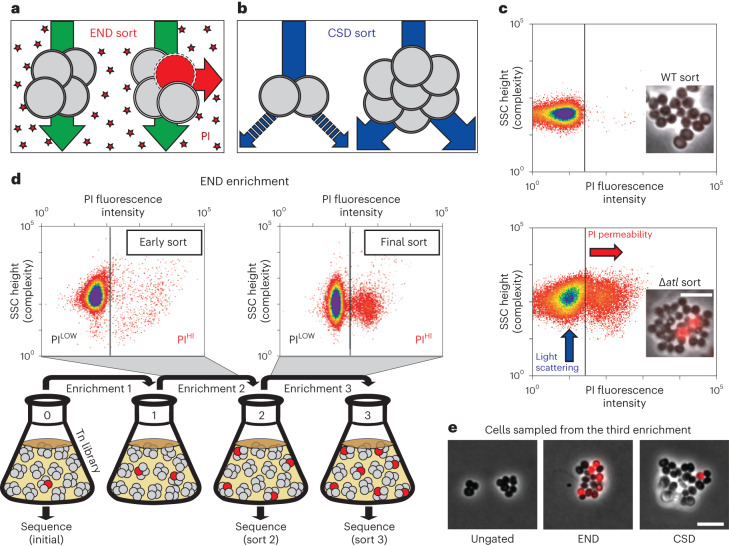
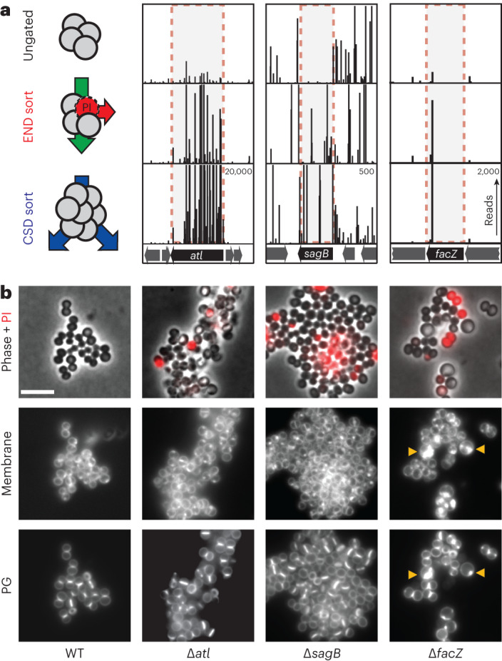
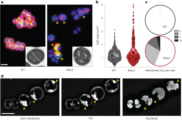
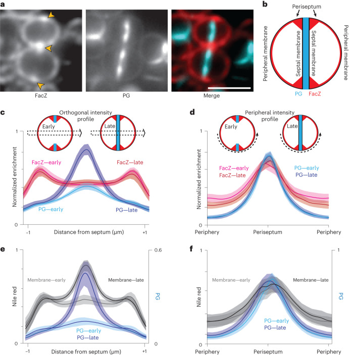
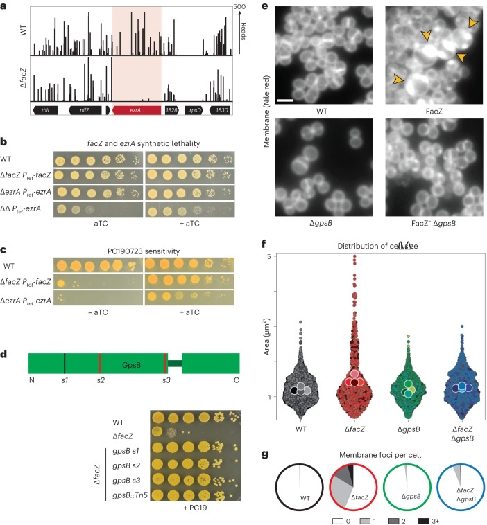
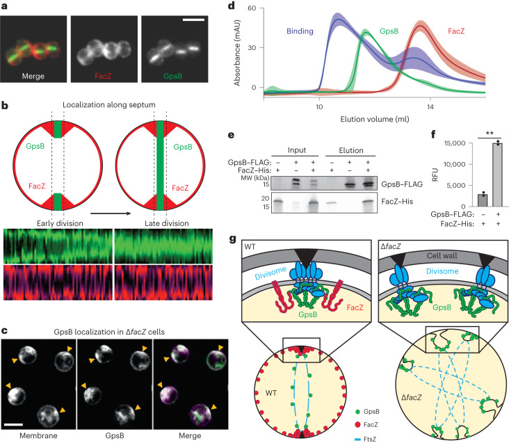
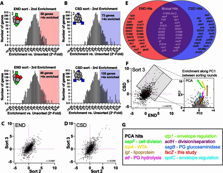
Staphylococcus aureus is a Gram-positive pathogen responsible for antibiotic-resistant infections. To identify vulnerabilities in cell envelope biogenesis that may overcome resistance, we enriched for S. aureus transposon mutants with defects in cell surface integrity or cell division by sorting for cells that stain with propidium iodide or have increased light-scattering properties, respectively. Transposon sequencing of the sorted populations identified more than 20 previously uncharacterized factors impacting these processes. Cells inactivated for one of these proteins, factor preventing extra Z-rings (FacZ, SAOUHSC_01855), showed aberrant membrane invaginations and multiple FtsZ cytokinetic rings. These phenotypes were suppressed in mutants lacking the conserved cell-division protein GpsB, which forms an interaction hub bridging envelope biogenesis factors with the cytokinetic ring in S. aureus. FacZ was found to interact directly with GpsB in vitro and in vivo. We therefore propose that FacZ is an envelope biogenesis factor that antagonizes GpsB function to prevent aberrant division events in S. aureus.


Conflict of interest statement

The authors declare no competing interests.

Figures

Fig. 1
Fig. 1. High-throughput FACS-based enrichments for mutants defective in envelope assembly.
a,b, Schematics showing the logic of the END (a) and CSD (b) enrichments. See text for details. c, FACS profiles monitoring light scattering (SSC height) and PI fluorescence for wild type (WT) and Δatl strains. Inset micrographs show phase contrast images with an overlay of PI staining in red (scale bar = 4 µm; the scale bar applies to all images in c). d, Schematic detailing the END enrichment workflow. An analogous CSD enrichment was performed in parallel (not shown). See text for details. Enrichment for PIHI mutants was examined by FACS profiling at each stage (top). e, The final cell populations (strain aTB015 [RN4220 ∆attB(f11)::Orf5 pTM378]) from the END and CSD sorts, as well as an ungated control, were imaged on 2% agar pads (scale bar = 4 μm; the scale bar applies to all images in e). PI staining (red) was overlaid on the phase contrast image.
Fig. 2
Fig. 2. Validation and initial characterization of screening hits.
a, Tn-seq profiles from three genomic loci showing enrichment of transposon insertions at the completion of the END and CSD sorting protocols relative to the control sort. Each vertical line represents a mapped insertion site, and the height of the line is the number of reads mapping to that site, which reflects the representation of the insertion mutant in the population. Profiles for each locus are scaled separately with the maximum number of reads indicated in the top right corner of the bottom profile. b, Representative images of WT and mutant cells (∆atl [aTP103], ∆sagB [aTB287] and ∆facZ [aTB251]) pulse labelled with sBADA to visualize PG synthesis (bottom row), stained with N,N,N-trimethyl-4-(6-phenyl-1,3,5-hexatrien-1-yl)-benzenaminium, 4-methylbenzenesulfonate (TMA–DPH) to label the cell membrane (‘Membrane’, middle row) and treated with PI to assess envelope permeability (‘Phase + PI’, top row). Yellow arrowheads highlight membrane and PG synthesis defects. The fluorescence intensity for each channel was scaled identically for all strains to facilitate direct comparison between images (scale bar = 4 µm; the scale bar applies to all images in b).
Fig. 3
Fig. 3. Analysis of morphological defects shown by ∆facZ cells.
a, Cells from WT and ∆facZ strains were pulse labelled with sBADA to visualize PG synthesis (green), washed three times with PBS to arrest growth and remove unincorporated sBADA, and then labelled for 5 min with the membrane stain Nile red (red). Cells were imaged on 2% agar pads containing DAPI to visualize the nucleoid (blue). Yellow arrowheads highlight cells with aberrant membranes and PG synthesis. Insets show cells from the same strains imaged by TEM. The bright spots in the ∆facZ mutant are consistent with the cell envelope inclusions, and probably represent cell envelope projections into the cell interior (Supplementary Fig. 3b). b, Violin plots showing the cell area of the indicated strains harbouring a cytoplasmic fluorescent protein. The results from four biological replicates were pooled, each shaded differently; small circles are individual measurements, and large circles are medians from each replicate (WT = 1,760 cells, ∆facZ = 701 cells; Supplementary Fig. 3). c, Quantification of aberrant membrane foci in TMA–DPH-labelled cells (n > 200 cells; Supplementary Fig. 3a). d, 3D-SIM microscopy of ∆facZ cells stained identically to those in a. Yellow arrowheads highlight the position of aberrant cell wall and membrane accumulations that exclude the nucleoid. Scale bars = 2 µm. The scale bars apply to all images in a and d. Source data
Fig. 4
Fig. 4. Localization of FacZ in dividing S. aureus.
a, Representative fluorescence images of WT cells expressing FacZ–mCherry stained with HADA to label PG (scale bar, 2 μm; scale bar applies to all images in a). The protein fusion (left) and PG label (centre) and a pseudo-coloured merged image are shown (FacZ–mCherry in red, HADA in blue) (Extended Data Fig. 5e–g). b, Schematic depicting the membrane localization of FacZ in a dividing S. aureus cell. c, Graphs of mean fluorescence intensity of HADA (light and dark blue) and FacZ–mCherry (light and dark red) collected along lines perpendicular to the septum of cells labelled as in a; shaded areas represent 95% confidence intervals. Central peaks show intensity at the septum, and peripheral peaks show intensity at the cell periphery. Cells were grouped into early division (cells with two HADA foci representing an unresolved septum) or late-stage division (cells with a single central HADA focus representing a resolved septum). HADA labelling is increased in the centre of late-stage-dividing cells (Welch’s t-test, P < 0.05). Localization of FacZ–mCherry does not change significantly as the septum resolves (Kolmogorov–Smirnov test, P > 0.05). d, Graphs of intensity profile scans measuring fluorescence intensity along peripheral arcs centred on the periseptum for early- and late-stage-dividing cells. e,f, Graphs of intensity profile scans of WT cells stained with HADA (light and dark blue) and Nile red (light and dark grey) imaged and analysed as in d and e, respectively. Perpendicular intensity profiles (c and e) were normalized from 0 to 1 for each cell. Peripheral intensity profiles (d and f) were interpolated using MATLAB, and intensity profiles were normalized from 0 to 1 for each fluorescence channel within each experiment, to facilitate comparison of cells of different sizes (c, n = 50 cells; d, n = 100 cells; e, n = 24 cells; f, n = 54 cells). Source data
Fig. 5
Fig. 5. Inactivation of facZ impairs cell division and is rescued by deletion of gpsB.
a, Transposon insertion profiles of the ezrA locus in strains aTB015 [WT] and aTB259 [∆facZ]. b, Spot titre of cultures of aTB003 [WT], aTB372 [∆facZ Ptet-facZ], aTP481 [∆ezrA Ptet-ezrA] and aTB378 [∆facZezrA Ptet-facZ]. Cells were normalized to OD600 = 1.0, serially diluted and spotted on TSA agar with or without an aTC inducer (50 ng ml−1). c, Spot titre of aTB003 [WT], aTB372 [∆facZ Ptet-facZ] and aTP481 [∆ezrA Ptet-ezrA] as in b except that the plates contained PC190723 (100 ng ml−1). d, Top: Diagram showing the location of mutations in gpsB that suppress the PC190723 sensitivity of a ∆facZ mutant. Suppressor mutations are mapped onto a diagram of the two folded domains of GpsB (lines indicate position of mutations, with red lines indicating mutations generating premature stop codons). Bottom: Cultures of strains aTB003 [WT], aTB251 [∆facZ], aTB453 [∆facZ gpsB-s1], aTB476 [∆facZ gpsB-s2], aTB478 [∆facZ gpsB-s3] and aTB497 [∆facZ gpsB::Tn] were OD normalized and spotted on TSA supplemented with PC190723 (100 ng ml−1) as in b. e, Representative fluorescence images of aTB003 [WT], aTB372 [∆facZ Ptet-facZ], aTB525 [∆gpsB] and aTB540 [∆gpsBfacZ Ptet-facZ]. Strains were grown to mid-log phase without induction of facZ, and membranes were stained with Nile red (Extended Data Fig. 6). Yellow arrowheads highlight membrane defects. Scale bar = 2 µm. The scale bar applies to all images in e. f,g, Cultures of aTB521 [WT], aTB527 [∆facZ], aTB529 [∆gpsB] and aTB542 [∆facZgpsB] constitutively expressing cytoplasmic red fluorescent protein from pKK30 were labelled with TMA–DPH in mid-log phase and imaged on 2% agarose pads. f, Violin plots showing the cell area of indicated strains based on cytoplasmic fluorescence (WT, n = 1,760 cells; ∆facZ, n = 701 cells; ∆gpsB, n = 1,544 cells; ∆facZgpsB, n = 1,217 cells). Cell shape was quantified with MicrobeJ (Methods). g, The number of aberrant membrane foci per cell was quantified for display in pie charts (WT, n = 791 cells; ∆facZ, n = 400 cells; ∆gpsB, n = 998 cells; ∆facZgpsB, n = 721 cells). Source data
Fig. 6
Fig. 6. FacZ interacts with GpsB and influences its localization.
a, Representative images of aTB517 [∆facZgpsB Ptet-gpsB-mNeon Pspac-facZ-mCherry] grown in the presence of aTC (50 ng ml−1) and IPTG (25 ng ml−1) and imaged by fluorescence microscopy (scale bar = 2 µm; the scale bar applies to all images in a). b, Graphs showing the fluorescence intensity of FacZ–mCherry (light and dark red) and GpsB–mNeon (light and dark green) along lines parallel with the septum. Cells were separated into two groups based on the GpsB–mNeon signal: cells with discontinuous GpsB foci were considered early-division cells (top left) whereas cells with a continuous GpsB–mNeon band at the septum were considered late-division cells (top right) (n ≥ 50 cells for each group). c, Representative deconvolved fluorescence images of GpsB–mNeon localization in ∆facZ cells (aTB519) labelled with Nile red (scale bar = 2 µm; the scale bar applies to all images in c). d, Gel filtration profiles of GpsB (1–75) alone (green), SUMO-3x-FacZ (127–146) alone (red) or a mixture of the two proteins (binding, blue). Elution profiles represent the average and standard deviation of six runs. e, Lysates were generated from strains expressing tagged variants of FacZ and GpsB [aTB411, 513 and 514]. Membrane fractions were solubilized, loaded onto α-FLAG (DYKDDDDK octamer) magnetic beads, washed and eluted. Input (membrane homogenate) and output (elution) were resolved by sodium dodecyl sulfate–polyacrylamide gel electrophoresis (SDS-PAGE) and blotted using α-His and α-FLAG antibodies (left). f, Fluorescence intensities of the FacZ bands from the blot in e and a replicate experiment (n = 2 biologically independent samples) were measured and relative fluorescence units (RFUs) were plotted (right) (**P = 0.0018). g, Schematic model of FacZ function within the divisome. Cell division is properly localized when FacZ is functional (left). In the absence of FacZ, GpsB is unregulated such that cell constriction is initiated at many sites (right). See text for details. Source data
Extended Data Fig. 1
Extended Data Fig. 1. CSD and END enrichment data.
(a, b) Histograms showing the relative enrichment of transposon insertions in genes following the second and third END (A) or CSD (B) sorting rounds. Colored boxes highlight genes with >4x enrichment in transposon insertions. (c, d) Scatterplots showing relative enrichment at each locus in sort 3 over relative enrichment at the same locus in sort 2 for both the END (C) and CSD (D) screens. Linear regression analysis indicates that relative enrichment is well-correlated between sorting rounds, suggesting enrichment is driven by phenotypic selection and not chance. (e) Venn diagram comparison of the 50 genes in which transposon insertions were most strongly-enriched in the CSD and END sorts. (f) Enrichment in the final CSD sort is moderately correlated with enrichment in the final END sort, consistent with the overlapping mutant populations isolated from the two screens. 2D-PCA of the final sorts identified a vector (PC1, red) that serves as a proxy for enrichment in both sorts, allowing ranking of mutual hits based on enrichment along this vector between rounds of sorting (PCA panel, and see Supplementary Tables 4–5). The PCA pop-out panel (right) shows relative enrichment at each genetic locus in round 2 (light gray) and round 3 (dark gray), rotated so that PC1 is vertical. Colored arrows show the change in enrichment between rounds of sorting for a subset of hits with known roles in envelope biogenesis. Upward movement in this vector space indicates enrichment in both screens. (g) A selection of hits identified in the PCA, many of which are known cell-envelope biogenesis genes. Text colors match with the arrow colors for the given genes in the PCA plot.
Extended Data Fig. 2
Extended Data Fig. 2. Validation and characterization of additional hits from the screen.
a) Volcano plots showing the relative enrichment of transposon insertions in each locus versus the p-value from Mann-Whitney U for the same locus. The final CSD sort (top) and final END sort (middle) are compared to an ungated control generated in parallel. Selected hits (bottom) are color coded and correspond to the colored circle in the plots. b) Representative images of two mutants identified in our screens, derived from the NTML Tn-mutant library. To assess END phenotype (top), mid-log phase cells were washed with PBS and incubated for 5 minutes with PI, then placed on pads containing PI and imaged. To assess CSD phenotype (bottom), mid-log phase cells were pulse-labeled with HADA to visualize peptidoglycan synthesis (PG) (middle row, blue), washed three times with PBS to arrest growth and remove incorporated HADA, and then labeled with WGA Alexa-594 to label teichoic acid (top row, red). Compared to the parental S. aureus USA300 strain [aTB001], Tn-inactivation of USA300 orthologs of SAOUHSC_01974 [aTB112] and SAOUHSC_02383 [aTB113], have increased PI staining and morphological defects (white arrowheads), consistent with END and CSD phenotypes. Scale bars: top = 2 µm, bottom = 1 µm. c) Table showing the fold-change of transposon insertions in the genes inactivated in (B) in the final round of the END and CSD sorts, as well as relative enrichment along PC1. The enrichment percentile of mutations in those genes compared to all mutants is given in parentheses. Source data
Extended Data Fig. 3
Extended Data Fig. 3. Inactivation of FacZ impairs cell division and morphogenesis.
a) Representative images showing membrane labeling and cytoplasmic fluorescence of WT [aTB523] and ∆facZ [aTB527] cells used to quantify morphological and membrane defects in Fig. 3b, c. Cells were grown overnight in the presence of Tmp (5 µg/ml) to maintain the RFP-bearing plasmid, then subcultured into medium free of antibiotics, and grown into mid-log phase. These cells were then labeled with TMA-DPH, imaged on M9 pads (2% agarose), and segmented on cytoplasmic fluorescence signal. Cell size measurements for violin plots (Fig. 3b) were automated using MicrobeJ; red arrowheads point to unusually large cells. For each cell identified by MicrobeJ, the number of aberrant membrane foci (blue arrowheads) was recorded manually (Fig. 3c). b) Z−stack of 3D-SIM reconstruction of ΔfacZ cells [aTB251] stained identically to Fig. 3 shows aberrant features. Red arrowheads follow membrane features (Nile Red, top) through Z−planes (left to right); features are continuous throughout the cell in the Z−dimension, and do not emerge in the middle of the cytoplasm, consistent with these membrane features being continuous invaginations of the cell membrane, rather than completely internalized structures. Analogous continuous features are apparent when imaging labeled cell wall (green arrowheads, sBADA, middle), and correspond to local exclusion of the nucleoid (blue arrowheads, DAPI, bottom), both of which also extend throughout all Z−slices. c) FtsZ-GFP was induced at low levels in exponentially-growing S. aureus to determine which cells were dividing and to determine the orientation of the division plane. In WT S. aureus [aTB219], most cells exhibited a single-ring, consistent with normal cell division. In cells depleted of FacZ [aTB390], aberrant FtsZ structures (defined as multiple Z-structures, drastically off-centre Z-structures, or diffuse cytoplasmic FtsZ-GFP signal) were apparent. A similar phenotype was observed in cells depleted for the division protein EzrA [aTB391]. d) Stacked bar graphs showing the frequency of aberrant FtsZ structures (black) and normal FtsZ structures (gray) from a representative experiment, with horizontal bars indicating significant differences (p-value < 0.001). Left: The division defect associated with inactivation of FacZ was largely rescued in the presence of aTC (25 ng/mL) to induce facZ expression (∆facZ Ptet-facZ vs. ∆ezrA Ptet-ezrA: p = 1.5 × 10−5, chi-squared test); WT: n = 129 cells, ∆ezrA Ptet-ezrA: n = 100 cells; ∆facZ Ptet-facZ: n = 114 cells. Right: Depletion of EzrA or FacZ causes division defects (WT vs. ∆ezrA Ptet-ezrA: p = 2.7 × 10−25; WT vs. ∆facZ Ptet-facZ: p = 1.2 × 10−27; ∆facZ Ptet-facZ vs. ∆ezrA Ptet-ezrA: p = 5.3 × 10−6, chi-squared test); WT: n = 105 cells, ∆ezrA Ptet-ezrA: n = 143 cells; ∆facZ Ptet-facZ: n = 100 cells. e) Micrographs showing localization of divisome PG synthases FtsW-GFP and GFP-Pbp1 expressed from a multicopy pLOW plasmid in WT and ∆facZ cells as described (see Methods). Left: FtsW-GFP localizes to the divisome in WT cells [aTB666] but mislocalizes to the envelope foci characteristic of ∆facZ mutants [aTB675]. Right: similar localization patterns are observed with GFP-Pbp1 in WT [aTB665] and ∆facZ cells [aTB673]. f) WT S. aureus with [aTB341] and without [aTB003] an integrated Ptet-facZ expression construct were imaged alongside ∆facZ cells with [aTB372] and without [aTB251] the same integrated facZ expression construct. Cells were grown to mid-log phase, treated with aTC (25 ng/mL) to induce FacZ expression from the Ptet promoter, and exposed to HADA to label active zones of PG insertion. Only ∆facZ cells lacking the complementing allele exhibited morphological defects (yellow arrowheads). g) Representative full fields of view corresponding to electron micrograph conditions displayed in Fig. 3a insets. (All scale bars = 2 µm). Source data
Extended Data Fig. 4
Extended Data Fig. 4. FacZ structural predictions and functionality of the FacZ-mCherry fusion.
a) Predicted membrane topology of FacZ (Protter). FacZ is predicted to have one transmembrane helix with a short extracellular N-terminal region. Most of the protein is predicted to extend into the cytoplasm, with a coiled-coil region (blue box) and a disordered C-terminal extension (green box). b) AlphaFold2 model of full-length FacZ, colored by pLDDT confidence score. Predicted domains and orientation in the plasma membrane are labeled. c) AlphaFold2-multimer predictions of FacZ assemblies from one copy to six copies. d) A ∆facZ strain harboring facZ-mCherry under control of a Ptet promoter [aTB373] was grown into mid-log phase with 0 or 50 ng/µl aTC, pulse-labeled with HADA as described, and imaged on PBS pads containing 2% agarose. Cells depleted of or overexpressing FacZ-mCherry (red) had aberrant sites of PG synthesis (yellow arrowheads), while aTC (50 ng/mL) induction of facZ-mCherry restored normal morphology and PG incorporation. Insets, bottom right: When FacZ-mCherry is expressed at levels that restore normal cell morphology to ∆facZ cells, the fluorescent fusion is enriched at periseptal regions, flanking actively-growing zones of septal PG synthesis (yellow arrowheads) (scale bar = 2 µm). e) Diagram showing two potential models for FacZ-mCherry localization at the periseptum. FacZ may be enriched at the highly-curved periseptal rim, or may be equally enriched along the curved peripheral surface and extend slightly into the flat septal membrane. f) Schematic showing regions of cell surface used to measure periseptal:peripheral fluorescence ratios. Periseptal regions were defined as the six pixels centred around the septum (the peak of HADA labeling in dividing cells). The peripheral regions were defined as the six pixels half the distance between the periseptum and the ends of hemispherical arcs along which fluorescence intensity was measured. These intermediate regions were chosen because the extreme ends of the hemispherical arcs could display increased membrane abundance due to previously-formed septa in cells with immature division planes, or nascent orthogonal septa in cells with completed division planes. Periseptal enrichment was measured by taking the ratio of periseptal fluorescence intensity to peripheral fluorescence intensity for FacZ-mCherry (2.10-fold periseptal enrichment) or Nile Red (1.79-fold periseptal enrichment), a non-specific membrane label (see Fig. 4). FacZ-mCherry periseptal enrichment was 1.18 times greater than Nile Red periseptal enrichment (Student’s t-test, p = 2.8 × 10-4). g) WT [aTB003], ∆facZ [aTB251], and ∆facZ cells expressing untagged facZ [aTB372] or facZ-mCherry [aTB373] under the control of the Ptet promoter were normalized to OD600 = 1.0, serially diluted, and spotted onto TSA agar plates with aTC (50 ng/mL) and with or without PC190723 (100 ng/mL) as indicated.
Extended Data Fig. 5
Extended Data Fig. 5. FacZ is required for normal envelope biogenesis and cell division in B. subtilis.
a) Dendrogram highlighting the distribution of FacZ (blue) in a broad range of bacterial species, made in AnnoTree, with large monophyletic phyla labeled. b) Dendrogram highlighting the distribution of FacZ orthologs within all monophyletic Firmicute species annotated in AnnoTree, with individual lines representing genera and major families labeled on the periphery of the dendrogram. c) Alignment of FacZ orthologs from selected Firmicutes, with proposed GpsB-interaction motif underlined. Colors indicate residue side-chain properties as standard for ClustalOmega alignments (red = small hydrophobic, blue = acidic, magenta = basic, green = hydroxyl/sulfhydryl/amin, gray = unusual/imino). Asterisk (*) indicates single fully-conserved residue; colon (:) indicates strong similarity. d-g) All B. subtilis microscopy was performed on exponentially-growing derivatives of strain PY79 imaged on agarose pads. All images were scaled identically (scale bar = 4 µm). D) WT [bDR2789] and ∆facZ [bTB039] cells that constitutively express cytoplasmic GFP were labeled with the membrane dye Nile Red. Yellow arrowheads highlight membrane invaginations (membrane, middle) that correspond to local depletions of cytoplasmic GFP (left). These phenotypes are similar to those associated with ∆facZ in S. aureus (Fig. 3). E) WT [bDR2229] and ∆facZ [bTB018] cells expressing FtsZ-GFP from an ectopic locus. Aberrant FtsZ structures are highlighted with yellow arrowheads. F) WT [bDR2637] cells (false-colored cyan) expressing RFP and ∆facZ [bTB040] cells (false-colored magenta) expressing BFP were co-cultured and imaged on the same pads, allowing for direct comparison of cell size. G) Cells were segmented using cytoplasmic fluorescence in MicrobeJ to compare cell morphology. Violin plots show cell length, cell width, and aspect ratio of WT and ∆facZ cells from a representative experiment (WT, n = 202 cells; ∆facZ, n = 68 cells). Dotted lines show median and solid lines show quartiles. h) Cartoon depicting the morphological differences between a typical WT and ∆facZ cell using the median parameters determined in this analysis (WT length = 2.86 µm, width = 0.916 µm; ∆facZ length = 3.97 µm, width = 1.00 µm). i) Spot titer of B. subtilis strains (WT [bDR2660], ∆facZ [bTB039], ∆gpsB [bTB044], and ∆facZgpsB [bTB041]) were grown into exponential phase as described, serially diluted, and plated on a range of PC190723 concentrations. Source data
Extended Data Fig. 6
Extended Data Fig. 6. ∆gpsB corrects the morphological defects of ∆facZ mutants.
a) Representative images of WT (aTB003), ∆gpsB (aTB525), FacZ- (aTB372, ∆facZ Ptet -facZ), and FacZ-gpsB (aTB540, ∆facZ Ptet -facZgpsB) grown into mid-log phase without induction of facZ, and pulse-labeled with sBADA to label active PG insertion and Nile Red to label cell membranes (see Fig. 5e). Inactivation of gpsB has minimal impact on cell morphology or localization of envelope probes. Depletion of FacZ causes characteristic envelope and morphological defects (yellow arrowheads), which are rescued by inactivation of gpsB (bar = 2 µm). b) Spot titers of the same strains imaged in panel A confirms that depletion of FacZ and/or inactivation of gpsB has negligible impact on cell viability. c) Growth curves of WT (aTB003), ∆facZ [aTB251], ∆gpsB [aTB492], and ∆facZgpsB [aTB497], grown at 30 C. Values are means of six biological replicates (error bars = 95% CI). Inactivation of FacZ or GpsB causes minor growth defects, while the double mutant is largely restored to WT growth rates. d) Median cell size and coefficient of variance of cell size (CoeV) of the indicated strains from replicated experiments (see pooled data in Fig. 5f). Graphs display mean values from four biological replicates, with individual replicate values included as circles, and bars showing standard error. Inactivation of gpsB alone has no significant impact on size or variability, but rescues defects caused by depletion of FacZ (Student’s t-test, p = 0.01). e–g) Cells were imaged to measure the localization of FacZ and GpsB in different stages of cell division (see Fig. 6a, b). E) Graphs showing the fluorescence intensity of FacZ-mCherry (light and dark red) and GpsB-mNeon (light and dark green) along lines parallel to the septum. Cells were separated into two groups based on the GpsB-mNeon signal: cells with discontinuous GpsB foci were considered early-division cells (top left) whereas cells with a continuous GpsB-mNeon band at the septum were considered late-division cells (top right). (n ≥ 50 cells for each group). F) Graph showing the linear regression of the inter-peak distance of FacZ (red) and GpsB (green) in dividing cells (error bars = 95% CI). Cells were sorted by decreasing inter-peak distance of GpsB, so that measurements are pairwise for individual cells. FacZ and GpsB overlap in early division, but FacZ is left behind at the periseptum as GpsB departs with the closing divisome. G) Violin plots of raw interpeak distance of FacZ in early and late division show no significant change in FacZ localization between early and late division (n = 50 cells). H) Spot titers testing the synthetic interactions of facZ, gpsB, and ezrA. FacZ [aTB378] and GpsB [aTB663] were inactivated separately and together [aTB679] in an EzrA depletion strain [aTB264], and spot titers prepared with or without aTC as in Fig. 4b. Inactivation of facZ and ezrA is synthetically lethal, while inactivation of gpsB and ezrA is not. Inactivation of gpsB somewhat, but not completely, restores growth in a ∆facZ EzrA- strain. I) A plasmid containing Ptet-gpsB was integrated into WT [aTB632] or ∆facZ S. aureus [aTB639], and cell viability was assayed by spot titer alongside their parental strains lacking Ptet-gpsB [aTB003 & aTB251] as described (see Methods, Fig. 4b). Source data
Extended Data Fig. 7
Extended Data Fig. 7. A C-terminal motif in FacZ is required for function and interaction with GpsB.
a, b) Gel filtration assays testing FacZ-GpsB interactions. Left: Binding assays performed with SUMO-3x-FacZ (127–146) containing the native binding motif (NRHYRR); Right: FacZ Binding assays performed with SUMO-3x-FacZ (127–146) containing an altered motif (NDHYDD). A) Gel filtration profiles of GpsB (1–75) alone (green), SUMO-3x-FacZ (127–146) alone (red), or a mixture of the two proteins (binding, blue). The sum of GpsB (1–75) alone (green) and SUMO-3x-FacZ (127-146) is shown in gray for display purposes. Elution profiles are representative of 6 runs. B) Representative SDS-PAGE gels of fractions from the size-exclusion column from runs with FacZ alone (top), GpsB alone (middle), and a mixture of both proteins (bottom). c) Diagram of FacZ C-terminus with proposed GpsB-interaction motif (yellow box) and the location of four arginine residues (bolded red R). d) Various R → D FacZ alleles were expressed from Plac on a pLOW vector in a ∆facZ background of strain RN4220, and then plated with or without PC19 as indicated. Disruption of R1 [aTB568], R2 [aTB570], or R3 [aTB572] alone had little impact on growth on PC19, while inactivation of all three together (3R → 3D) [aTB565] did not enable growth on PC19. Disruption of R4 [aTB574], outside of the proposed binding motif, had no impact on growth on PC19. e) Western blot of native FacZ [aTB347], FacZ-6x-His [aTB349], and FacZDDD (3R → 3D) [aTB651] expressed from a pLOW vector. f) Full blots of FacZ-His and GpsB-FLAG pulldowns from two representative replicates (see Fig. 6e, f). Source data

Update of

References

    1. Silhavy TJ, Kahne D, Walker S. The bacterial cell envelope. Cold Spring Harb. Perspect. Biol. 2010;2:a000414. doi: 10.1101/cshperspect.a000414. - DOI - PMC - PubMed
    1. Koch AL. Growth and form of the bacterial cell wall. Am. Sci. 1990;78:327–341.
    1. Loomba PS, Taneja J, Mishra B. Methicillin and vancomycin resistant S. aureus in hospitalized patients. J. Glob. Infect. Dis. 2010;2:275–283. doi: 10.4103/0974-777X.68535. - DOI - PMC - PubMed
    1. Rohs PDA, Bernhardt TG. Growth and division of the peptidoglycan matrix. Annu. Rev. Microbiol. 2021;75:315–336. doi: 10.1146/annurev-micro-020518-120056. - DOI - PubMed
    1. Oshida T, et al. A Staphylococcus aureus autolysin that has an N-acetylmuramoyl-L-alanine amidase domain and an endo-B-N-acetylglucosaminidase domain: cloning, sequence analysis, and characterization. Proc. Natl Acad. Sci. USA. 1994;92:285–289. doi: 10.1073/pnas.92.1.285. - DOI - PMC - PubMed