. 2024 Mar 5;22(1):241.
doi: 10.1186/s12967-024-04973-7.

Comparing anti-tumor and anti-self immunity in a patient with melanoma receiving immune checkpoint blockade

Affiliations

Comparing anti-tumor and anti-self immunity in a patient with melanoma receiving immune checkpoint blockade

Shuming Chen et al. J Transl Med. .

Abstract

Background: Tumor regression following immune checkpoint blockade (ICB) is often associated with immune-related adverse events (irAEs), marked by inflammation in non-cancerous tissues. This study was undertaken to investigate the functional relationship between anti-tumor and anti-self immunity, to facilitate irAE management while promoting anti-tumor immunity.

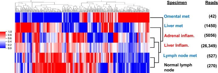
Methods: Multiple biopsies from tumor and inflamed tissues were collected from a patient with melanoma experiencing both tumor regression and irAEs on ICB, who underwent rapid autopsy. Immune cells infiltrating melanoma lesions and inflamed normal tissues were subjected to gene expression profiling with multiplex qRT-PCR for 122 candidate genes. Subsequently, immunohistochemistry was conducted to assess the expression of 14 candidate markers of immune cell subsets and checkpoints. TCR-beta sequencing was used to explore T cell clonal repertoires across specimens.

Results: While genes involved in MHC I/II antigen presentation, IFN signaling, innate immunity and immunosuppression were abundantly expressed across specimens, irAE tissues over-expressed certain genes associated with immunosuppression (CSF1R, IL10RA, IL27/EBI3, FOXP3, KLRG1, SOCS1, TGFB1), including those in the COX-2/PGE2 pathway (IL1B, PTGER1/EP1 and PTGER4/EP4). Immunohistochemistry revealed similar proportions of immunosuppressive cell subsets and checkpoint molecules across samples. TCRseq did not indicate common TCR repertoires across tumor and inflammation sites, arguing against shared antigen recognition between anti-tumor and anti-self immunity in this patient.

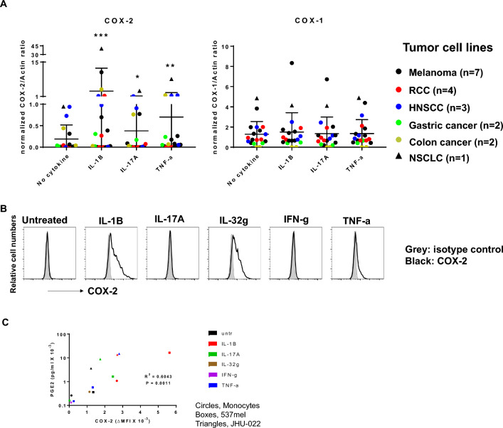
Conclusions: This comprehensive study of a single patient with melanoma experiencing both tumor regression and irAEs on ICB explores the immune landscape across these tissues, revealing similarities between anti-tumor and anti-self immunity. Further, it highlights expression of the COX-2/PGE2 pathway, which is known to be immunosuppressive and potentially mediates ICB resistance. Ongoing clinical trials of COX-2/PGE2 pathway inhibitors targeting the major COX-2 inducer IL-1B, COX-2 itself, or the PGE2 receptors EP2 and EP4 present new opportunities to promote anti-tumor activity, but may also have the potential to enhance the severity of ICB-induced irAEs.

Keywords: COX-2; Gene expression profiling (GEP); Immune checkpoint blockade (ICB); Immune related adverse events (irAEs); Melanoma; Rapid autopsy; Tumor infiltrating lymphocytes (TILs).


Conflict of interest statement

LCC is a consultant for Amgen, Bristol Myers Squibb, and Mallinckrodt, and receives research funding from Bristol Myers Squibb. RAA receives research support from Bristol Myers Squibb and RAPT Therapeutics, and serves on advisory boards or consults for AstraZeneca, Bristol Myers Squibb, and Merck SD. EJL is an advisor/consultant for Bristol Myers Squibb, CareDX, Eisai, Genentech, HUYA Bioscience International, Immunocore, Instil Bio, Merck, Natera, Nektar, Novartis, Oncosec, Pfizer, Rain Therapeutics, Regeneron, Replimune, and Sanofi, and has research funding from Bristol Myers Squibb, Haystack Oncology, Merck, Regeneron, and Sanofi. DMP and SLT receive consulting fees from Amgen, Bristol Myers Squibb, Compugen, Dragonfly Therapeutics, Janssen Pharmaceuticals, Normunity, PathAI, RAPT Therapeutics, Regeneron, Takeda Pharmaceuticals, and Tizona LLC; receive research grants from Bristol Myers Squibb, Compugen, and Immunomic Therapeutics; have stock options or stock in Atengen Inc., DNAtrix, Dracen, Dragonfly Therapeutics, ManaT Bio, RAPT Therapeutics, and Tizona LLC; and have patents related to T-cell regulatory molecules including LAG-3, and the treatment of MSI-high cancers with anti-PD-1. JMT reports research grants and consulting fees from Akoya Biosciences and Bristol Myers Squibb; consulting for AstraZeneca, Compugen, Genentech, GlaxoSmithKline, Lunaphore, Merck, Regeneron and Roche; and an institutional patent filed on machine learning for scoring pathologic response to immunotherapy. TLM, AS, FS, J-WS, LAC-R, IRM, PS, AEB, JSD, QCZ, and JEH declare no potential conflicts of interest.

Figures

Fig. 1
Fig. 1
Clinical timeline and tissue specimens. A, Melanoma course, occurrence of irAEs, and specimens collected from patient MA-6. The top timeline shows tissue collections; the middle timeline shows melanoma status and systemic therapies (not including focal radiotherapy); the bottom timeline shows the irAEs and their pharmacologic management. Not included in the bottom timeline is the onset of grade 2 adrenal insufficiency of clinically unclear etiology, treated with hydrocortisone replacement. IFN-a, adjuvant interferon alfa. B, Tissue specimens from patient MA-6 that were used in this study. Biopsy and surgical specimens were collected pre-mortem, others were collected at autopsy. Green boxes, assays performed; black boxes, assays not done. IHC immunohistochemistry; met, metastasis, qRT-PCR quantitative real-time reverse transcription polymerase chain reaction, TCRseq T-cell receptor sequencing
Fig. 2
Fig. 2
Similarities and differences in gene expression by immune cells infiltrating inflamed tissues vs melanoma metastases. A, LCM of immune cells infiltrating regions of normal tissue inflammation and cancer in a post-mortem liver specimen. Yellow circles, dissected areas containing inflammatory infiltrates in the non-cancerous portion of the liver (specimen I-5); green circles, dissected areas containing tumor infiltrating immune cells in a metastatic melanoma deposit (specimen T-4). H&E, hematoxylin and eosin staining. Black bars, 100 µm. B, Unsupervised hierarchical clustering of PTPRC-normalized Ct values reveals similarities in expression of many genes across all samples. Expression of 122 candidate genes was evaluated with multiplex qRT-PCR. PTPRC-normalized Cts (Ctgene – CtPTPRC) were clustered and visualized by heat map using Java TreeView. Pink or green colors indicate genes expressed abundantly or poorly, respectively, in the samples. Clustering reveals a high degree of similarity between a lymph node metastasis and a normal lymph node specimen, but does not differentiate the 6 remaining specimens including 2 melanoma metastases and 4 non-cancerous inflamed tissues. C, Volcano plot reveals groups of genes with related functions upregulated in 4 inflamed normal tissues vs 3 tumor specimens. PTPRC-normalized Ct values were used to calculate fold changes of gene expression. Red dots in the upper left region of the plot indicate genes significantly upregulated (fold change magnitude ≥ 2.0, and Welch’s t-test 2-sided p-value ≤ 0.10) in inflamed normal tissues. Genes related to immunosuppressive cell types and cytokines are indicated by magenta boxes, Tregs by blue boxes, COX-2/PGE2 pathway by green boxes, and lymphocyte activation by peach boxes. Black dots in the lower half of the plot indicate genes without differential expression. No genes were upregulated in tumor samples. Genes that were undetectable in any specimen (IL4, IL17A, IL22, and IL23A) were omitted
Fig. 3
Fig. 3
DeepTCR analysis reveals relatedness of T-cell repertoires in two inflammation samples. Summarized counts of productive TCR clones that translate into the same amino acid sequences were used for DeepTCR analysis. Unsupervised clustering of 256 normalized structural concepts is shown. Samples with less than 500 reads should be interpreted with caution. Blue text, tumor samples; red text, inflamed normal tissues
Fig. 4
Fig. 4
COX-2 expression by tumor and immune cells correlates with PGE2 secretion. A, In human tumor cells, cytokines significantly induce COX-2 but not COX-1 expression across cancer types. 19 tumor lines from 6 cancer types were cultured with the indicated cytokines for 1 day. COX proteins were detected by Western blotting. Band intensities were quantified and normalized as described in Methods. Means ± SD are indicated. Wilcoxon signed-rank test, 2-sided comparisons to no cytokine, *p = 0.030, **p = 0.005, ***p = 0.001. Similar results were observed with intracellular flow cytometry (not shown). B, Cytokine induction of COX-2 in human monocytes. Peripheral blood monocytes from a normal donor were exposed to the indicated cytokines in vitro for 1 day. COX-2 expression was assessed by intracellular FACS. Data representative of 2 experiments with monocytes from 3 normal donors. C, COX-2 expression by tumor cells and monocytes correlates with PGE2 secretion. 537mel, JHU-022 and monocytes were treated with the indicated cytokines for 1 day. COX-2 expression was quantified by intracellular FACS and PGE2 secretion was measured by ELISA. Pearson correlation, 2-sided p-value

References

    1. Kawakami Y, Eliyahu S, Delgado CH, Robbins PF, Sakaguchi K, Appella E, et al. Identification of a human melanoma antigen recognized by tumor-infiltrating lymphocytes associated with in vivo tumor rejection. Proc Natl Acad Sci USA. 1994;91(14):6458–6462. doi: 10.1073/pnas.91.14.6458. - DOI - PMC - PubMed
    1. Topalian SL, Drake CG, Pardoll DM. Immune checkpoint blockade: a common denominator approach to cancer therapy. Cancer Cell. 2015;27(4):450–461. doi: 10.1016/j.ccell.2015.03.001. - DOI - PMC - PubMed
    1. Weber JS, Hodi FS, Wolchok JD, Topalian SL, Schadendorf D, Larkin J, et al. Safety profile of nivolumab monotherapy: a pooled analysis of patients with advanced melanoma. J Clin Oncol. 2017;35(7):785–792. doi: 10.1200/JCO.2015.66.1389. - DOI - PubMed
    1. Makohon-Moore AP, Lipson EJ, Hooper JE, Zucker A, Hong J, Bielski CM, et al. The genetic evolution of treatment-resistant cutaneous, acral, and uveal melanomas. Clin Cancer Res. 2021;27(5):1516–1525. doi: 10.1158/1078-0432.CCR-20-2984. - DOI - PMC - PubMed
    1. Duffield AS, Ascierto ML, Anders RA, Taube JM, Meeker AK, Chen S, et al. Th17 immune microenvironment in Epstein-Barr virus-negative Hodgkin lymphoma: implications for immunotherapy. Blood Adv. 2017;1(17):1324–1334. doi: 10.1182/bloodadvances.2017007260. - DOI - PMC - PubMed

Publication types