. 2024 Mar 5;12(1):32.
doi: 10.1186/s40364-024-00576-y.

Combined TP53 status in tumor-free resection margins and circulating microRNA profiling predicts the risk of locoregional recurrence in head and neck cancer

Affiliations

Combined TP53 status in tumor-free resection margins and circulating microRNA profiling predicts the risk of locoregional recurrence in head and neck cancer

Federica Ganci et al. Biomark Res. .

Erratum in

Abstract

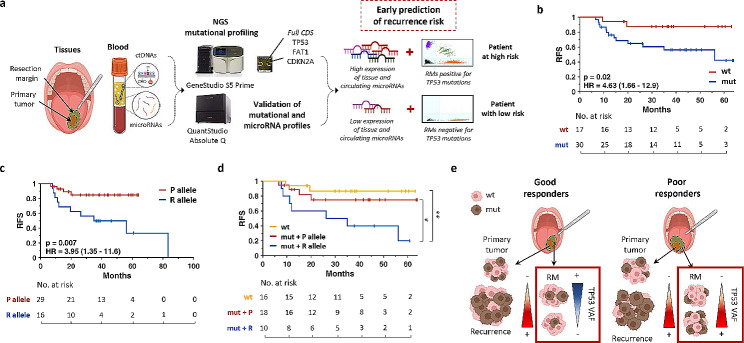
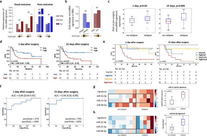
Locoregional recurrences represent a frequently unexpected problem in head and neck squamous cell carcinoma (HNSCC). Relapse often (10-30%) occurs in patients with histologically negative resection margins (RMs), probably due to residual tumor cells or hidden pre-cancerous lesions in normal mucosa, both missed by histopathological examination. Therefore, definition of a 'clean' or tumor-negative RM is controversial, demanding for novel approaches to be accurately explored. Here, we evaluated next generation sequencing (NGS) and digital PCR (dPCR) as tools to profile TP53 mutational status and circulating microRNA expression aiming at scoring the locoregional risk of recurrence by means of molecular analyses. Serial monitoring of these biomarkers allowed identifying patients at high risk, laying the ground for accurate tracking of disease evolution and potential intensification of post-operative treatments. Additionally, our pipeline demonstrated its applicability into the clinical routine, being cost-effective and feasible in terms of patient sampling, holding promise to accurately (re)-stage RMs in the era of precision medicine.

Keywords: HNSCC; Liquid biopsy; Local recurrence; Resection margins; TP53; microRNA profiling.


Conflict of interest statement

The authors declare that they have no known competing financial interests or personal relationships that could influence this paper. However, it has to be stated that both the custom mutational panel including the full coding sequence of TP53, CDKN2A and FAT1 and the 4-microRNAs signature have been patented by B.G., G.F., S.F., (#102020000017896) and by B.G., G.F., M.V and P.R., (#102020000019024), respectively.

Figures

Fig. 1
Fig. 1
Molecular profiling of RMs and patient outcome. (a) Model of liquid biopsy (LB) and tissues analyses for early prediction of recurrence. TP53 mutational status and expression of the prognostic microRNAs signature have been assessed in resection margins (RMs) and LB samples from representative HNSCC patients by NGS, qPCR and dPCR. The combination of TP53 status and microRNAs expression in histologically tumor-free RMs and in sera samples taken at different time points (i.e., before and after surgery) may early predict tumor persistence or the risk of local recurrence in HNSCC. (b-c) Kaplan-Meier (KM) analyses of RMs according to (b) TP53 mutational status (wt or mutated, red or blue, respectively) or (c) the TP53 p.P72R polymorphism (P or R allele, red or blue, respectively). P72-positive RMs includes patients with P allele VAF > 75% while R72-positive RMs contains samples harboring heterozygous P/R or homozygous R alleles. CI values (95%) are shown within parenthesis. (d) Merged KM analyses resulting from TP53 mutational status and P72R polymorphism. (e) Representative model of TP53 abundance (VAF, brown cells) dynamics in tumor and resection margins. CDS: coding sequence; HR: hazard ratio; RFS: recurrence-free survival; VAF: variant allele frequency; wt: wild type; mut = mutated. *: p = 0.06; **: p = 0.006
Fig. 2
Fig. 2
Prognostic value of circulating microRNAs and TP53 mutational status of matched RMs. (a) A 4-prognostic microRNA signature was assessed on RMs collected at disease onset (RMp) or relapse (RMr) and compared with tumor relapse. Patients were stratified according to their clinical outcome. Relative expression of microRNAs is shown. Dynamics of TP53 VAF is indicated. (b) The same microRNA signature was assessed on tissues from pt#3 (left) and pt#2 (right). For pt#3, the RM of the first primitive tumor (RMp#1) developed on the palatine tonsil, on which he recurred, was compared with the RM of the second primitive tumor (RMp#2) on floor of the mouth, on which the patient has ever not recurred. For pt#2, RMp has been compared with the PHE lesion, developed one year before the first recurrence. Relative expression of microRNAs is shown. Dynamics of TP53 VAF is indicated. (c-d) Box plot and KM analysis showing the diagnostic and prognostic value of our circulating microRNA signature to early predict local recurrence. microRNAs expression of sera collected at 1 day or 15 days post-surgery has been normalized to microRNAs expression of matched pre-surgery sera. (e-f) KM and ROC curve analyses according to mutational status of RMs and microRNAs signature expression at 1 day (left) or 15 days post-surgery (right). For each KM, HR value and the relative confident interval (CI) 95% has been indicated. (g) Supervised clustering (left) analysis representing the expression of the 3 prognostic microRNAs in normal tissues from 13 HNSCC patients cultured with CM from Cal27 cells according to patient’s outcome. Colors represent folds of modulation of CM vs RPMI. Box plot (right) representing the expression level of miR-21-3p and miR-21-5p significantly (p=0.02) up-regulated in histologically tumor-free tissues from n=5 recurrent patients vs n=8 patients with no evidence of disease (NED) for at least 36 months cultured with CM as compared to the same tissues cultured with RPMI. (h) Supervised clustering (left) analysis representing the expression of the 3 prognostic microRNAs in normal tissues from 13 HNSCC patients cultured with RPMI medium according to their clinical outcome. Box plot (right) showing miR-96-5p up-regulation (p=0.04) in histologically tumor-free tissues from n=5 recurrent patients vs n=8 patients with NED for at least 36 months, cultured in the presence of RPMI. Raw data of miRNAs expression and the relative patient outcome are available in Suppl. Tables, sheet 5-6. HR: hazard ratio; NED: no evidence of the disease; Rec: recurrence; RFS: recurrence-free survival.

Similar articles

Cited by

References

    1. Brouwer de Koning SG, Schaeffers A, Schats W, van den Brekel MWM, Ruers TJM, Karakullukcu MB. Assessment of the deep resection margin during oral cancer surgery: a systematic review. Eur J Surg Oncol. 2021;47(9):2220–32. doi: 10.1016/j.ejso.2021.04.016. - DOI - PubMed
    1. Marret G, Bièche I, Dupain C, Borcoman E, du Rusquec P, Ricci F, et al. Genomic alterations in Head and Neck squamous cell carcinoma: level of evidence according to ESMO Scale for clinical actionability of molecular targets (ESCAT) JCO Precision Oncol. 2021;5:215–26. doi: 10.1200/PO.20.00280. - DOI - PubMed
    1. Cho J, Johnson DE, Grandis JR. Therapeutic implications of the Genetic Landscape of Head and Neck Cancer. Semin Radiat Oncol. 2018;28(1):2–11. doi: 10.1016/j.semradonc.2017.08.005. - DOI - PMC - PubMed
    1. Comprehensive genomic characterization Of head and neck squamous cell carcinomas. Nature. 2015;517(7536):576–82. doi: 10.1038/nature14129. - DOI - PMC - PubMed
    1. Moe SE, Erland FA, Fromreide S, Lybak S, Brydoy M, Dongre HN et al. The TP53 Codon 72 Arginine Polymorphism Is Found with Increased TP53 Somatic Mutations in HPV(-) and in an Increased Percentage among HPV(+) Norwegian HNSCC Patients. Biomedicines. 2023;11(7). - PMC - PubMed