. 2024 Jul 16;123(14):2110-2121.
doi: 10.1016/j.bpj.2024.03.004. Epub 2024 Mar 5.

Distinct potassium channel types in brain capillary pericytes

Affiliations

Distinct potassium channel types in brain capillary pericytes

Maria Sancho et al. Biophys J. .

Abstract

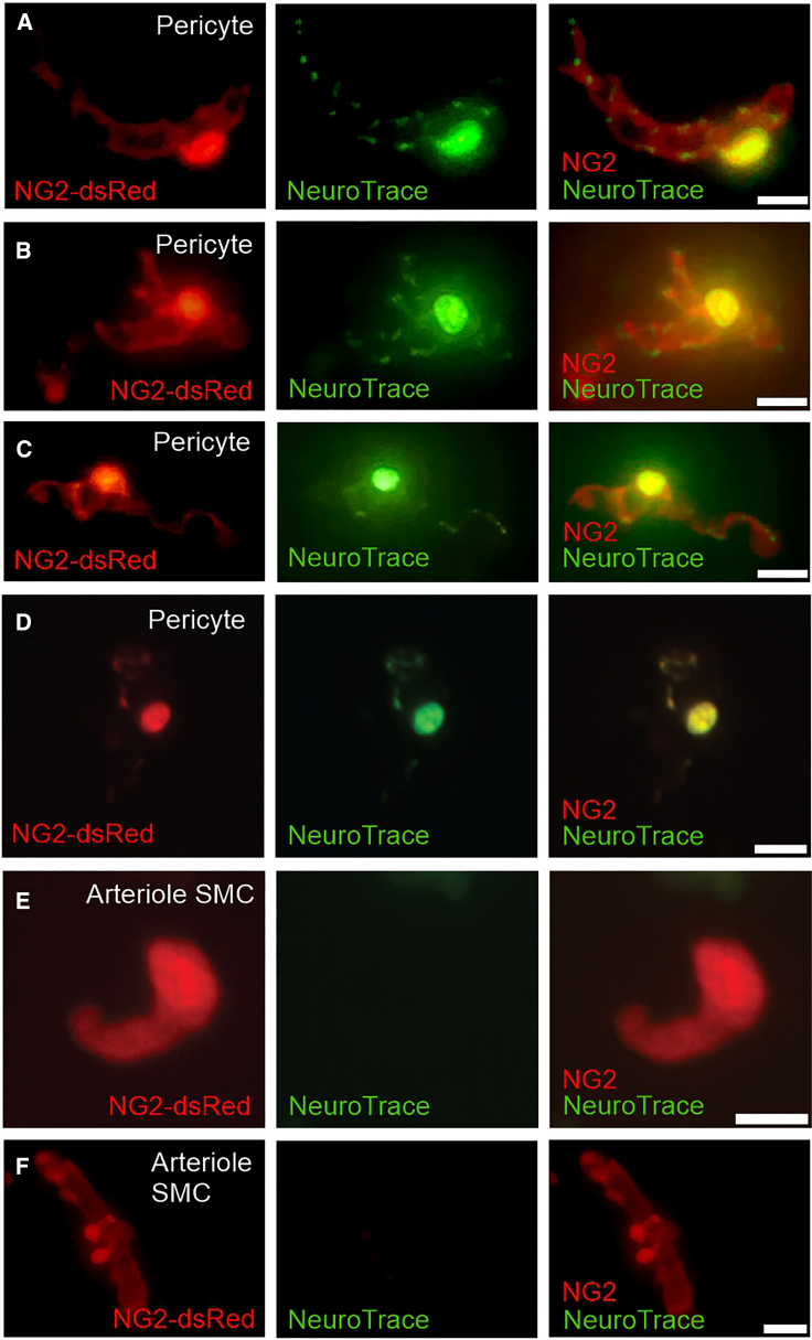
Capillaries, composed of electrically coupled endothelial cells and overlying pericytes, constitute the vast majority of blood vessels in the brain. The most arteriole-proximate three to four branches of the capillary bed are covered by α-actin-expressing, contractile pericytes. These mural cells have a distinctive morphology and express different markers compared with their smooth muscle cell (SMC) cousins but share similar excitation-coupling contraction machinery. Despite this similarity, pericytes are considerably more depolarized than SMCs at low intravascular pressures. We have recently shown that pericytes, such as SMCs, possess functional voltage-dependent Ca2+ channels and ATP-sensitive K+ channels. Here, we further investigate the complement of pericyte ion channels, focusing on members of the K+ channel superfamily. Using NG2-DsRed-transgenic mice and diverse configurations of the patch-clamp technique, we demonstrate that pericytes display robust inward-rectifier K+ currents that are primarily mediated by the Kir2 family, based on their unique biophysical characteristics and sensitivity to micromolar concentrations of Ba2+. Moreover, multiple lines of evidence, including characteristic kinetics, sensitivity to specific blockers, biophysical attributes, and distinctive single-channel properties, established the functional expression of two voltage-dependent K+ channels: KV1 and BKCa. Although these three types of channels are also present in SMCs, they exhibit distinctive current density and kinetics profiles in pericytes. Collectively, these findings underscore differences in the operation of shared molecular features between pericytes and SMCs and highlight the potential contribution of these three K+ ion channels in setting pericyte membrane potential, modulating capillary hemodynamics, and regulating cerebral blood flow.


Conflict of interest statement

Declaration of interests The authors declare no competing interests.

Figures

Figure 1
Figure 1
Experimental approach for isolating native pericytes from brain capillaries for use in patch-clamp electrophysiology. Pericytes were isolated from NG2-DsRed-BAC transgenic mouse brains by mechanical disruption and enzymatic dissociation. A small portion of the somatosensory cortex of the mouse brain was dissected and cut into small fragments using sharp scissors. The tissue fragments were then transferred to isolation solution and exposed to a two-step digestion process consisting of a 17-min incubation in isolation solution containing enzyme 1 (37°C) followed by a 12-min incubation with enzyme 2. The brain suspension was then triturated to yield single cells and incubated for an additional 12 min at 37°C. The cell suspension was filtered through a 62-μm nylon mesh and diluted by adding 3 mL of cold isolation solution. The resulting single brain capillary pericytes, identified by their DsRed fluorescence, were then ready for patch-clamp analyses.
Figure 2
Figure 2
NeuroTrace 500/525 dye labels freshly isolated NG2-expressing capillary pericytes. Images of freshly isolated brain capillary pericytes were captured from NG2-DsRed-BAC transgenic mice using a digital camera attached to the microscope (AC, F) or by z stack imaging using anupright spinning-disk confocal microscope (D and E). (AD) Robust overlap between the DsRed fluorescence of NG2-expressing capillary pericytes and NeuroTrace 500/525 staining. NeuroTrace labeling appeared bright and intense in the cell soma and along the processes of the pericytes, displaying a punctuate pattern. (E and F) Ring-like parenchymal arteriole NG2-positive SMCs with absent NeuroTrace dye labeling. Each image is representative of images acquired from at least five isolated pericytes from at least three different mice. Scale bars, 10 μm (AE) and 20 μm (F).
Figure 3
Figure 3
Brain capillary pericytes express functional strong inwardly rectifying K+ (Kir2) channels. (A) Whole-cell K+ currents recorded in isolated pericytes in the absence (black trace) and presence (red trace) of 100 μM Ba2+ showing the large inward component at voltages negative to EK (−23 mV) and its effective block by Ba2+. A modest Ba2+-insensitive outward component was present at more depolarized voltages (positive to EK). Cells were bathed in high [K+], and currents were elicited by a 400-ms voltage ramp from −140 to +40 mV (upper inset). (B) Ba2+-subtracted currents (green trace), revealing the classic profile of a strongly rectifying K+ current. (C) Summary data comparing peak (−140 mV) inward current density (pA/pF) of control, Ba2+ (100 μM), and Ba2+-sensitive currents (n = 15 cells from seven mice). Dotted line represents zero current level. Values are presented as means ± SD (p < 0.05; paired t-test). External and internal [K+] were 60 and 140 mM, respectively.
Figure 4
Figure 4
Functional voltage-gated K+ (KV) channels are present in brain capillary pericytes. Original traces of whole-cell K+ currents recorded from an isolated brain pericyte before (A; black trace) and after (B; red trace) treatment with 4-AP (1 mM). Currents were elicited by voltage steps from −70 to +70 mV from a holding potential of −50 mV. (C) 4-AP-sensitive currents (dark yellow trace) obtained after subtracting the original recordings plotted in (A) and (B) indicating the presence of functional KV channels in brain pericytes. (D) Original traces of whole-cell currents elicited by a voltage step to +70 mV from holding potential of −50 mV in the absence (black trace) and presence (red trace) of the KV1.2/1.5 inhibitor 4-AP (1 mM). (E) Whole-cell current-voltage relationship, measured at peak outward current at each test potential corresponding to control, 4-AP-treated, and 4-AP-sensitive currents (n = 8 cells from three mice). (F) Activation time constants (τ activation) obtained from an exponential fit of individual voltage-evoked (20-mV pulses) current recordings obtained before (black trace) and after (red trace) application of 1 mM 4-AP (n = 8 cells from three mice). (G) Steady-state activation properties of KV currents (n = 8 cells from three mice). The half-maximal activation voltage (V0.5) was determined from a fit of the data to the Boltzmann equation. Dotted line represents zero current level. Values are presented as means ± SD (p < 0.05; Wilcoxon matched-pairs signed-rank test). External and internal [K+] were 6 and 140 mM, respectively.
Figure 5
Figure 5
Brain capillary pericytes possess large-conductance Ca2+-activated K+ (BKCa) channels. (A) Representative traces of whole-cell K+ currents recorded from an isolated brain pericyte before (black trace) and after (purple trace) application of 1 μM paxilline. Dark pink trace indicates paxilline-sensitive currents obtained by subtracting currents before and after paxilline treatment. Currents were elicited by voltage steps from −70 to +70 mV from a holding potential of −50 mV. (B) Summary data showing peak outward current at each holding potential corresponding to control, paxilline-treated, and paxilline-sensitive currents (n = 9 cells from three mice). (C) Original traces of whole-cell currents recorded from a native brain pericyte before (black trace) and after (dark green trace) application of 100 nM iberiotoxin (IbTx). Light green trace indicates IbTx-sensitive currents obtained by subtracting currents before and after IbTx application. (D) Whole-cell current-voltage relationship, measured at peak outward current at each voltage, corresponding to control, IbTx-treated, and IbTx-sensitive currents (n = 6 cells from three mice). (E) Perforated patch-clamp traces (left) and summary data showing STOCs frequency (middle) and amplitude (right) in the absence and the presence of paxilline (1 μM) (n = 10 cells from four mice). Currents were recorded at a holding potential of −20 mV. (F) Original traces (left) and averaged data for STOC frequency (middle) and amplitude (right) before and after treatment with IbTx (100 nM) (n = 8 cells from three mice). Currents were recorded at a holding potential of −20 mV. Dotted line represents zero current level. Values are presented as means ± SD (p < 0.05; Wilcoxon matched-pairs signed-rank test). External and internal [K+] were 6 and 140 mM, respectively.
Figure 6
Figure 6
Single-channel properties of large-conductance Ca2+-activated K+ (BKCa) channels expressed in brain capillary pericytes. (A) Representative single-channel recordings from brain pericytes, obtained using the cell-attached configuration and symmetrical 140/140 mM bath (extra-) and pipette (intracellular) solutions. Single-channel events were recorded from a range of applied potentials from 0 to +100 mV (in 20-mV increments). The averaged open probabilities (NPo) are displayed for each holding potential. C, closed state; O1, O2, and O3, open states. (B) Plot of unitary current-voltage relationship based on averaged unitary currents measured at 0 mV (n = 8 cells from five mice), +20 mV (n = 8 cells from five mice), +40 mV (n = 9 cells from five mice), +60 mV (n = 9 cells from five mice), +80 mV (n = 9 cells from five mice), and +100 mV (n = 12 from five mice). The single-channel conductance, obtained from the slope of the linear fit, was determined to be 227.6 pS. Values are presented as means ± SD. Dotted line in (B) represents zero unitary current level.

Comment in

References

    1. Gould I.G., Tsai P., et al. Linninger A. The capillary bed offers the largest hemodynamic resistance to the cortical blood supply. J. Cerebr. Blood Flow Metabol. 2017;37:52–68. - PMC - PubMed
    1. Attwell D., Buchan A.M., et al. Newman E.A. Glial and neuronal control of brain blood flow. Nature (London, U. K.) 2010;468:232–243. - PMC - PubMed
    1. Knot H.J., Nelson M.T. Regulation of arterial diameter and wall [Ca2+] in cerebral arteries of rat by membrane potential and intravascular pressure. J. Physiol. 1998;508:199–209. - PMC - PubMed
    1. Gonzales A.L., Klug N.R., et al. Nelson M.T. Contractile pericytes determine the direction of blood flow at capillary junctions. Proc. Natl. Acad. Sci. USA. 2020;117:27022–27033. - PMC - PubMed
    1. Nystoriak M.A., O’Connor K.P., et al. Wellman G.C. Fundamental increase in pressure-dependent constriction of brain parenchymal arterioles from subarachnoid hemorrhage model rats due to membrane depolarization. Am. J. Physiol. Heart Circ. Physiol. 2011;300:H803–H812. - PMC - PubMed

Publication types

Substances

LinkOut - more resources