. 2024 May;68(5):165-178.
doi: 10.1111/1348-0421.13121. Epub 2024 Mar 6.

N-glycosylation of the SARS-CoV-2 spike protein at Asn331 and Asn343 is involved in spike-ACE2 binding, virus entry, and regulation of IL-6

Affiliations

N-glycosylation of the SARS-CoV-2 spike protein at Asn331 and Asn343 is involved in spike-ACE2 binding, virus entry, and regulation of IL-6

Tuhin Das et al. Microbiol Immunol. 2024 May.

Abstract

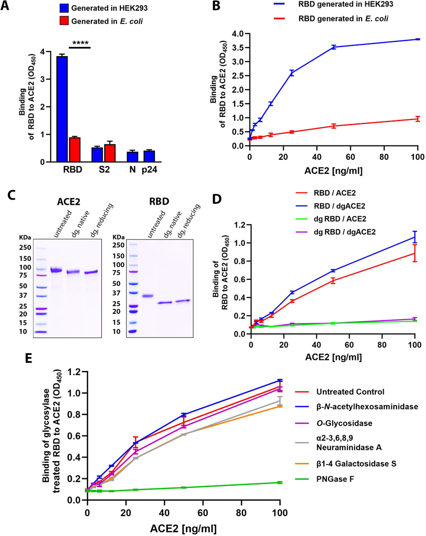
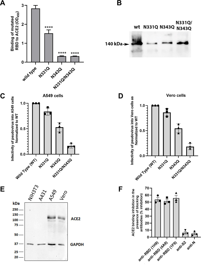
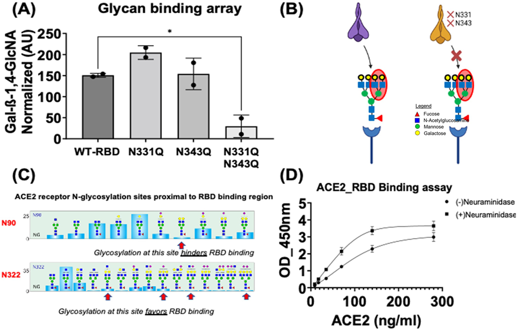
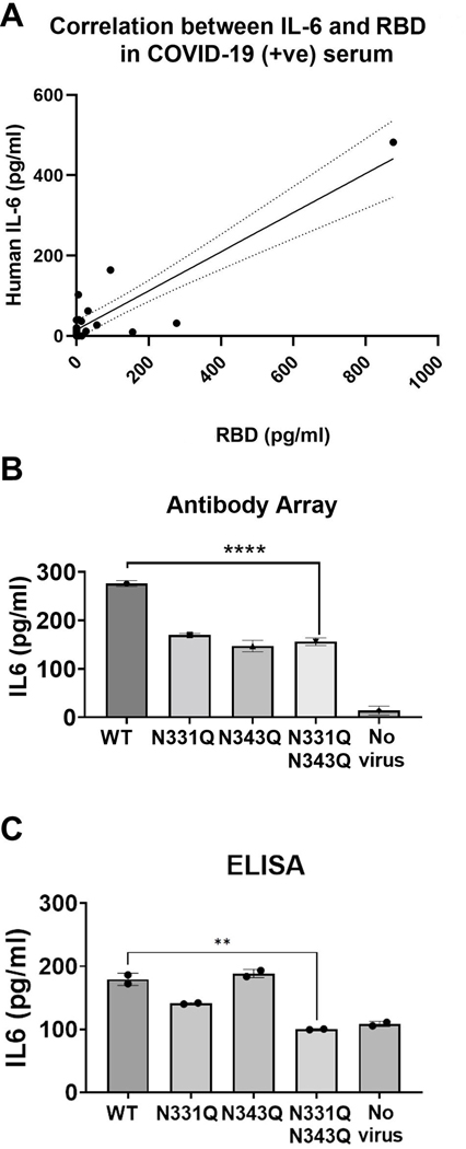
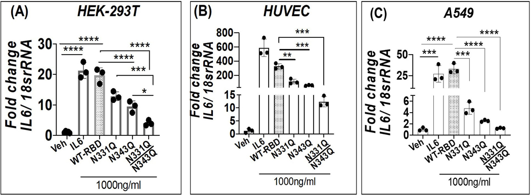
The coronavirus disease 2019 (COVID-19) pandemic is an ongoing global public health crisis. The causative agent, the SARS-CoV-2 virus, enters host cells via molecular interactions between the viral spike protein and the host cell ACE2 surface protein. The SARS-CoV-2 spike protein is extensively decorated with up to 66 N-linked glycans. Glycosylation of viral proteins is known to function in immune evasion strategies but may also function in the molecular events of viral entry into host cells. Here, we show that N-glycosylation at Asn331 and Asn343 of SARS-CoV-2 spike protein is required for it to bind to ACE2 and for the entry of pseudovirus harboring the SARS-CoV-2 spike protein into cells. Interestingly, high-content glycan binding screening data have shown that N-glycosylation of Asn331 and Asn343 of the RBD is important for binding to the specific glycan molecule G4GN (Galβ-1,4 GlcNAc), which is critical for spike-RBD-ACE2 binding. Furthermore, IL-6 was identified through antibody array analysis of conditioned media of the corresponding pseudovirus assay. Mutation of N-glycosylation of Asn331 and Asn343 sites of the spike receptor-binding domain (RBD) significantly reduced the transcriptional upregulation of pro-inflammatory signaling molecule IL-6. In addition, IL-6 levels correlated with spike protein levels in COVID-19 patients' serum. These findings establish the importance of RBD glycosylation in SARS-CoV-2 pathogenesis, which can be exploited for the development of novel therapeutics for COVID-19.

Keywords: ACE2; COVID‐19; IL‐6; N‐glycosylation; SARS‐CoV‐2; receptor‐binding domain; spike protein.


Conflict of interest statement

Author Contributions: RPH, TD, SL, HT conceived and designed the experiments. TD, SL, HT, JF, SD, SH, SX, GFH, BA and BZ performed all experiments. TD, SL, HT, SD, RPH analyzed all data. AS, LEP, PA, JL contributed intellectually and assisted in figure design. TD, VSJ and RPH wrote the manuscript.

Conflict of Interest: Tuhin Das, Shuhong Luo, Hao Tang, Jinamin Fang, Yingqing Mao, Haw-Han Yen, Steven Huang, Valerie Sloane Jones, Shehuo Xie, Gordon Huang, Jinqiao Lu, Blake Anderson, Benyue Zhang and Ruo-Pan Huang are employees of and have a financial stake in RayBiotech, Inc.

Figures

Figure 1:
Figure 1:. N-linked glycosylation of RBD protein is required for binding to ACE2
(A) Binding activity of SARS-CoV-2 RBD and S2 proteins derived from HEK293 cells or from E. coli. Binding was measured using an ACE2 binding assay, and nucleocapsid protein (N) and HIV p24 (P24) were used as negative controls. (B) Binding activity of RBD expressed either in HEK293 cells or in E. coli to titrated concentrations of ACE2. (C) Detection of purified and deglycosylated recombinant RBD and ACE2 proteins by SDS-PAGE. From left to right: untreated proteins, proteins deglycosylated (dg) under native conditions, proteins deglycosylated under reducing conditions. (D) Detection of RBD and ACE2 binding using the ACE2 binding assay under conditions where either or both RBD and ACE2 were deglycosylated (dg). (E) RBD was deglycosylated with individual enzymes: PNGase F, O-glycosidase, α2–3,6,8,9 neuraminidase A, β1–4 galactosidase S, or β-N-acetylhexosaminidasef, prior to the ACE2 binding assay. Statistical significance was tested by T test for all experiments. ****=p<0.0001. n≥3 for all experiments.
Figure 2:
Figure 2:. Glycosylation of RBD at Asn343 is essential for interaction with ACE2
(A) Detection of the binding of RBD wild type, or its N331Q, N343Q and a N331Q/N343Q mutated derivatives to ACE2 using the ACE2 binding assay. 40ng/ml ACE2 was used in all assays. (B) Immunoblot analysis of recombinant SARS-CoV-2 RBD wild type or N331Q, N343Q and N331Q/N343Q double mutated derivatives expressed within purified pseudovirus particles propagated in BHK21 cultured cells. (C) Detection of luciferase activity in A549 cultured cells infected with pseudovirus harboring S1 wild type, or its N331Q, N343Q and a N331Q/N343Q mutated derivatives. (D) Detection of luciferase activity in Vero cultured cells infected with pseudovirus harboring S1 wild type, or its N331Q, N343Q and a N331Q/N343Q mutated derivatives. (E) ACE2 expression level (protein) in three cell lines HEK293, Vero and A549, were detected by immunoblot. (F) The ACE2 binding was measured in presence of blocking antibodies including anti-RBD (1H9; 4A9; 1F9), anti-S2, and anti-N to assess the specificity of ACE2 binding to RBD. Statistical significance was tested by T test for all experiments. ****=p<0.001. n=4/5 for all experiments.
Figure 3:
Figure 3:. Identification of glycan binding epitope in the N-Glycan mutant site of RBD protein
(A) Identification of glycan recognition domains in the N331Q and N343Q mutants of RBD protein using a glycan array. Data represents two independent experiments. Data were analyzed using unpaired Tukey’s test *p < 0.02, for statistical significance. (B) Schematic model of interaction inhibition in between double mutant (N331Q and N343Q) RBD mutant and WT RBD protein. Adapted from BioRender.com (2021). (C) N-Glycan distribution on sites N90 and N322 of human ACE2 receptor which are proximal to RBD binding region of S1 protein (red arrows show the glycans with terminal galactose). Modified from Shajahan A et al., with permission [23]. The glycan structure shown is drawn using the SNFG nomenclature, where the blue squares represent N-acetylglucosamine, the yellow circles represent galactose, green circles represent mannose and red triangle represents fucose [23, 55]. (D) RBD-ACE2 binding increased in the presence of ACE2 treated with neuraminidase for 1hr at 37°C in a dose-dependent manner (n=2 and duplicate for each set).
Figure 4:
Figure 4:. Glycosylation mutants of RBD do not induce IL-6 expression.
(A) Correlation between IL-6 and RBD in COVID-19-positive sera. Serum samples from COVID-positive patients were analyzed by ELISA for IL-6 and RBD (n=21). IL-6 was also measured in conditioned media from the pseudovirus assay (A549 cells) by (B) antibody array and (C) sandwich ELISA; all runs were performed in at least duplicate. Data were analyzed using one-way ANOVA (****p < 0.0001) and unpaired Tukey’s test **p < 0.0075 for statistical significance.
Figure 5:
Figure 5:. N-Glycan mutations at N331 & N343 of RBD protein did not induce IL-6 expression (A-C).
Gene expression data for IL-6 mRNA was normalized with respect to 18sRNA (housekeeping control). Cell cytotoxicity was assessed in each cell line of the corresponding experiment in parallel using LDH measurements. LDH fold changes were normalized with corresponding protein amounts quantified in the cellular lysates using BCA assay method (mg/ml). Each data represents the mean and SE from the n=3 independent experiments performed in duplicate. Data were analyzed using one-way ANOVA with Tukey’s test **p < 0.005, ***p < 0.001, and ****p < 0.0005 for statistical significance.
Figure 6:
Figure 6:
Proposed model: Mutations in N-glycosylation sites at N331 and N343 residues of RBD protein inhibit S-ACE2 binding, IL-6 expression and cytotoxicity.

Similar articles

Cited by

References

    1. Dong E, Du H, Gardner L. An interactive web-based dashboard to track COVID-19 in real time. Lancet Infect Dis. 2020;20(5):533–4. - PMC - PubMed
    1. Grant OC, Montgomery D, Ito K, Woods RJ. Analysis of the SARS-CoV-2 spike protein glycan shield reveals implications for immune recognition. Sci Rep. 2020;10(1):14991. - PMC - PubMed
    1. Beigel JH, Tomashek KM, Dodd LE, Mehta AK, Zingman BS, Kalil AC, et al. Remdesivir for the Treatment of Covid-19 - Final Report. N Engl J Med. 2020;383(19):1813–26. - PMC - PubMed
    1. Chapter Payne S. 17 - Family Coronaviridae. In: Payne S, editor. Viruses: From Understanding to Investigation: Academic Press; 2017. p. 149–58.
    1. Ksiazek TG, Erdman D, Goldsmith CS, Zaki SR, Peret T, Emery S, et al. A novel coronavirus associated with severe acute respiratory syndrome. N Engl J Med. 2003;348(20):1953–66. - PubMed