

In Vitro Chondrogenic Differentiation of Human Adipose-Derived Stem Cells by Diacerein
- PMID: 38444710
- PMCID: PMC10912900
- DOI: 10.5812/ijpr-137803
In Vitro Chondrogenic Differentiation of Human Adipose-Derived Stem Cells by Diacerein
Abstract
Background: Tissue engineering is the application system that tries to restore damaged tissues by different approaches, such as cellular therapy, application of cell differential factors, and various materials. One of the important goals in tissue engineering is to guide stem cells directly to the desired tissue, and researchers tried to utilize different molecules as effective factors to improve this technique.
Objectives: This study aims to demonstrate the effects of diacerein, a slow-acting drug for the treatment of osteoarthritis, on mesenchymal stem cell proliferation and evaluate its potential in the chondrogenesis process.
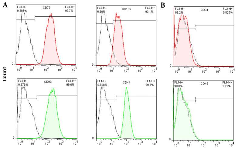
Methods: Stem cells were isolated from adipose tissue, characterized by flow cytometry, and cells were treated with 10-5M diacerein for three weeks. Chondrogenic gene expression of SOX9, COL2A1, ACAN, and TGFB1 were analyzed by qRT-PCR and immunocytochemistry techniques.
Results: Our results showed that diacerein increased the expression of the following genes involved in chondrogenesis: SOX9 (2.9-fold, P < 0.00), COL2A1 (2.2-fold, P < 0.00), ACAN (2.7-fold, P < 0.00), and TGFB1 (2.6-fold, P < 0.00). Immunocytochemistry results also showed increased production of collagen type II as the main protein marker for chondrocytes.
Conclusions: We observed that diacerein alone could initiate and enhance chondrogenesis, and it can be used as a differentiation factor for stem cells to chondrocyte besides its ability to inhibit IL-1β. Knowing the actual function of diacerein, it could be a good candidate for the treatment of osteoarthritis.
Keywords: Adipose Tissue; Cartilage Disease; Chondrogenesis; Diacerein; Mesenchymal Stem Cells.
Copyright © 2023, Honarpardaz et al.
Conflict of interest statement
The authors declare no conflict of interest.
Figures


Similar articles
-
Centrifugal gravity-induced BMP4 induces chondrogenic differentiation of adipose-derived stem cells via SOX9 upregulation.Stem Cell Res Ther. 2016 Dec 8;7(1):184. doi: 10.1186/s13287-016-0445-6. Stem Cell Res Ther. 2016. PMID: 27931264 Free PMC article.
-
The effect of two- and three-dimensional cell culture on the chondrogenic potential of human adipose-derived mesenchymal stem cells after subcutaneous transplantation with an injectable hydrogel.Cell Transplant. 2011;20(10):1575-88. doi: 10.3727/096368910X557191. Epub 2011 Feb 3. Cell Transplant. 2011. PMID: 21294960
-
Hyaluronic acid facilitates chondrogenesis and matrix deposition of human adipose derived mesenchymal stem cells and human chondrocytes co-cultures.Acta Biomater. 2017 Apr 1;52:130-144. doi: 10.1016/j.actbio.2017.01.064. Epub 2017 Jan 25. Acta Biomater. 2017. PMID: 28131943
-
Characterization of human adipose-derived stem cells and expression of chondrogenic genes during induction of cartilage differentiation.Clinics (Sao Paulo). 2012;67(2):99-106. doi: 10.6061/clinics/2012(02)03. Clinics (Sao Paulo). 2012. PMID: 22358233 Free PMC article.
-
Melatonin rescued interleukin 1β-impaired chondrogenesis of human mesenchymal stem cells.Stem Cell Res Ther. 2018 Jun 14;9(1):162. doi: 10.1186/s13287-018-0892-3. Stem Cell Res Ther. 2018. PMID: 29898779 Free PMC article.
References
-
- Huh JE, Koh PS, Seo BK, Park YC, Baek YH, Lee JD, et al. Mangiferin reduces the inhibition of chondrogenic differentiation by IL-1beta in mesenchymal stem cells from subchondral bone and targets multiple aspects of the Smad and SOX9 pathways. Int J Mol Sci. 2014;15(9):16025–42. doi: 10.3390/ijms150916025. - DOI - PMC - PubMed
LinkOut - more resources
Full Text Sources
Research Materials
Miscellaneous