. 2024 Feb 20:12:1316048.
doi: 10.3389/fcell.2024.1316048. eCollection 2024.

Prdm15 acts upstream of Wnt4 signaling in anterior neural development of Xenopus laevis

Affiliations

Prdm15 acts upstream of Wnt4 signaling in anterior neural development of Xenopus laevis

Ernestine Saumweber et al. Front Cell Dev Biol. .

Abstract

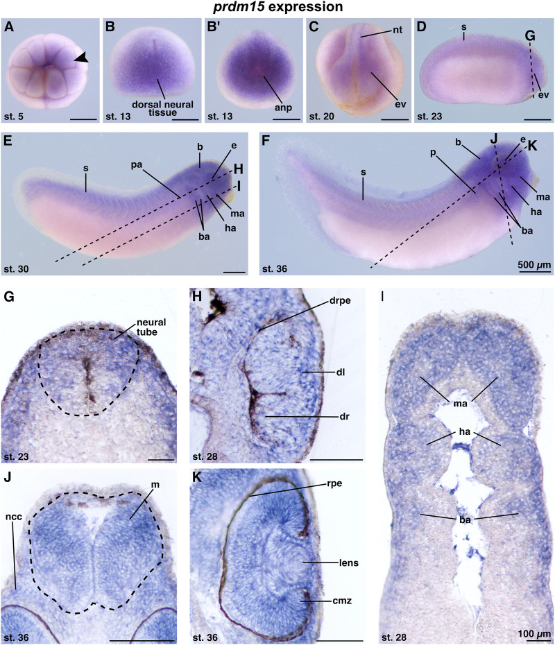
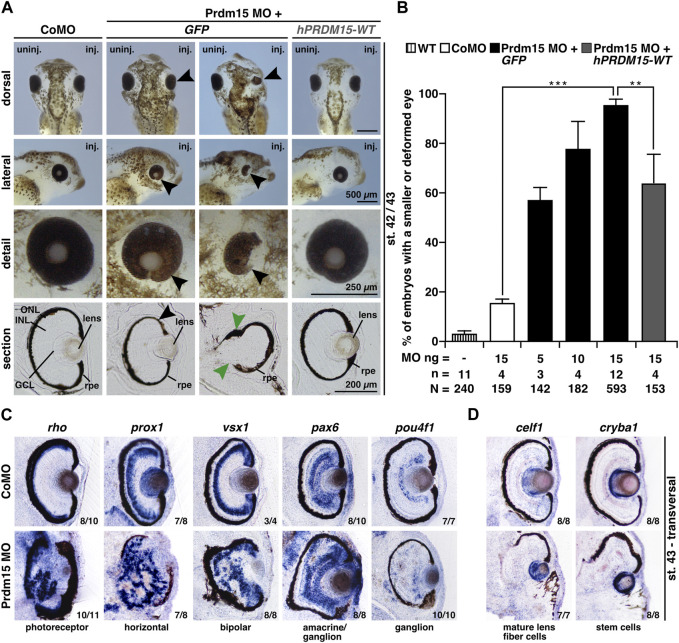
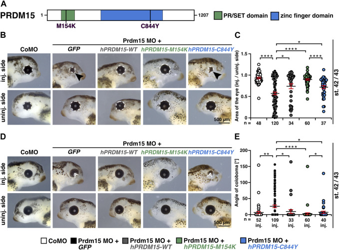
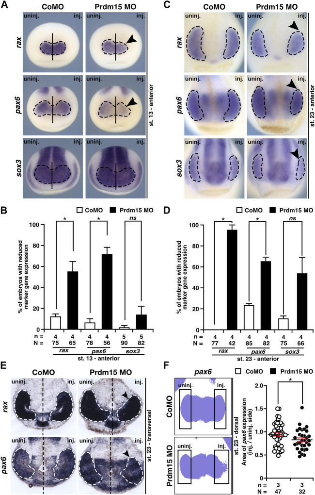
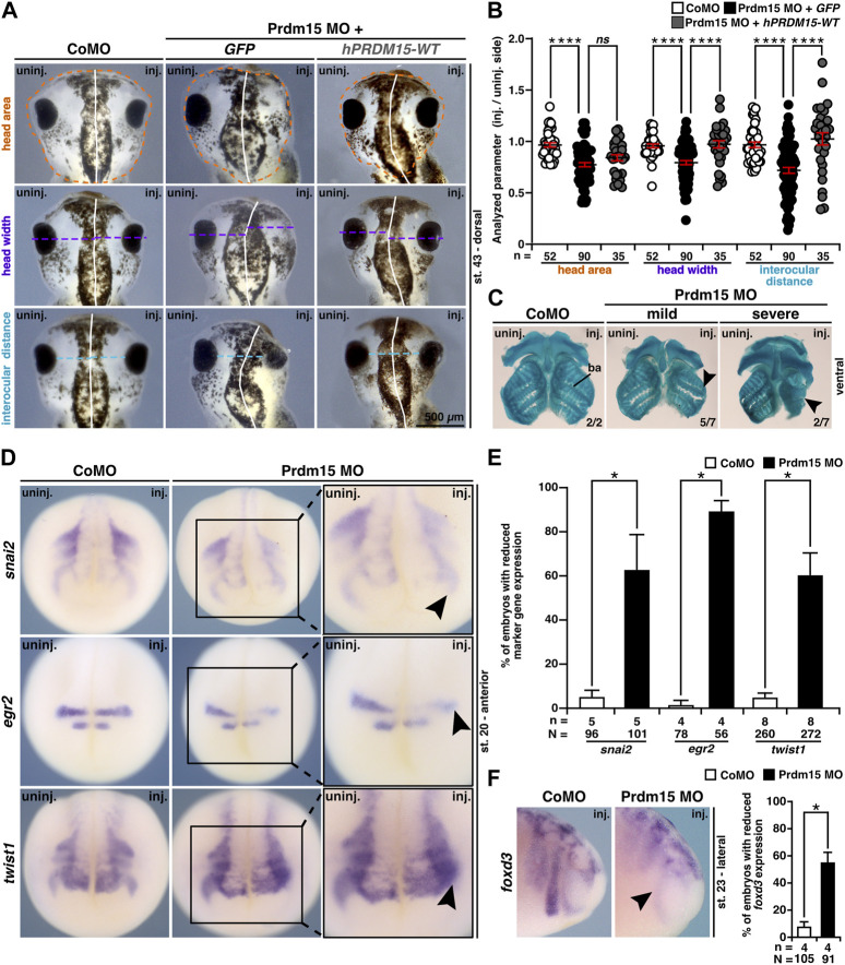
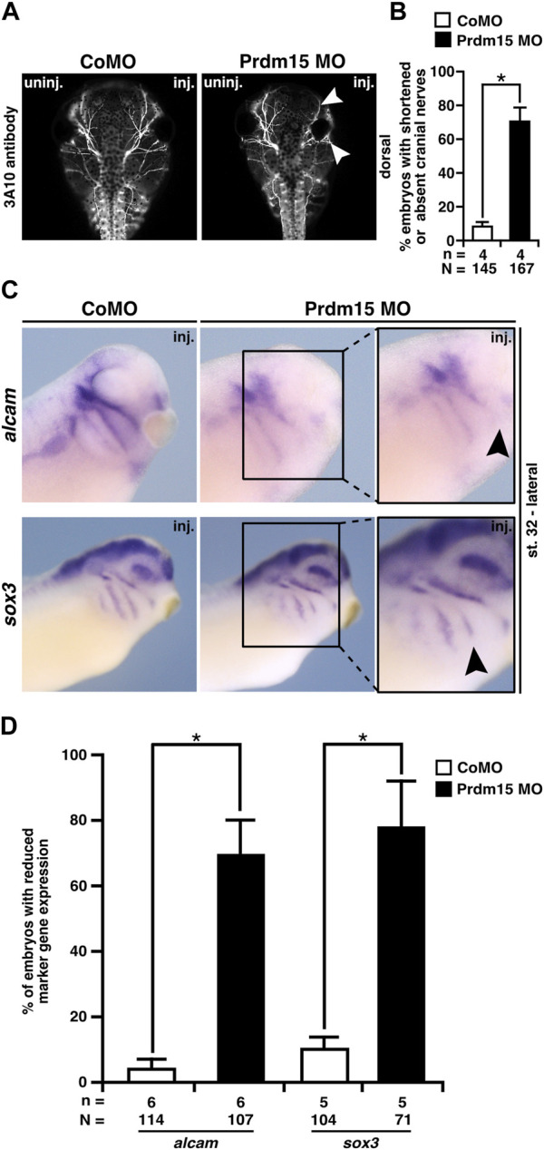
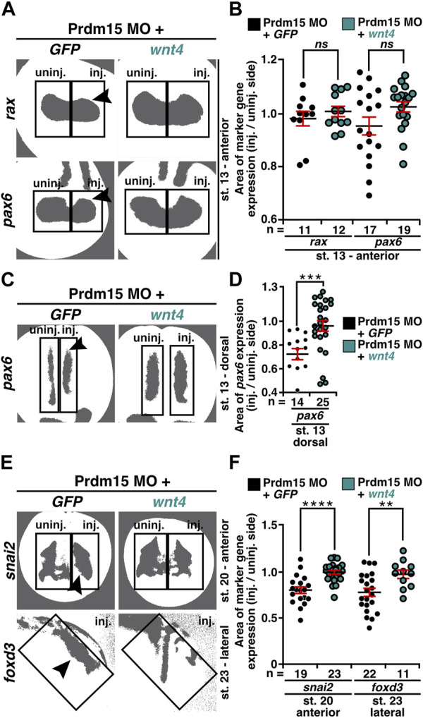
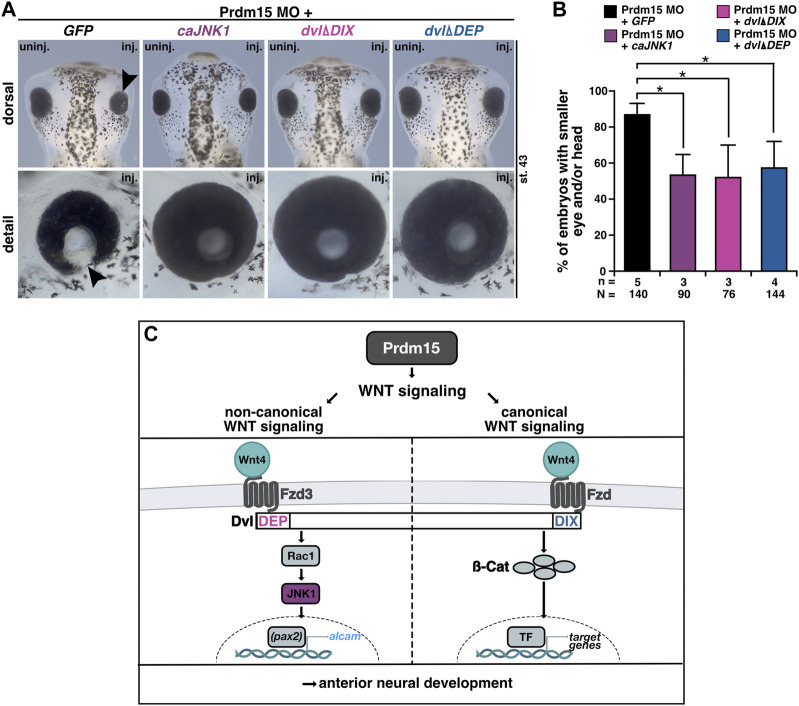
Mutations in PRDM15 lead to a syndromic form of holoprosencephaly (HPE) known as the Galloway-Mowat syndrome (GAMOS). While a connection between PRDM15, a zinc finger transcription factor, and WNT/PCP signaling has been established, there is a critical need to delve deeper into their contributions to early development and GAMOS pathogenesis. We used the South African clawed frog Xenopus laevis as the vertebrate model organism and observed that prdm15 was enriched in the tissues and organs affected in GAMOS. Furthermore, we generated a morpholino oligonucleotide-mediated prdm15 knockdown model showing that the depletion of Prdm15 leads to abnormal eye, head, and brain development, effectively recapitulating the anterior neural features in GAMOS. An analysis of the underlying molecular basis revealed a reduced expression of key genes associated with eye, head, and brain development. Notably, this reduction could be rescued by the introduction of wnt4 RNA, particularly during the induction of the respective tissues. Mechanistically, our data demonstrate that Prdm15 acts upstream of both canonical and non-canonical Wnt4 signaling during anterior neural development. Our findings describe severe ocular and anterior neural abnormalities upon Prdm15 depletion and elucidate the role of Prdm15 in canonical and non-canonical Wnt4 signaling.

Keywords: GAMOS; HPE; Prdm15; Wnt signaling; Xenopus laevis; disease modeling.


Conflict of interest statement

The authors declare that the research was conducted in the absence of any commercial or financial relationships that could be construed as a potential conflict of interest. The authors have declared that they were an editorial board member of Frontiers at the time of submission. This had no impact on the peer review process and the final decision.

Figures

FIGURE 1
FIGURE 1
Prdm15 is expressed during X. laevis development in disease relevant tissues of GAMOS. Spatiotemporal expression pattern of prdm15 visualized by whole-mount in situ hybridization (WMISH). Embryonic stages and scale bars are indicated in each panel. Upper part: WMISH with exterior view. Scale bars are equivalent to 500 µm. Black dashed lines represent section planes. Lower part: WMISH following sections. Scale bars are equivalent to 100 µm. (A) Vegetal view of X. laevis embryos at stage 5 shows a prdm15 expression in the animal pole (black arrowhead). At stage 13, prdm15 is strongly expressed in the dorsal neural tissue (B, dorsal view) and the anterior neural plate (anp) (B′, anterior view). (C) At stage 20, prdm15 is expressed in the neural tube (nt) and in the eye vesicle (ev). (D) The lateral view at stage 23 shows prdm15 transcripts in the somites (s) and the developing eye, more precisely in the eye vesicle (ev). (E, F) Prdm15 expression is detected in lateral views from stages 28 to 36 in the somites (s), the eye (e), the embryonic kidney [pronephric anlage (pa), pronephros (p)], the brain (B), and in the mandibular (ma), hyoid (ha), and branchial arches (ba). (G) The transversal section shows an expression of prdm15 in the neural tube (nt). The horizontal sections (H, I) reveal prdm15 transcripts in the developing retinal pigmented epithelium (rpe), developing lens, and the mandibular (ma), hyoid (ha), and branchial arches (ba). (J) The transversal section shows prdm15 expression in the mesencephalon (m) and neural crest cells (ncc). (K) In later stages, prdm15 is expressed in the eye, more detailed in the lens and ciliary marginal zone (cmz) by horizontal sectioning. Abbreviations: anp, anterior neural plate; b, brain; ba, branchial arch; cmz, ciliary marginal zone; dl, developing lens; dr, developing retina: drpe, developing retinal pigmented epithelium; e, eye; ev, eye vesicle; ha, hyoid arch; m, mesencephalon; ma, mandibular arch; µm, micrometer; ncc, neural crest cells; nt, neural tube; p, pronephros; pa, pronephric anlage; prdm15, PR-domain zinc finger protein 15; rpe, retinal pigmented epithelium; s, somite; st., stage; WMISH, whole-mount in situ hybridization.
FIGURE 2
FIGURE 2
Prdm15 knockdown leads to a severe eye phenotype that is rescued by human PRDM15 wild-type (WT) RNA. (A) Unilateral knockdown (KD) of Prdm15 results in microphthalmia compared to control MO (CoMO), while co-injection of human full-length PRDM15-WT RNA rescues the eye phenotype during X. laevis eye development. The lateral and detailed views of the embryo show the eye in more detail and the sections specify the lamination of the eye. Black arrowheads point to smaller and deformed eyes, and green arrowheads point to the disturbed and thickened retinal pigmented epithelium (rpe). Representative embryos are shown. (B) Statistical evaluation of smaller and deformed eyes as indicated in (A). (C) Transversal vibratome sections after whole-mount in situ hybridization (WMSH) of Prdm15 MO–injected embryos show a severe eye phenotype in contrast to CoMO-injected embryos. Specific marker genes for retina cell layers are used for specific cell populations of the retina as described in the main text. Most of the cell types are disorganized and displaced. (D) Lens-specific marker genes celf1 and cryba1 are also affected, showing a smaller expression size, but the organization of the cells is unaffected upon Prdm15 KD. Abbreviations: celf1, CUGBP elav-like family member 1; CoMO, control morpholino oligonucleotide; cryba1, crystallin beta A1; GCL, ganglion cell layer; GFP, green fluorescent protein; hPRDM15, human PRDM15; INL, inner nuclear cell layer; inj., injected; MO, morpholino oligonucleotide; n, number of independent experiments; N, number of analyzed embryos in total; ng, nanogram; ONL, outer nuclear cell layer; pax6, paired box 6; pou4f1, POU class 4 homeobox 1; Prdm15, PR-domain zinc finger protein 15; prox1, prospero homeobox 1; rho, rhodopsin; rpe, retinal pigmented epithelium; st., stage; uninj., un-injected; vsx1, visual system homeobox 1; WT, wild type. Error bars indicate standard errors of the means. **, p ≤ 0.01; ***, p ≤ 0.001.
FIGURE 3
FIGURE 3
Prdm15 knockdown is rescued by human PRDM15 RNA with a PR/SET domain mutation but only partially by the PRDM15 RNA with a zinc finger mutation. (A) Schematic overview of the human PRDM15 protein. The PR/SET and zinc finger domains are shown. The positions of the mutated proteins in the PR/SET [p.M154K (c.461T>A); Mann et al., 2021; Mzoughi et al., 2020] and zinc finger domain [p.C844Y (c.2531G>A); Mann et al., 2021; Mzoughi et al., 2020] in GAMOS patients are indicated. (B) Co-injection of human full-length PRDM15-WT RNA rescues microphthalmia phenotype. While co-injection of human PRDM15 RNA with a PR/SET variant (hPRDM15-M154K) identified in affected individuals also rescues the eye phenotype in most embryos, human Prdm15 RNA with a variant in the zinc finger domain (hPRDM15-C844Y) identified in affected individuals only partially rescues the Prdm15 MO–mediated eye phenotype. Black arrowheads point to smaller and deformed eyes. Representative embryos are shown. (C) Statistical evaluation of the eye area as illustrated in (B) (white dashed line: measured eye area; injected vs un-injected side). (D) Co-injection of human full-length PRDM15-WT RNA rescues the coloboma phenotype. Co-injection of hPRDM15-M154K RNA rescues the coloboma phenotype in most embryos; hPRDM15-C844Y RNA with a variant in the zinc finger domain could partially rescue the Prdm15 MO–mediated coloboma phenotype. White angle shows the measured angle of the colobomas. Representative embryos are shown. (E) Statistical evaluation of the coloboma phenotype as illustrated in (D) (white angle: measured coloboma angle). Abbreviations: C844Y, cysteine-844-tyrosine; CoMO, control morpholino oligonucleotide; GFP, green fluorescent protein; hPRDM15, human PRDM15; inj., injected; M154K, methionine-154-lysine; MO, morpholino oligonucleotide; n, number of independent experiments; Prdm15, PR-domain zinc finger protein 15; st., stage; uninj., un-injected; WT, wild type. Error bars indicate standard errors of the means. *, p ≤ 0.05; **, p ≤ 0.01; ****, p ≤ 0.0001.
FIGURE 4
FIGURE 4
Prdm15 is necessary for proper eye development in X. laevis. (A) In stage 13, during eye field induction, the anterior expression (black dashed line) of the eye-specific marker genes rax and pax6 is reduced (black arrowheads) in Prdm15 MO–injected embryos visualized by whole-mount in situ hybridization (WMISH). By contrast, the pan-neural marker gene sox3 and CoMO injection did not alter the expression on the injected side. (B) Statistical evaluation of embryos with reduced marker gene expression as described in (A). (C) Prdm15 knockdown (KD) interferes with eye-specific marker gene expression (black dashed line) of rax, pax6, and sox3 in stage 23. Anterior views show a reduced expression in the developing eye (black arrowheads). (D) Statistical evaluation of embryos with reduced marker gene expression as described in (C). (E) Prdm15 KD influences eye-specific rax and pax6 expressions. Transversal vibratome sections after WMISH with rax and pax6 confirm the eye phenotype showing a smaller and reduced marker gene expression area (black arrowheads). (F) Statistical evaluation of embryos with pax6 expression area as analyzed by ImageJ2 (Rueden et al., 2017) shows a significantly reduced expression area in Prdm15 MO–injected embryos (black arrowhead). Abbreviations: CoMO, control morpholino oligonucleotide; inj., injected; KD, knockdown; MO, morpholino oligonucleotide; n, number of independent experiments; N, number of analyzed embryos in total; ns, non-significant; pax6, paired box 6; Prdm15, PR-domain zinc finger protein 15; rax, retina and anterior neural fold homeobox; sox3, SRY-box transcription factor 3; st., stage; uninj., un-injected; WMISH, whole-mount in situ hybridization. Error bars indicate standard errors of the means. ns, p > 0.05; *, p ≤ 0.05.
FIGURE 5
FIGURE 5
Prdm15 is required for head development in X. laevis. (A) Unilateral KD of Prdm15 leads to a microcephaly phenotype compared to control MO (CoMO)–injected embryos. Co-injection of human full-length PRDM15-WT RNA rescues the head phenotype. The dorsal views of representative stage 43 embryos are shown. Dashed lines indicate the measured area and length. (B) Statistical evaluation of smaller and deformed heads as indicated in (A) reveals a significantly smaller head area (orange), head width (purple), and interocular distance (light blue), while co-injection of hPRDM15 RNA rescues almost all Prdm15 MO–induced phenotypes. (C) Ventral view of Alcian blue–stained and dissected cranial cartilages from control and Prdm15 morphants (stage 45). Prdm15 KD results in a mild and severe phenotype showing narrowed or deformed cartilage structures (black arrowheads), especially at the branchial arch (ba). (D) Anterior view of CoMO-injected and Prdm15 MO–injected embryos (stage 20) after whole-mount in situ hybridization (WMISH) with NCC-specific marker genes during NCC migration such as snai2, egr2, and twist1 show a reduced marker gene expression on Prdm15 MO–injected side (black arrowheads). (E) Statistical evaluation of NCC-specific marker genes' expression as illustrated in (D) reveals a significant reduction in marker gene expression upon Prdm15 MO KD. (F) Lateral view of CoMO-injected and Prdm15 MO–injected embryos (stage 23) after WMISH with the NCC marker foxd3 shows a reduced expression on the Prdm15 MO–injected side (black arrowhead). Statistical evaluation of foxd3 expression reveals a significant reduction in its expression upon Prdm15 MO KD. Abbreviations: CoMO, control morpholino oligonucleotide; egr2, early growth response 2; foxd3, forkhead box D3; inj., injected; hPRDM15, human PRDM15; MO, morpholino oligonucleotide; n, number of independent experiments; N, number of analyzed embryos in total; ns, non-significant; Prdm15, PR-domain zinc finger protein 15; snai2, snail family transcriptional repressor 2; st., stage; twist1, twist family bHLH transcription factor 1; uninj., un-injected; WMISH, whole-mount in situ hybridization; WT, wild type. Error bars indicate standard errors of the means. ns, p > 0.05; *, p ≤ 0.05; ****, p ≤ 0.0001.
FIGURE 6
FIGURE 6
Prdm15 MO injection hinders proper development of the cranial nerves and placodes. (A) Dorsal views of control MO-injected and Prdm15 MO–injected embryos show a shortened or decreased branching of cranial nerves (white arrowheads) upon Prdm15 KD visualized by 3A10 antibody staining. (B) Statistical evaluation of embryos with shortened or absent branching of cranial nerves as illustrated in (A). (C) Lateral view of stage 32 embryos reveals a reduction (black arrowheads) of the respective expression upon Prdm15 MO KD in both marker genes of the lateral placodes (alcam and sox3) using whole-mount in situ hybridization. (D) Statistical evaluation of alcam and sox3 expression revealed a significant reduction in its expression upon Prdm15 KD as illustated in (C). Abbreviations: alcam, activated leukocyte cell adhesion molecule; inj., injected; MO, morpholino oligonucleotide; n, number of independent experiments; N, number of analyzed embryos in total; Prdm15, PR-domain zinc finger protein 15; sox3, SRY-box transcription factor 3; uninj., un-injected. Error bars indicate standard errors of the means. *, p ≤ 0.05.
FIGURE 7
FIGURE 7
Prdm15 acts upstream of Wnt4. (A) Anterior view at stage 20 and dorsal view at stage 28 of Prdm15 MO–injected embryos show a reduced expression of wnt4 (black arrowheads) in comparison to the un-injected side, as analyzed by the expression area and intensity of wnt4 after whole-mount in situ hybridization (WMISH). (B) Statistical analysis of embryos with reduced wnt4 expression as indicated in (A) analyzed with a light microscope. (C) Statistical analysis of the expression area of wnt4 as described in (A) using a computer-based approach. (D) Statistical analysis of the mean intensity of the expression area of wnt4 as described in (A) using a computer-based approach. The statistical analysis of wnt4 expression reveals a significantly reduced wnt4 expression except for the analysis of wnt4 in stage 20 upon Prdm15 knockdown. (E) Anterior view of stage 20 and lateral view of stage 28 embryos. Prdm15 MO–injected embryos show a reduction in alcam expression on the injected side (black arrowheads), as analyzed by expression area (G) and intensity (H) using WMISH. (F) Statistical analysis of embryos with reduced alcam expression analyzed with a light microscope. (G) Statistical analysis of the expression area using a computer-based approach. (H) Statistical analysis of the mean intensity of the expression area of alcam using a computer-based approach reveals a significantly reduced alcam expression upon Prdm15 knockdown. (I) Dorsal and lateral views of stage 43 embryos show rescue of the Prdm15 MO–induced head and eye phenotype by wnt4 RNA. Black arrowhead points to the reduced and deformed eye size upon Prdm15 MO injection. (J) Statistical evaluation of the embryos with smaller eye and/or head as illustrated in (I). Abbreviations: alcam, activated leukocyte cell adhesion molecule; CoMO, control morpholino oligonucleotide; GFP, green fluorescent protein; inj., injected; MO, morpholino oligonucleotide; n, number of independent experiments; N, number of analyzed embryos in total; ns, non-significant; Prdm15, PR-domain zinc finger protein 15; st., stage; uninj., un-injected; WMISH, whole-mount in situ hybridization; wnt4, wnt family member 4. Error bars indicate standard errors of the means. ns, p > 0.05; *, p ≤ 0.05; **, p ≤ 0.01; ****, p ≤ 0.0001.
FIGURE 8
FIGURE 8
Co-injection of wnt4 RNA rescues the Prdm15 MO–induced reduced gene expression. (A) Anterior view of stage 13 embryos injected with Prdm15 MO in combination with wnt4 RNA refers to the measured rax and pax6 expression area. Black arrowheads point to the reduced gene expression. Wnt4 RNA could partially rescue the Prdm15 MO–induced reduced marker gene expression. (B) Statistical evaluation of the marker gene expression area of rax and pax6 as described in (A). (C) Dorsal view of stage 13 embryos injected with Prdm15 MO in combination with wnt4 RNA illustrates the measured pax6 expression area. Black arrowhead points to the reduced gene expression. Co-injection of wnt4 RNA restores the Prdm15 MO–induced pax6 expression. (D) Statistical evaluation of the marker gene expression area as described in (C) shows rescue of the dorsal pax6 expression by wnt4 RNA. (E) Anterior and lateral views of stage 20 and stage 23 embryos, respectively, injected with Prdm15 MO in combination with wnt4 RNA illustrate the measured snai2 and foxd3 expression area. Black arrowheads point to the reduced gene expression. Co-injection of wnt4 RNA rescues the Prdm15 MO–induced reduced marker gene expression. (F) Statistical evaluation of snai2 and foxd3 expression area as described in (E) shows a significant rescue of the expression area of snai2 and foxd3. Abbreviations: foxd3, forkhead box D3; GFP, green fluorescent protein; inj., injected; MO, morpholino oligonucleotide; n, number of independent experiments; ns, non-significant; pax6, paired box 6; Prdm15, PR-domain zinc finger protein 15; rax, retina and anterior neural fold homeobox; snai2, snail family transcriptional repressor 2; st., stage; uninj., un-injected; wnt4, wnt family member 4. Error bars indicate standard errors of the means. ns, p > 0.05; **, p ≤ 0.01; ***, p ≤ 0.001; ****, p ≤ 0.0001.
FIGURE 9
FIGURE 9
Prdm15 influences canonical and non-canonical Wnt signaling upstream of Wnt4. (A) Dorsal and detailed view of stage 43 embryos injected with Prdm15 MO in combination with GFP RNA or the constitutive active JNK1 (caJNK1) RNA or the two different mutated disheveled versions (dvlΔDIX, dvlΔDEP). The co-injection of Prdm15 MO with caJNK1 and the dvlΔDIX-mutated version as well as the dvlΔDEP-mutated version shows rescue of the severe Prdm15 MO–induced phenotype in contrast to the negative control GFP (black arrowhead). (B) Statistical evaluation of embryos with smaller eye or head as described in (A) shows a rescue with caJNK1, dvlΔDIX, and dvlΔDEP RNA. (C) Schematic overview of Prdm15 and WNT signaling. Prdm15 acts upstream of Wnt4 and influences the expression of wnt4 and alcam through the non-canonical WNT/PCP signaling pathway possibly via the transcription factor pax2. Furthermore, the canonical Wnt signaling pathway is also affected downstream of Prdm15. Abbreviations: alcam, activated leukocyte cell adhesion molecule; ß-cat, β-catenin; caJNK1, constitutive active JNK 1; DEP, disheveled Egl-10 and pleckstrin; DIX, disheveled Axin; dvl, disheveled; Fzd, frizzled; Fzd3, frizzled3; GFP, green fluorescent protein; inj., injected; JNK1, c-Jun N-terminal kinase 1; MO, morpholino oligonucleotide; n, number of independent experiments; N, number of analyzed embryos in total; pax2, paired box 2; Prdm15, PR-domain zinc finger protein 15; Rac1, Rac1 family small GTPase 1; st., stage; TF, transcription factor; uninj., un-injected; WNT, wingless-type MMTV integration site family member; wnt4, wnt family member 4. Error bars indicate standard errors of the means. *, p ≤ 0.05.

Similar articles

Cited by

References

    1. Al-Rakan M. A., Abothnain M. D., Alrifai M. T., Alfadhel M. (2018). Extending the ophthalmological phenotype of Galloway-Mowat syndrome with distinct retinal dysfunction: a report and review of ocular findings. BMC Ophthalmol. 18, 147–218. 10.1186/s12886-018-0820-4 - DOI - PMC - PubMed
    1. Bernard P., Fleming A., Lacombe A., Harley V. R., Vilain E. (2008). Wnt4 inhibits beta-catenin/TCF signalling by redirecting beta-catenin to the cell membrane. Biol. Cell 100, 167–177. 10.1042/BC20070072 - DOI - PMC - PubMed
    1. Braun D. A., Rao J., Mollet G., Schapiro D., Daugeron M.-C., Tan W., et al. (2017). Mutations in KEOPS-complex genes cause nephrotic syndrome with primary microcephaly. Nat. Genet. 49, 1529–1538. 10.1038/ng.3933 - DOI - PMC - PubMed
    1. Bugner V., Tecza A., Gessert S., Kühl M. (2011). Peter Pan functions independently of its role in ribosome biogenesis during early eye and craniofacial cartilage development in Xenopus laevis . Development 138, 2369–2378. 10.1242/dev.060160 - DOI - PubMed
    1. Cavodeassi F. (2014). Integration of anterior neural plate patterning and morphogenesis by the Wnt signaling pathway. Dev. Neurobiol. 74, 759–771. 10.1002/dneu.22135 - DOI - PubMed

LinkOut - more resources