

White matter network underlying semantic processing: evidence from stroke patients
- PMID: 38444912
- PMCID: PMC10914445
- DOI: 10.1093/braincomms/fcae058
White matter network underlying semantic processing: evidence from stroke patients
Abstract
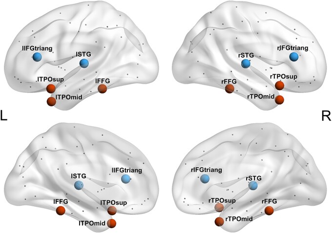
The hub-and-spoke theory of semantic representation fractionates the neural underpinning of semantic knowledge into two essential components: the sensorimotor modality-specific regions and a crucially important semantic hub region. Our previous study in patients with semantic dementia has found that the hub region is located in the left fusiform gyrus. However, because this region is located within the brain damage in patients with semantic dementia, it is not clear whether the semantic deficit is caused by structural damage to the hub region itself or by its disconnection from other brain regions. Stroke patients do not have any damage to the left fusiform gyrus, but exhibit amodal and modality-specific deficits in semantic processing. Therefore, in this study, we validated the semantic hub region from a brain network perspective in 79 stroke patients and explored the white matter connections associated with it. First, we collected data of diffusion-weighted imaging and behavioural performance on general semantic tasks and modality-specific semantic tasks (assessing object knowledge on form, colour, motion, sound, manipulation and function). We then used correlation and regression analyses to examine the association between the nodal degree values of brain regions in the whole-brain structural network and general semantic performance in the stroke patients. The results revealed that the connectivity of the left fusiform gyrus significantly predicted general semantic performance, indicating that this region is the semantic hub. To identify the semantic-relevant connections of the semantic hub, we then correlated the white matter integrity values of each tract connected to the left fusiform gyrus separately with performance on general and modality-specific semantic processing. We found that the hub region accomplished general semantic processing through white matter connections with the left superior temporal pole, middle temporal gyrus, inferior temporal gyrus and hippocampus. The connectivity between the hub region and the left hippocampus, superior temporal pole, middle temporal gyrus, inferior temporal gyrus and parahippocampal gyrus was differentially involved in object form, colour, motion, sound, manipulation and function processing. After statistically removing the effects of potential confounding variables (i.e. whole-brain lesion volume, lesion volume of regions of interest and performance on non-semantic control tasks), the observed effects remained significant. Together, our findings support the role of the left fusiform gyrus as a semantic hub region in stroke patients and reveal its crucial connectivity in the network. This study provides new insights and evidence for the neuroanatomical organization of semantic memory in the human brain.
Keywords: modality-specific connection; semantic hub; semantic processing; stroke; white matter network.
© The Author(s) 2024. Published by Oxford University Press on behalf of the Guarantors of Brain.
Conflict of interest statement
The authors report no competing interests.
Figures


Similar articles
-
White matter basis for the hub-and-spoke semantic representation: evidence from semantic dementia.Brain. 2020 Apr 1;143(4):1206-1219. doi: 10.1093/brain/awaa057. Brain. 2020. PMID: 32155237 Free PMC article.
-
Left Anterior Temporal Lobe and Bilateral Anterior Cingulate Cortex Are Semantic Hub Regions: Evidence from Behavior-Nodal Degree Mapping in Brain-Damaged Patients.J Neurosci. 2017 Jan 4;37(1):141-151. doi: 10.1523/JNEUROSCI.1946-16.2016. J Neurosci. 2017. PMID: 28053037 Free PMC article.
-
Neural substrates of amodal and modality-specific semantic processing within the temporal lobe: A lesion-behavior mapping study of semantic dementia.Cortex. 2019 Nov;120:78-91. doi: 10.1016/j.cortex.2019.05.014. Epub 2019 Jun 12. Cortex. 2019. PMID: 31280071
-
The neural basis of semantic cognition: converging evidence from neuropsychology, neuroimaging and TMS.Cortex. 2013 Mar;49(3):611-25. doi: 10.1016/j.cortex.2012.10.008. Epub 2012 Nov 13. Cortex. 2013. PMID: 23260615 Review.
-
A meta-analysis of fMRI studies on Chinese orthographic, phonological, and semantic processing.Neuroimage. 2012 Oct 15;63(1):381-91. doi: 10.1016/j.neuroimage.2012.06.047. Epub 2012 Jul 1. Neuroimage. 2012. PMID: 22759996 Review.
References
-
- Tulving E. Organization of memory. Academic Press; 1972.
-
- Chapman CA, Hasan O, Schulz PE, Martin RC. Evaluating the distinction between semantic knowledge and semantic access: Evidence from semantic dementia and comprehension-impaired stroke aphasia. Psychon Bull Rev. 2020;27(4):607–639. - PubMed
-
- Gardner HE, Lambon Ralph MA, Dodds N, Jones T, Ehsan S, Jefferies E. The differential contributions of pFC and temporo-parietal cortex to multimodal semantic control: Exploring refractory effects in semantic aphasia. J Cogn Neurosci. 2012; 24(4):778–793. - PubMed
-
- Han Z, Ma Y, Gong G, He Y, Caramazza A, Bi Y. White matter structural connectivity underlying semantic processing: Evidence from brain damaged patients. Brain. 2013;136(10):2952–2965. - PubMed
-
- Kumar AA. Semantic memory: A review of methods, models, and current challenges. Psychon Bull Rev. 2021;28(1):40–80. - PubMed
LinkOut - more resources
Full Text Sources