

Integration of Augmented Reality Into Glioma Resection Surgery: A Case Report
- PMID: 38445166
- PMCID: PMC10914376
- DOI: 10.7759/cureus.53573
Integration of Augmented Reality Into Glioma Resection Surgery: A Case Report
Abstract
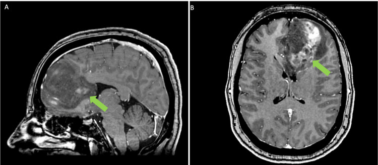
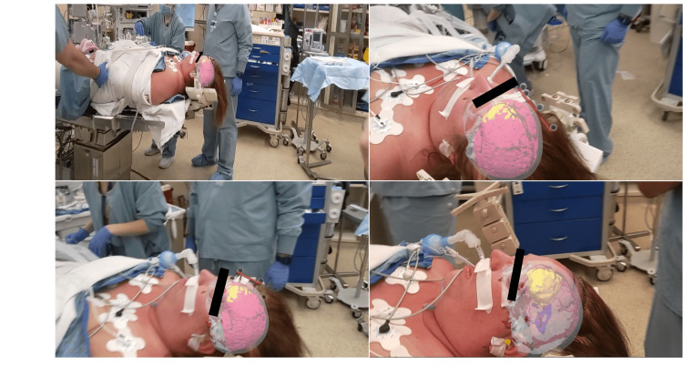
Augmented reality (AR) is an exciting technology that has garnered considerable attention in the field of neurosurgery. Despite this, clinical use of this technology is still in its infancy. An area of great potential for this technology is the ability to display 3D anatomy overlaid with the patient to assist with presurgical and intraoperative decision-making. A 39-year-old woman presented with headaches and was experiencing what was described as a whooshing sound. MRI revealed the presence of a large left frontal mass involving the genu of the corpus callosum, with heterogeneous enhancement and central hemorrhagic necrosis, confirmed to be a glioma. She underwent a craniotomy with intraoperative MRI for resection. An augmented reality system was used to superimpose 3D holographic anatomy onto the patient's head for surgical planning. This report highlights a new AR technology and its immediate application to cranial neurosurgery. It is critical to document new uses of this technology as the field continues to integrate AR as well as other next-generation technologies into practice.
Keywords: augmented reality; cranial neurosurgery; glioblastoma; medical device; mixed reality.
Copyright © 2024, Hunt et al.
Conflict of interest statement
The authors have declared that no competing interests exist.
Figures
References
-
- A review of newly diagnosed glioblastoma. Oronsky B, Reid TR, Oronsky A, Sandhu N, Knox SJ. https://www.frontiersin.org/articles/10.3389/fonc.2020.574012. Front Oncol. 2020;10:574012. - PMC - PubMed
-
- Chystaya Y, Poyade M, Rea PM, et al. Vol. 10. Cham: Springer International Publishing; 2022. Application of AR and 3D Technology for Learning Neuroanatomy; p. 1007.
-
- Application of mixed reality in medical training and surgical planning focused on minimally invasive surgery. Sánchez-Margallo JA, de Miguel CP, Anzules RA, Sánchez-Margallo FM. https://www.frontiersin.org/articles/10.3389/frvir.2021.692641 Front Virtual Real. 2021;2
-
- Augmented reality in education and training. Lee K. Tech Trends. 2012;56:13–21.
Publication types
LinkOut - more resources
Full Text Sources
Research Materials