. 2025 Jan 20;138(2):193-204.
doi: 10.1097/CM9.0000000000002978. Epub 2024 Mar 6.

Therapeutic role of miR-26a on cardiorenal injury in a mice model of angiotensin-II induced chronic kidney disease through inhibition of LIMS1/ILK pathway

Affiliations

Therapeutic role of miR-26a on cardiorenal injury in a mice model of angiotensin-II induced chronic kidney disease through inhibition of LIMS1/ILK pathway

Weijie Ni et al. Chin Med J (Engl). .

Abstract

Background: Chronic kidney disease (CKD) is associated with common pathophysiological processes, such as inflammation and fibrosis, in both the heart and the kidney. However, the underlying molecular mechanisms that drive these processes are not yet fully understood. Therefore, this study focused on the molecular mechanism of heart and kidney injury in CKD.

Methods: We generated an microRNA (miR)-26a knockout (KO) mouse model to investigate the role of miR-26a in angiotensin (Ang)-II-induced cardiac and renal injury. We performed Ang-II modeling in wild type (WT) mice and miR-26a KO mice, with six mice in each group. In addition, Ang-II-treated AC16 cells and HK2 cells were used as in vitro models of cardiac and renal injury in the context of CKD. Histological staining, immunohistochemistry, quantitative real-time polymerase chain reaction (PCR), and Western blotting were applied to study the regulation of miR-26a on Ang-II-induced cardiac and renal injury. Immunofluorescence reporter assays were used to detect downstream genes of miR-26a, and immunoprecipitation was employed to identify the interacting protein of LIM and senescent cell antigen-like domain 1 (LIMS1). We also used an adeno-associated virus (AAV) to supplement LIMS1 and explored the specific regulatory mechanism of miR-26a on Ang-II-induced cardiac and renal injury. Dunnett's multiple comparison and t -test were used to analyze the data.

Results: Compared with the control mice, miR-26a expression was significantly downregulated in both the kidney and the heart after Ang-II infusion. Our study identified LIMS1 as a novel target gene of miR-26a in both heart and kidney tissues. Downregulation of miR-26a activated the LIMS1/integrin-linked kinase (ILK) signaling pathway in the heart and kidney, which represents a common molecular mechanism underlying inflammation and fibrosis in heart and kidney tissues during CKD. Furthermore, knockout of miR-26a worsened inflammation and fibrosis in the heart and kidney by inhibiting the LIMS1/ILK signaling pathway; on the contrary, supplementation with exogenous miR-26a reversed all these changes.

Conclusions: Our findings suggest that miR-26a could be a promising therapeutic target for the treatment of cardiorenal injury in CKD. This is attributed to its ability to regulate the LIMS1/ILK signaling pathway, which represents a common molecular mechanism in both heart and kidney tissues.


Conflict of interest statement

None.

Figures
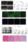
Figure 1
Figure 1
miR-26a KO aggravates Ang-II infusion-induced cardiac damage. (A) M-mode echocardiograms showing left ventricular dimensions. (B) Real-time PCR analysis of ANP, BNP, and β-MHC mRNA levels (n = 6). (C) Representative immunofluorescence staining of WGA (green) in heart. Scale bars: 40 μm. (D) Representative immunofluorescence staining of CD68 (green) in heart. Scale bars: 40 μm. (E) Representative Western blot figure showing the levels of IL-1β, IL-18, α-SMA, and FN in the heart of mice. (F) Semi-quantitative statistical analysis of IL-1β, IL-18, α-SMA, and FN protein levels for Western blotting (n = 6). (G) Representative Masson’s staining of heart. Scale bars: 20 μm. Data are presented as mean ± SD. Ang: Angiotensin; ANP: Atrial natriuretic peptide; BNP: Brain natriuretic peptide; CD: Cluster of Differentiation; Ctrl: Control; DAPI: 4′,6-diamidino-2-phenylindole; FN: Fibronectin; GAPDH: Glyceraldehyde-3-phosphate dehydrogenase; IL: Interleukin; KO: Knockout; miR: MicroRNA; PCR: Polymerase chain reaction; SD: Standard deviation; WGA: Wheat germ agglutinin; WT: Wild type; α-SMA: α-smooth muscle actin; β-MHC: β-myosin heavy chain.
Figure 2
Figure 2
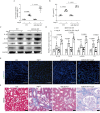
miR-26a KO aggravates Ang-II infusion-induced kidney damage. (A) 24-h urinary protein quantity at the 4th week post Ang-II infusion in WT and miR-26a KO mice (n = 6). (B) SCr levels at the 4th week of Ang-II infusion in WT and miR-26a-KO mice (n = 6). (C) Representative Western blot figure showing the levels of IL-1β, IL-18, α-SMA, and FN in the kidney of mice. (D) Semi-quantitative statistical analysis of IL-1β, IL-18, α-SMA, and FN protein levels by Western blotting test (n = 6). (E) Representative immunofluorescence staining of CD68 (green) in kidney. Scale bars: 40 μm. (F) Representative Masson’s staining of kidney. Scale bars: 20 μm. Data are presented as mean ± SD. Ang: Angiotensin; CD: Cluster of differentiation; Ctrl: Control; DAPI: 4′,6-diamidino-2-phenylindole; FN: Fibronectin; GAPDH: Glyceraldehyde-3-phosphate dehydrogenase; IL: Interleukin; KO: Knockout; miR: MicroRNA; SCr: Serum creatinine; SD: Standard deviation; WT: Wild type; α-SMA: α-smooth muscle actin.
Figure 3
Figure 3
ILK signaling was activated after Ang-II infusion in WT and miR-26a-KO mice. (A) Representative Western blotting figure showing the levels of ILK and LIMS1 in the heart of mice. (B) Semi-quantitative statistical analysis of ILK and LIMS1 protein levels in the heart for Western blotting results (n = 6). (C) Representative Western blotting figure showing the levels of ILK and LIMS1 in the kidney of mice. (D) Semi-quantitative statistical analysis of ILK and LIMS1 protein levels in the kidney for Western blotting results (n = 6). (E) Representative IHC staining of ILK and LIMS1 in the heart and kidney. Scale bars: 40 μm. (F & G) Co-immunoprecipitation demonstrated an interaction between LIMS1 and ILK. Heart or kidney tissue lysates were immunoprecipitated with specific antibody against LIMS1, followed by immunoblotting with antibodies against ILK. Data are presented as mean ± SD. Ang: Angiotensin; Ctrl: Control; GAPDH: Glyceraldehyde-3-phosphate dehydrogenase; IB: Immunoblotting; IHC: Immunohistochemistry; ILK: Integrin-linked kinase; IP: Immunoprecipitation; KO: Knockout; LIMS1: LIM and senescent cell antigen-like domain 1; miR: MicroRNA; SD: Standard deviation; WT: Wild type.
Figure 4
Figure 4
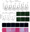
Inhibition of LIMS1 significantly improved Ang-II-induced heart injury. (A) Semi-quantitative statistical analysis of LIMS1, ILK, IL-1β, IL-18, α-SMA and FN protein levels in the heart for Western blotting results (n = 6). (B) Real-time PCR analysis of ANP, BNP and β-MHC mRNA levels (n = 6). (C) Representative immunofluorescence staining of WGA (green) in heart. Scale bars: 40 μm. (D) Representative immunofluorescence staining of CD68 (green) in heart. Scale bars: 40 μm. (E) Representative Masson’s staining of heart. Scale bars: 20 μm. Data are presented as mean ± SD. Ang: Angiotensin; ANP: Atrial natriuretic peptide; BNP: Brain natriuretic peptide; CD: Cluster of Differentiation; Ctrl: Control; DAPI: 4′,6-diamidino-2-phenylindole; FN: Fibronectin; IL: Interleukin; ILK: Integrin-linked kinase; KO: Knockout; LIMS1: LIM and senescent cell antigen-like domain 1; miR: MicroRNA; SCr: Serum creatinine; SD: Standard deviation; WGA: Wheat germ agglutinin; WT: Wild type; α-SMA: α-smooth muscle actin; β-MHC: β-myosin heavy chain.
Figure 5
Figure 5
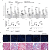
Inhibition of LIMS1 significantly improved Ang-II-induced kidney injury. (A) Semi-quantitative statistical analysis of LIMS1, ILK, IL-1β, IL-18, α-SMA and FN protein levels in kidney for Western blotting results (n = 6). (B) 24-h urinary protein quantity at the 4th week of Ang-II infusion in WT and miR-26a-KO mice (n = 6). (C) SCr levels at the 4th week of Ang-II infusion in WT and miR-26a KO mice (n = 6). (D) Representative immunofluorescence staining of CD68 (green) in kidney. Scale bars: 40 μm. (E) Representative Masson’s staining of kidney. Scale bars: 20 μm. Data are presented as mean ± SD. Ang: Angiotensin; CD: Cluster of Differentiation; Ctrl: Control; DAPI: 4′,6-diamidino-2-phenylindole; FN: Fibronectin; IL: Interleukin; ILK: Integrin-linked kinase; KO: Knockout; LIMS1: LIM and senescent cell antigen-like domain 1; miR: MicroRNA; SCr: Serum creatinine; SD: Standard deviation; WT: Wildtype; α-SMA: α-smooth muscle actin.
Figure 6
Figure 6
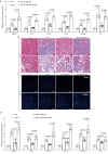
Overexpression of miR-26a significantly improved Ang-II-induced cardiorenal injury. (A) Semi-quantitative statistical analysis of LIMS1, ILK, IL-1β, IL-18, α-SMA and FN protein levels in heart for Western blotting results (n = 6). (B) Representative Masson’s staining of heart and kidney. Scale bars: 20 μm. (C) Representative immunofluorescence staining of CD68 (green) in heart and kidney. Scale bars: 40 μm. (D) Semi-quantitative statistical analysis of LIMS1, ILK, IL-1β, IL-18, α-SMA and FN protein levels in kidney for Western blotting results (n = 6). Data are presented as mean ± SD. Ang: Angiotensin; CD: Cluster of differentiation; Ctrl: Control; DAPI: 4′,6-diamidino-2-phenylindole; FN: Fibronectin; IL: Interleukin; ILK: Integrin-linked kinase; KO: Knockout; LIMS: LIM and senescent cell antigen-like domain 1; miR: MicroRNA; SCr: Serum creatinine; SD: Standard deviation; α-SMA: α-smooth muscle actin.

Similar articles

References

    1. Kalantar-Zadeh K, Jafar TH, Nitsch D, Neuen BL, Perkovic V. Chronic kidney disease. Lancet 2021;398:786–802. doi: 10.1016/S0140-6736(21)00519-5. - PubMed
    1. Ruiz-Ortega M, Rayego-Mateos S, Lamas S, Ortiz A, Rodrigues-Diez RR. Targeting the progression of chronic kidney disease. Nat Rev Nephrol 2020;16:269–288. doi: 10.1038/s41581-019-0248-y. - PubMed
    1. Treiber T, Treiber N, Meister G. Regulation of microRNA biogenesis and its crosstalk with other cellular pathways. Nat Rev Mol Cell Biol 2019;20:5–20. doi: 10.1038/s41580-018-0059-1. - PubMed
    1. Li H, Yang T, Fei Z. miR‑26a‑5p alleviates lipopolysaccharide‑induced acute lung injury by targeting the connective tissue growth factor. Mol Med Rep 2021;23:5. doi: 10.3892/mmr.2020.11643. - PMC - PubMed
    1. Cheng C Chen X Wang Y Cheng W Zuo X Tang W, et al. . MSCs‑derived exosomes attenuate ischemia-reperfusion brain injury and inhibit microglia apoptosis might via exosomal miR-26a-5p mediated suppression of CDK6. Mol Med 2021;27:67. doi: 10.1186/s10020-021-00324-0. - PMC - PubMed