. 2024 May;81(5):e51-e62.
doi: 10.1161/HYPERTENSIONAHA.123.22024. Epub 2024 Mar 6.

Ovariectomy-Induced Arterial Stiffening Differs From Vascular Aging and Is Reversed by GPER Activation

Affiliations

Ovariectomy-Induced Arterial Stiffening Differs From Vascular Aging and Is Reversed by GPER Activation

Isabella M Kilanowski-Doroh et al. Hypertension. 2024 May.

Abstract

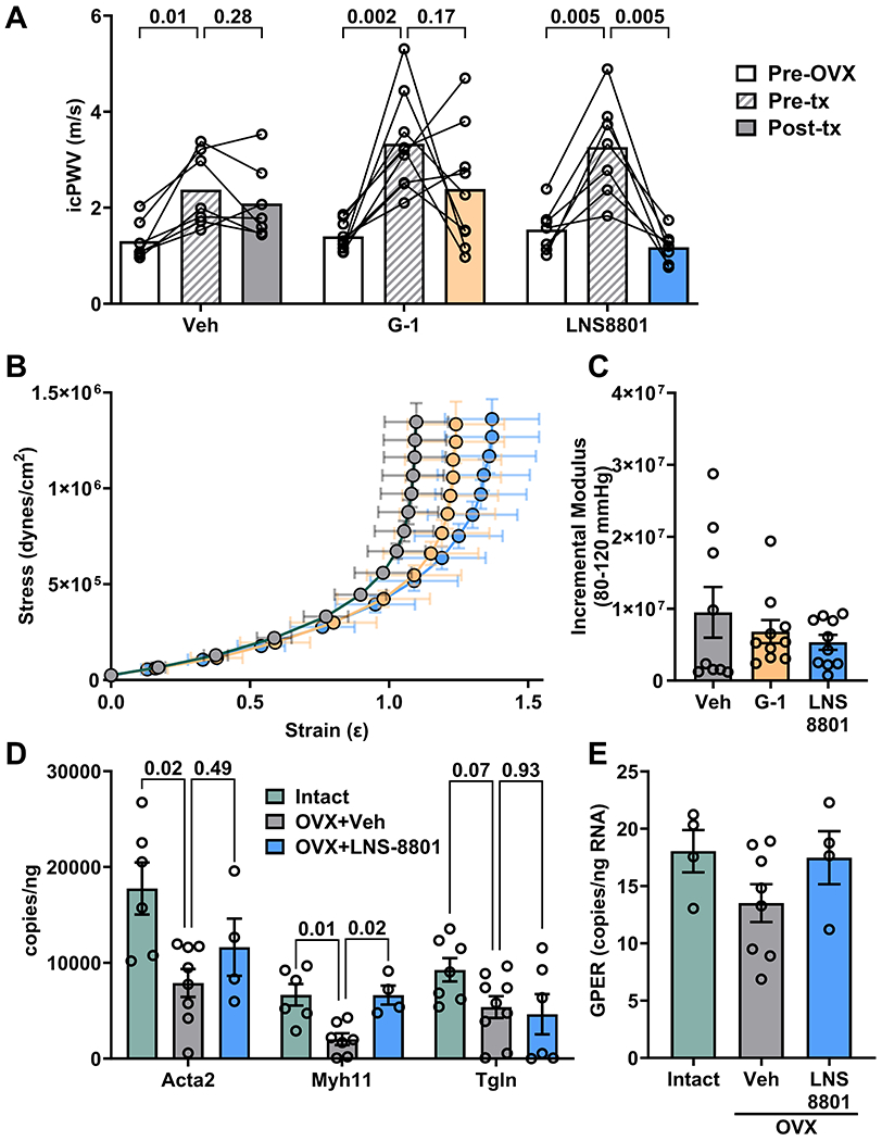
Background: Arterial stiffness is a cardiovascular risk factor and dramatically increases as women transition through menopause. The current study assessed whether a mouse model of menopause increases arterial stiffness in a similar manner to aging and whether activation of the G-protein-coupled estrogen receptor could reverse stiffness.

Methods: Female C57Bl/6J mice were ovariectomized at 10 weeks of age or aged to 52 weeks, and some mice were treated with G-protein-coupled estrogen receptor agonists.

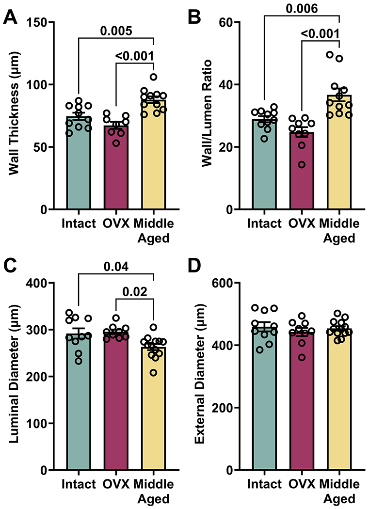
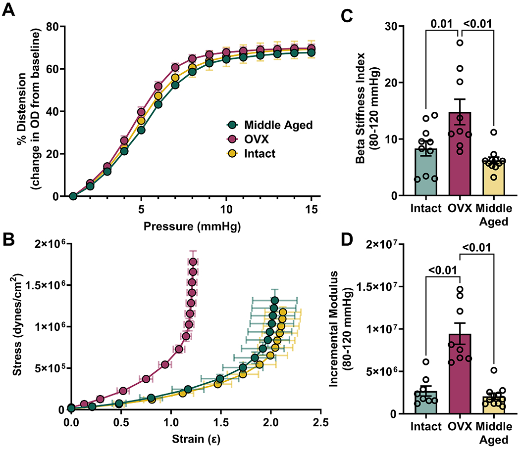
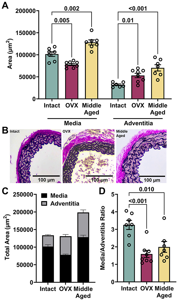
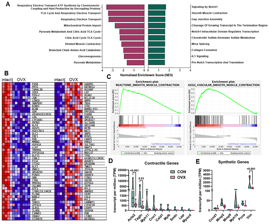
Results: Ovariectomy and aging increased pulse wave velocity to a similar extent independent of changes in blood pressure. Aging increased carotid wall thickness, while ovariectomy increased material stiffness without altering vascular geometry. RNA-sequencing analysis revealed that ovariectomy downregulated smooth muscle contractile genes. The enantiomerically pure G-protein-coupled estrogen receptor agonist, LNS8801, reversed stiffness in ovariectomy mice to a greater degree than the racemic agonist G-1. In summary, ovariectomy and aging induced arterial stiffening via potentially different mechanisms. Aging was associated with inward remodeling, while ovariectomy-induced material stiffness independent of geometry and a loss of the contractile phenotype.

Conclusions: This study enhances our understanding of the impact of estrogen loss on vascular health in a murine model and warrants further studies to examine the ability of LNS8801 to improve vascular health in menopausal women.

Keywords: aging; estrogens; menopause; mice; vascular stiffness.


Conflict of interest statement

Disclosures C.A. Natale is an employee and shareholder of Linnaeus Therapeutics and listed as inventor on patents related to this work. The other authors report no conflicts.

Figures

Figure 1.
Figure 1.
Impact of OVX and aging on PWV and blood pressure. Values are mean ± SEM, N=10-11 per group. (A) Intracarotid PWV (icPWV) and (B) Aortic PWV (aPWV) were significantly increased in both OVX and middle-aged groups compared with intact controls, one-way ANOVA, P=0.007 and P=0.01. (C) Systolic blood pressure (SBP) was different by one-way ANOVA, P=0.01, due to a difference between the OVX and middle-aged groups. (D) Pulse pressure was not different between groups, one-way ANOVA, P=0.14. (E) Blood pressure recorded by radiotelemetry also shows no difference between intact and OVX, analyzed by multiple unpaired t-tests.
Figure 2.
Figure 2.. Impact of OVX and aging on vascular geometry.
Values are mean ± SEM, N=10-11 per group. (A) Wall thickness and (B) wall-to-lumen ratio were significantly increased in middle-aged but not OVX mice, one-way ANOVA, both P<0.001. (C) Luminal diameter was significantly decreased in middle-aged mice, one-way ANOVA, P=0.009. (D) External diameter was not different between groups, one-way ANOVA, P=0.61. Significant findings from Bonferroni’s multiple comparisons tests are indicated on each graph.
Figure 3.
Figure 3.. Impact of OVX and aging on ex vivo arterial stiffness.
Values are mean ± SEM, N=10-11 per group. (A) There were no significant differences in carotid distensibility between groups over the range of physiological pressures, two-way ANOVA, Pgroup=0.38. (B) A leftward shift of the OVX stress-strain curve indicates increased material stiffness. (C) Beta stiffness index and (D) incremental modulus calculated between the physiological range of 80-120 mmHg shows that OVX but not aging increases material stiffness, one-way ANOVA, both P<0.01. Significant findings from Bonferroni’s multiple comparisons tests are indicated on each graph.
Figure 4.
Figure 4.. Impact of Aging and OVX on Aortic Wall Composition.
Values are mean ± SEM, N=10-11 per group. (A) The area of media and adventitia analyzed separately by one-way ANOVA were both P<0.001. Medial area was significantly decreased in OVX but increased with aging while adventitial area was increased by both OVX and aging. (B) Representative images stained with Verhoeff-Van Gieson stain are shown below the graph. (C) Summed data show the contribution of each layer to overall cross-sectional area. (D) The media to adventitia ratio is significantly reduced by both OVX and middle-age, one-way ANOVA, P<0.001. Significant findings from Bonferroni’s multiple comparisons tests are indicated on each graph.
Figure 5.
Figure 5.. Gene set enrichment (GSEA) analysis of intact and OVX aortas.
Bulk RNA-Seq was performed on aortas collected from young adult female mice after OVX for 8 weeks compared to aortas from intact mice (control). (A) Reactome enrichment analysis showing the top 10 enriched gene sets in both conditions, all with nominal p value < 0.05. A positive normalized enrichment score (NES) indicates enrichment in intact controls (green), while a negative NES indicates enrichment in OVX (purple). (B) Heat map of the top 50 genes for each phenotype. Expression values are represented as a range from red (high expression) to blue (low expression). (C) Both Reactome and KEGG gene analysis for gene sets associated with smooth muscle cell contraction show enrichment in intact control aortas. (D) Selected contractile genes showed significant downregulation by OVX, two-way ANOVA, Povx=0.002. (E) OVX also decreased expression of synthetic genes, two-way ANOVA, Povx=0.049. Significant findings from Bonferroni’s multiple comparisons tests are indicated on each graph.
Figure 6.
Figure 6.. Impact of GPER activation on PWV.
Values are mean ± SEM, N=8-10 per group. (A) First data point in each group is PWV before OVX, second PWV is 8 weeks post-OVX, and third PWV was after two-week drug treatment. Repeated measures ANOVA showed a significant effect of time, P<0.001, and the results of Holm-Sidak multiple comparisons test are shown on the graph. (B) A rightward shift in the stress-strain curve was observed for both G-1 and LNS-8801 treatment groups. (C) Incremental modulus calculated between 80-120 mmHg was not altered, one-way ANOVA, P=0.60. (D) Contractile gene expression was different between groups, two-way ANOVA, Pgene<0.001 and Pgroup=0.002. (F) GPER mRNA in aortic tissue was unaffected by OVX or treatment, one-way ANOVA, P=0.27. Bonferroni’s multiple comparisons tests are indicated on each graph.

Update of

Similar articles

Cited by

References

    1. Laurent S, Boutouyrie P, Asmar R, Gautier I, Laloux B, Guize L, Ducimetiere P, Benetos A. Aortic Stiffness Is an Independent Predictor of All-Cause and Cardiovascular Mortality in Hypertensive Patients. Hypertension. 2001;37:1236–1241. doi: doi:10.1161/01.HYP.37.5.1236 - DOI - PubMed
    1. Natoli AK, Medley TL, Ahimastos AA, Drew BG, Thearle DJ, Dilley RJ, Kingwell BA. Sex Steroids Modulate Human Aortic Smooth Muscle Cell Matrix Protein Deposition and Matrix Metalloproteinase Expression. Hypertension. 2005;46:1129–1134. doi: doi:10.1161/01.HYP.0000187016.06549.96 - DOI - PubMed
    1. Ogola BO, Zimmerman MA, Clark G, Abshire CM, Gentry KM, Miller K, Lindsey SH. New insights into arterial stiffening: does sex matter? Am J Physiol Heart Circ Physiol. 2018;315:H1073–H1087. - PMC - PubMed
    1. Oh YS, Berkowitz DE, Cohen RA, Figueroa CA, Harrison DG, Humphrey JD, Larson DF, Leopold JA, Mecham RP, Ruiz-Opazo N, et al. A special report on the NHLBI initiative to study cellular and molecular mechanisms of arterial stiffness and its association with hypertension. Circulation Research. 2017;121:1216–1218. doi: 10.1161/CIRCRESAHA.117.311703 - DOI - PMC - PubMed
    1. Mammoto A, Matus K, Mammoto T. Extracellular Matrix in Aging Aorta. Front Cell Dev Biol. 2022;10:822561. doi: 10.3389/fcell.2022.822561 - DOI - PMC - PubMed