

Characterization of the tumor microenvironment in the mouse oral cancer (MOC1) model after orthotopic implantation in the buccal mucosa
- PMID: 38445546
- PMCID: PMC11003840
- DOI: 10.1002/hed.27722
Characterization of the tumor microenvironment in the mouse oral cancer (MOC1) model after orthotopic implantation in the buccal mucosa
Abstract
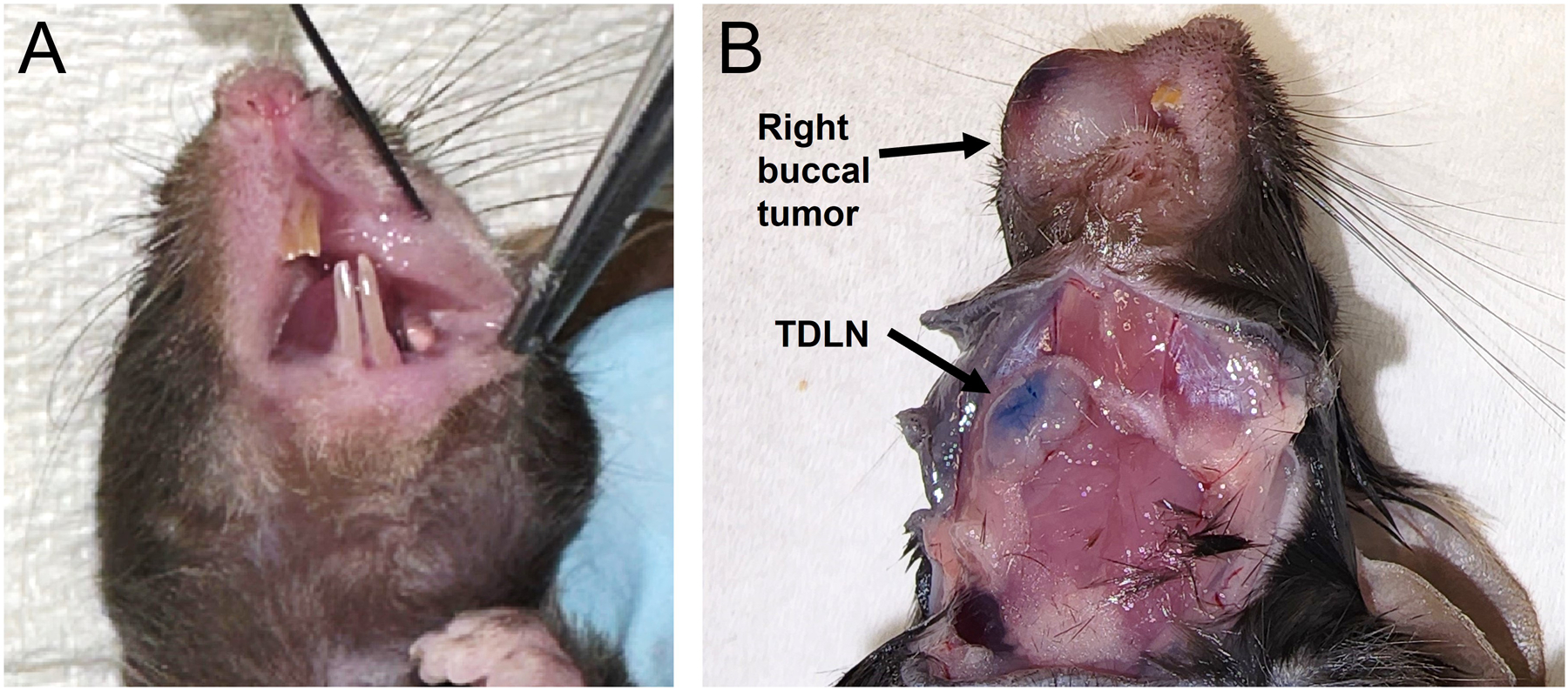
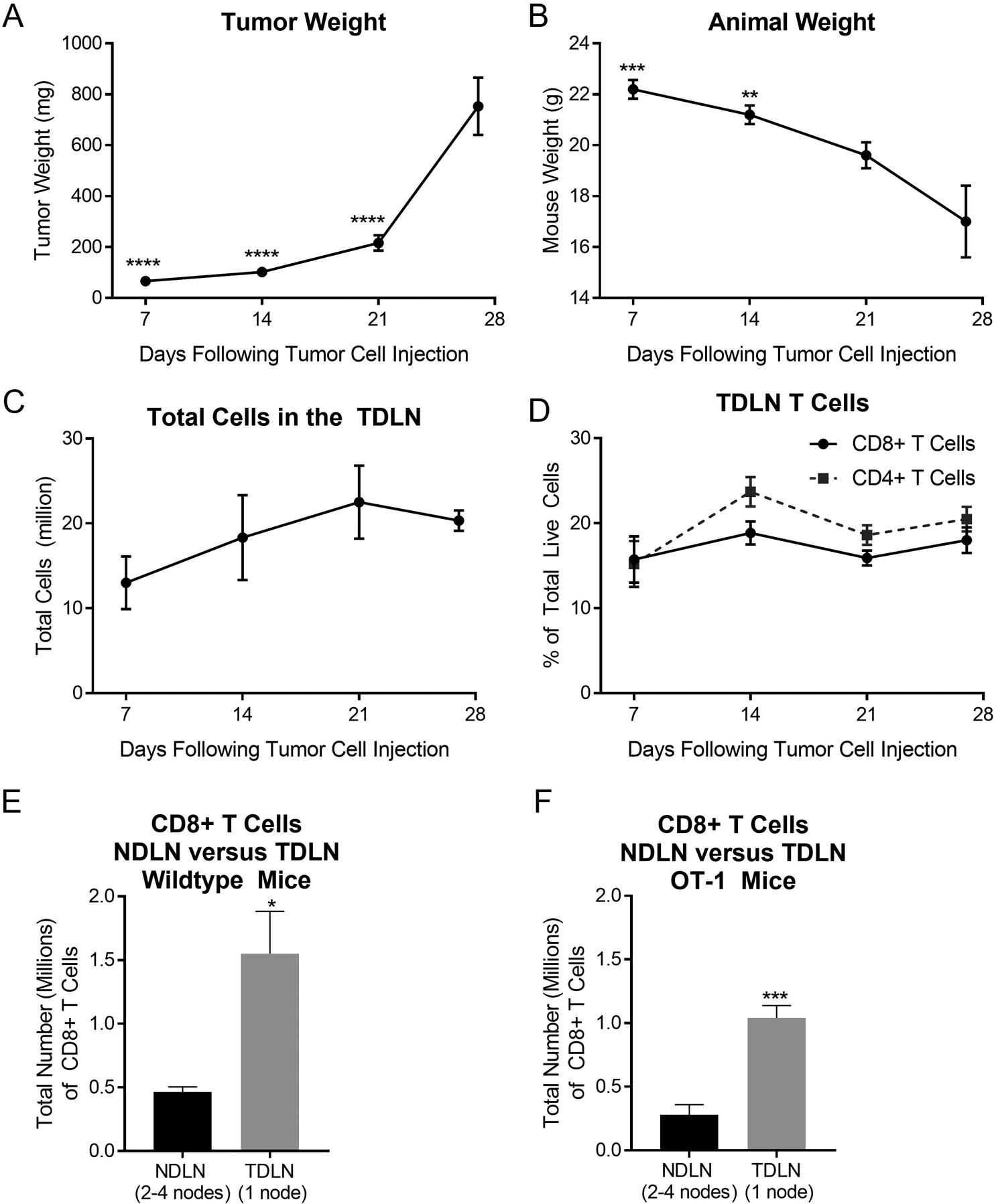
Background: Preclinical models are invaluable for studies of head and neck cancer. There is growing interest in the use of orthotopic syngeneic models, wherein cell lines are injected into the oral cavity of immunocompetent mice. In this brief report, we describe injection of mouse oral cancer 1 (MOC1) cells into the buccal mucosa and illustrate the tumor growth pattern, lymph node response, and changes in the tumor immune microenvironment over time.
Methods: MOC1 cells were injected into the buccal mucosa of C57BL6 mice. Animals were sacrificed at 7, 14, 21, or 27 days. Tumors and lymph nodes were analyzed by flow cytometry.
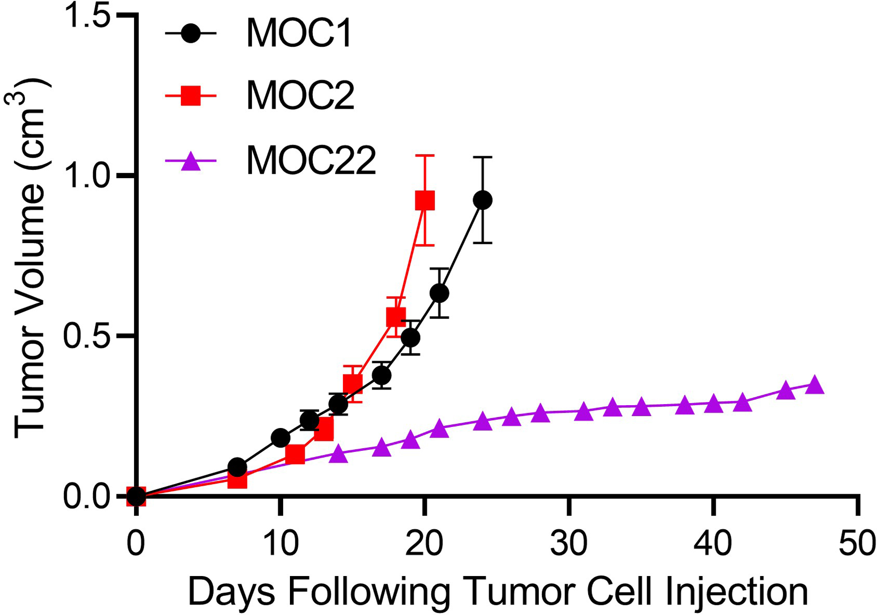
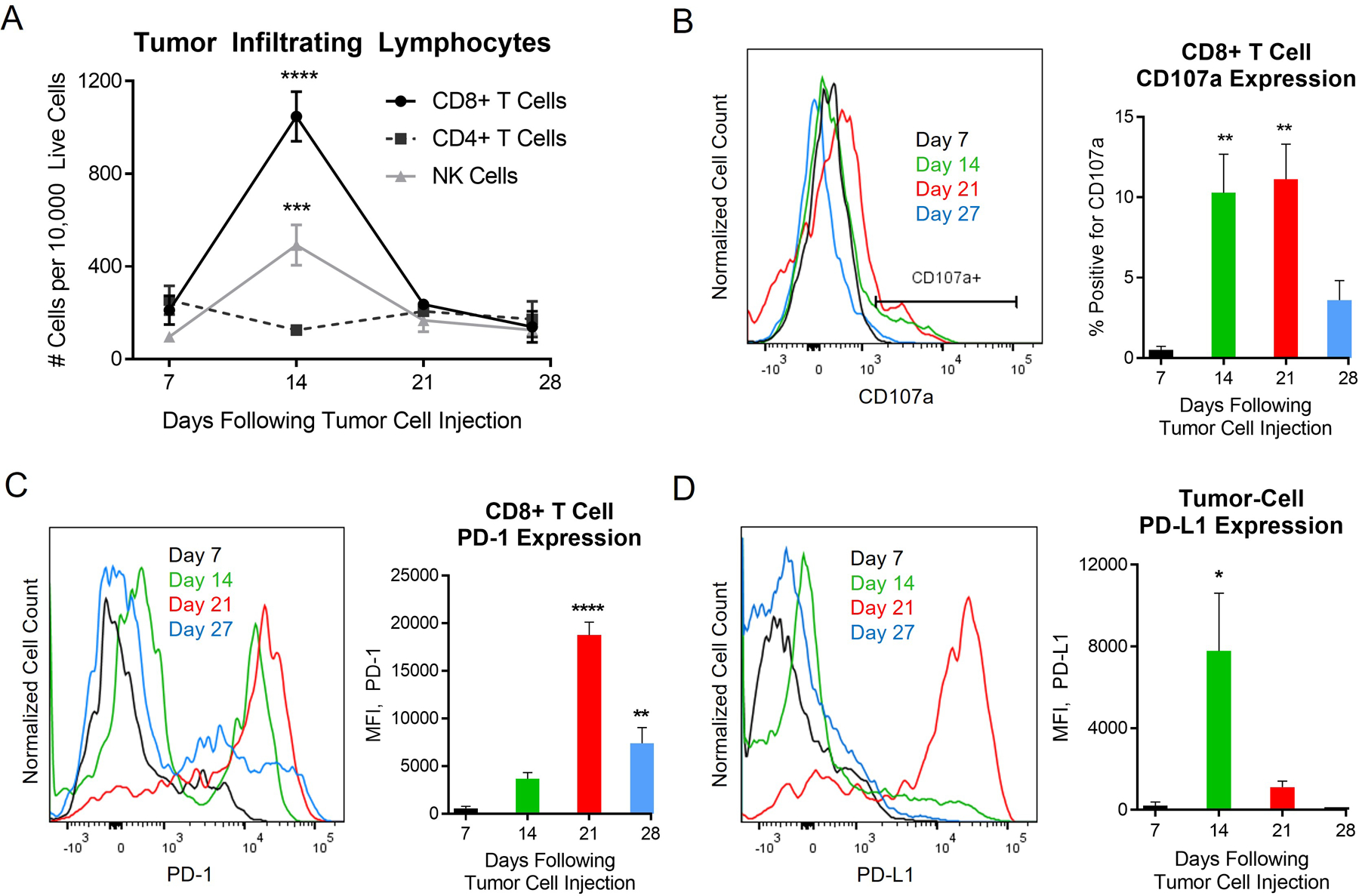
Results: All mice developed tumors by day 7 and required euthanasia for tumor burden and/or weight loss by day 27. Lymph node mapping showed that these tumors reliably drain to a submandibular lymph node. The proportion of intratumoral CD8+ T cells decreased over time, while neutrophilic myeloid cells increased dramatically. Growth of orthotopic MOC2 and MOC22 also showed similar growth patterns versus published data in flank tumors.
Conclusions: When used orthotopically in the buccal mucosa, the MOC1 model induces a robust lymph node response and distinct pattern of immune cell infiltration, with peak immune infiltration by day 14.
Keywords: head and neck cancer; mouse model; oral cancer; orthotopic model; syngeneic mouse model.
© 2024 Wiley Periodicals LLC.
Conflict of interest statement
Conflict of Interest: The authors declare no conflict of interest.
Figures

Similar articles
-
Orthotopic injection of an established syngeneic mouse oral cancer cell line (MOC1) induces a robust draining lymph node response.bioRxiv [Preprint]. 2024 Jan 14:2024.01.12.575399. doi: 10.1101/2024.01.12.575399. bioRxiv. 2024. PMID: 38260311 Free PMC article. Preprint.
-
Enhanced oral versus flank lymph node T cell response parallels anti-PD1 efficacy in head and neck cancer.Oral Oncol. 2024 May;152:106795. doi: 10.1016/j.oraloncology.2024.106795. Epub 2024 Apr 9. Oral Oncol. 2024. PMID: 38599127 Free PMC article.
-
MHC-I and PD-L1 Expression is Associated with Decreased Tumor Outgrowth and is Radiotherapy-inducible in the Murine Head and Neck Squamous Cell Carcinoma Model MOC1.Mol Imaging Biol. 2024 Oct;26(5):835-846. doi: 10.1007/s11307-024-01934-w. Epub 2024 Jul 15. Mol Imaging Biol. 2024. PMID: 39009951 Free PMC article.
-
The mouse oral carcinoma (MOC) model: A 10-year retrospective on model development and head and neck cancer investigations.Oral Oncol. 2022 Sep;132:106012. doi: 10.1016/j.oraloncology.2022.106012. Epub 2022 Jul 9. Oral Oncol. 2022. PMID: 35820346 Free PMC article. Review.
-
Model for mediastinal lymph node metastasis produced by orthotopic intrapulmonary implantation of lung cancer cells in mice.Hum Cell. 1999 Dec;12(4):197-204. Hum Cell. 1999. PMID: 10834106 Review.
Cited by
-
Statin Drugs Are Associated With Response to Immune Checkpoint Blockade in Recurrent/Metastatic Head and Neck Cancer.Cancer Med. 2025 Mar;14(5):e70718. doi: 10.1002/cam4.70718. Cancer Med. 2025. PMID: 40052634 Free PMC article.
-
Timely administration of drug combination improves chemoimmunotherapy of an immune-cold tumor.J Control Release. 2025 May 10;381:113579. doi: 10.1016/j.jconrel.2025.02.075. Epub 2025 Feb 27. J Control Release. 2025. PMID: 40023227
References
Publication types
MeSH terms
Grants and funding
LinkOut - more resources
Full Text Sources
Medical
Research Materials
