. 2024 May 29;36(6):2359-2374.
doi: 10.1093/plcell/koae074.

ENHANCER OF SHOOT REGENERATION1 promotes de novo root organogenesis after wounding in Arabidopsis leaf explants

Affiliations

ENHANCER OF SHOOT REGENERATION1 promotes de novo root organogenesis after wounding in Arabidopsis leaf explants

Kyounghee Lee et al. Plant Cell. .

Abstract

Plants have an astonishing ability to regenerate new organs after wounding. Here, we report that the wound-inducible transcription factor ENHANCER OF SHOOT REGENERATION1 (ESR1) has a dual mode of action in activating ANTHRANILATE SYNTHASE ALPHA SUBUNIT1 (ASA1) expression to ensure auxin-dependent de novo root organogenesis locally at wound sites of Arabidopsis (Arabidopsis thaliana) leaf explants. In the first mode, ESR1 interacts with HISTONE DEACETYLASE6 (HDA6), and the ESR1-HDA6 complex directly binds to the JASMONATE-ZIM DOMAIN5 (JAZ5) locus, inhibiting JAZ5 expression through histone H3 deacetylation. As JAZ5 interferes with the action of ETHYLENE RESPONSE FACTOR109 (ERF109), the transcriptional repression of JAZ5 at the wound site allows ERF109 to activate ASA1 expression. In the second mode, the ESR1 transcriptional activator directly binds to the ASA1 promoter to enhance its expression. Overall, our findings indicate that the dual biochemical function of ESR1, which specifically occurs near wound sites of leaf explants, maximizes local auxin biosynthesis and de novo root organogenesis in Arabidopsis.


Conflict of interest statement

Conflict of interest statement. None declared.

Figures

Figure 1.
Figure 1.
ESR1 enhances de novo root organogenesis. A) De novo root organogenesis from leaf explants of Col-0, esr1-D, and Pro35S:MYC-ESR1. Leaf explant–derived adventitious roots were photographed at 13 days after wounding. Scale bars, 5 mm. B) Quantification of the capacity of adventitious root formation, shown as a percentage of explants with adventitious roots. C) De novo root organogenesis from leaf explants of Col-0, esr1-2, and ProWIND1:WIND1-SRDX. Leaf explant–derived adventitious roots were photographed at 10 days after wounding. Scale bars, 5 mm. D) Quantification of leaf explants with adventitious roots from (C). In (A and C), third and fourth leaves of 2-week-old seedlings were excised and incubated on phytohormone-free MS medium at 22 °C in the dark to induce adventitious root formation. In (B and D), the box and whisker plots show the distribution of the biological replicates. The bottom and top of each box are the first (lower) and third (higher) quartiles, and the band in the box is the median value. The ends of the whiskers represent 1.5 interquartile range of lower and higher quartiles. Each dot represents the average for one biological replicate (n > 7 for each genotype in each biological replicate). Statistically significant differences were determined using one-way analysis of variance (ANOVA), followed by Newman–Keuls's post hoc test. Different letters indicate significant differences (P < 0.05).
Figure 2.
Figure 2.
ESR1 directly represses JAZ5 specifically in the midvein after wounding. A) Expression profiling of genes involved in JA signaling in wild-type and esr1-2 leaf explants. Third and fourth leaves of 2-week-old seedlings were excised and incubated on phytohormone-free MS medium at 22 °C for 6 h. The distal and proximal regions from the wound site of leaf explants were separately harvested (see Supplementary Fig. S2). B)JAZ5 expression in wild-type and Pro35S:MYC-ESR1 leaf explants. Leaf explants were incubated on phytohormone-free MS medium at 22 °C for 24 h. Whole leaf explants were harvested for gene expression analysis. In (A, B), transcript accumulation was analyzed by RT-qPCR. eIF4a (At3g13920) was used as an internal control. C) Structure of the JAZ5 locus. Gray lines above the labels (A, B, C, D) indicate regions amplified by quantitative real-time PCR (qPCR) following chromatin immunoprecipitation (ChIP). Red arrowhead represents GCC-box motif, which is the putative binding site of ESR1 transcription factor. Black boxes indicate exons. D) Enrichment of ESR1 at the JAZ5 locus. Leaf explants were incubated on phytohormone-free MS medium at 22 °C for 24 h. Whole leaf explants were harvested for ChIP-qPCR analysis. Values obtained from control plants (Col-0) were set to 1 after normalization against eIF4a. E) Spatial expression of JAZ5 in ProJAZ5:JAZ5-GFP-GUS transgenic leaf explants incubated on phytohormone-free MS medium at 22 °C for up to 24 h. The lower panels show magnified images of the midvein near the wound site. Scale bars, 1 mm. F) Spatial expression of ESR1 in ProESR1:ESR1-GFP transgenic leaf explants incubated on phytohormone-free MS medium at 22 °C for up to 24 h. Calcofluor white was used to stain cell walls. Scale bars, 200 μm. G) De novo root organogenesis from leaf explants of ProESR1:JAZ5. Leaf explant–derived adventitious roots were photographed at 10 days after wounding. Scale bars, 5 mm. H) Quantification of the capacity of adventitious root formation, shown as a percentage of explants with adventitious roots. The box and whisker plots show the distribution of the biological replicates. The bottom and top of each box are the first (lower) and third (higher) quartiles, and the band in the box is the median value. The ends of the whiskers represent 1.5 interquartile range of lower and higher quartiles. Each dot represents the average for one biological replicate (n > 10 for each genotype in each biological replicate). Data in (A, B, D) represent means ± SEM of three biological replicates. Asterisks indicate statistically significant differences (*P < 0.05, **P < 0.01, ***P < 0.001; Student's t-test).
Figure 3.
Figure 3.
ESR1 physically interacts with HDACs. A) De novo root organogenesis from leaf explants treated with dimethyl sulfoxide (DMSO) (TSA solvent control) or trichostatin A (TSA). Third and fourth leaves of 2-week-old Col-0 seedlings were excised and incubated on phytohormone-free MS medium with DMSO or 1 μM TSA at 22 °C in the dark. Leaf explant–derived adventitious roots were photographed at 12 days after wounding. Scale bars, 5 mm. B) Quantification of the capacity of adventitious root formation from explants in (A), shown as a percentage of explants with adventitious roots. The box and whisker plots show the distribution of the biological replicates. The bottom and top of each box are the first (lower) and third (higher) quartiles, and the band in the box is the median value. The ends of the whiskers represent 1.5 interquartile range of lower and higher quartiles. Each dot represents the average for one biological replicate (n > 13 for each genotype in each biological replicate). DAW, days after wounding. C)JAZ5 expression in leaf explants treated with DMSO or TSA. Leaf explants were incubated on phytohormone-free MS medium at 22 °C for 24 h. Whole leaf explants were harvested for gene expression analysis. Transcript levels were analyzed by RT-qPCR. eIF4a (At3g13920) was used as an internal control. Data represent means ± SEM of three biological replicates. Asterisks indicate statistically significant differences (*P < 0.05; Student's t-test). D) Yeast two-hybrid (Y2H) assays performed using HDACs fused to the GAL4 DNA-binding domain (BD) and ESR1 fused to the GAL4 transcriptional activation domain (AD). Interaction between the indicated proteins was determined by cell growth on selective medium. −LWHA: synthetic defined (SD) medium lacking Leu, Trp, His, and Ade; −LW: SD medium lacking Leu and Trp. GAL4 was used as a positive control (P). E) Expression of ESR1 and HDAC genes upon wounding. Gene expression patterns were analyzed based on REGENOMICS (Bae et al. 2022). The color code indicates Z-scores computed by scaling TPM expression values.
Figure 4.
Figure 4.
HDA6 represses JAZ5 specifically in the wounded region. A) HDA6 protein accumulation upon wounding. Third and fourth leaves of 2-week-old hda6-6 ProHDA6:HDA6-GFP seedlings were excised and incubated on phytohormone-free MS medium at 22 °C for up to 3 days. Whole leaf explants were harvested for immunoblot analysis. C, Coomassie blue-stained gel. B) Relative protein abundance of HDA6. Statistically significant differences were determined using one-way analysis of variance (ANOVA), followed by Newman–Keuls's post hoc test. Different letters indicate significant differences (P < 0.05). Error bars indicate SE. C) Spatial expression pattern of HDA6 in hda6-6 ProHDA6:HDA6-GFP leaf explants cultured on phytohormone-free MS medium at 22 °C for up to 24 h. Calcofluor white was used to stain cell walls. Scale bars, 200 μm. D) De novo root organogenesis from Col-0 and hda6-6 leaf explants. Leaf explant–derived adventitious roots were photographed at 10 days after wounding. Scale bars, 5 mm. E) Quantification of the capacity of adventitious root formation, shown as a percentage of explants with adventitious roots. The box and whisker plots show the distribution of the biological replicates. The bottom and top of each box are the first (lower) and third (higher) quartiles, and the band in the box is the median value. The ends of the whiskers represent 1.5 interquartile range of lower and higher quartiles. Each dot represents the average for one biological replicate (n > 8 for each genotype in each biological replicate). F)JAZ5 expression in Col-0 and hda6-6 leaf explants. Leaf explants were incubated on phytohormone-free MS medium at 22 °C for 6 h. The distal and proximal regions from the wound site of leaf explants were harvested for RT-qPCR analysis using eIF4a (At3g13920) as an internal control. G) Co-immunoprecipitation (Co-IP) assays performed using 2-week-old Pro35S:MYC-ESR1 × Pro35S:HDA6-GFP homozygous F3 transgenic seedlings. Total proteins were immunoprecipitated with anti-GFP antibody. Epitope-tagged proteins were detected immunologically using anti-GFP and anti-MYC antibodies. IP, immunoprecipitation. H) ChIP-qPCR analysis showing the enrichment of HDA6 at the JAZ5 locus in Pro35S:HDA6-GFP leaf explants. I) qPCR analysis showing the enrichment of HDA6 at the JAZ5 locus in Pro35S:HDA6-GFP and esr1-2 Pro35S:HDA6-GFP leaf explants. In (H and I), leaf explants were incubated on phytohormone-free MS medium at 22 °C for 24 h. Whole leaf explants were harvested for ChIP-qPCR. The labels (A, B, C, D) mean regions amplified by quantitative PCR (qPCR) following chromatin immunoprecipitation (ChIP) shown in (H). Values obtained from control plants were set to 1 after normalization against eIF4a. J) Abundance of H3 acetylation (H3ac) at the JAZ5 locus in Col-0 and hda6-6 leaf explants. K) H3ac abundance at the JAZ5 locus in Col-0 and the esr1-2 mutant. In (J and K), leaf explants were incubated on phytohormone-free MS medium at 22 °C for 6 h. The proximal region from the wound site of leaf explants was harvested for ChIP using anti-H3ac antibody. The labels (A, B, C, D) mean regions amplified by quantitative PCR (qPCR) following chromatin immunoprecipitation (ChIP) shown in (H). Data in (F, H–K) represent means ± SEM of three biological replicates. Asterisks indicate statistically significant differences (*P < 0.05, **P < 0.01; Student's t-test).
Figure 5.
Figure 5.
ESR1 and HDACs interdependently repress JAZ5 expression. A) De novo root organogenesis from Pro35S:MYC-ESR1 leaf explants treated with dimethyl sulfoxide (DMSO) alone or with trichostatin A (TSA). Third and fourth leaves of 2-week-old seedlings were excised and incubated on phytohormone-free MS medium with DMSO or 0.2 μM TSA at 22 °C in the dark to induce adventitious root formation. Leaf explant–derived adventitious roots were photographed at 7 and 14 days after wounding (DAW). Scale bars, 5 mm. B) Quantification of the capacity of adventitious root formation, shown as a percentage of explants with adventitious roots. C)JAZ5 expression in Col-0 and Pro35S:MYC-ESR1 leaf explants treated with DMSO or TSA. Leaf explants were incubated on phytohormone-free MS medium with or without 0.2 μM TSA at 22 °C for 24 h. Whole leaf explants were harvested for RT-qPCR analysis, using eIF4a (At3g13920) as an internal control. D) De novo root organogenesis from leaf explants of Col-0, hda6-6, Pro35S:MYC-ESR1, and hda6-6 Pro35S:MYC-ESR1 seedlings after incubation on phytohormone-free MS medium at 22 °C for 13 days. Scale bars, 5 mm. E) Percentages of leaf explants with adventitious roots. F) De novo root organogenesis from leaf explants of Col-0, Pro35S:MYC-ESR1, and Pro35S:MYC-ESR1 × Pro35S:HDA6-GFP seedlings after incubation on phytohormone-free MS medium for 7 days. Scale bars, 1 mm. G) Quantification of the portion of leaf explants containing different numbers of adventitious roots. The color keys indicate the number of adventitious roots formed. H)JAZ5 expression in Col-0, Pro35S:MYC-ESR1, Pro35S:HDA6-GFP, and Pro35S:MYC-ESR1 × Pro35S:HDA6-GFP seedlings. Leaf explants were incubated on phytohormone-free MS medium at 22 °C for 24 h. Whole leaf explants were harvested for gene expression analysis. I) Proposed model for the role of the ESR1–HDA6 complex in de novo root organogenesis. ESR1 represses JAZ5 upon wounding through its interaction with HDA6 to promote de novo root organogenesis. In (B, E, G), the box and whisker plots show the distribution of the biological replicates. The bottom and top of each box are the first (lower) and third (higher) quartiles, and the band in the box is the median value. The ends of the whiskers represent 1.5 interquartile range of lower and higher quartiles. Each dot represents the average for one biological replicate (n > 8 for each genotype in each biological replicate). Data in (C, H) represent means ± SEM of three biological replicates. In (B, H), asterisks indicate statistically significant differences (*P < 0.05, **P < 0.01; Student's t-test). In (C, E), statistically significant differences were determined using one-way analysis of variance (ANOVA), followed by Newman–Keuls's post hoc test. Different letters indicate significant differences (P < 0.05).
Figure 6.
Figure 6.
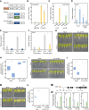
ESR1 activates ASA1 expression via two modes. A–C) Transient expression assays using Arabidopsis mesophyll protoplasts. The ASA1 promoter was cloned into the β-GLUCURONIDASE (GUS) reporter plasmid, which was transiently co-transfected with the indicated effector constructs into Arabidopsis protoplasts (A), and GUS activities were measured. B, C). Activity derived from the firefly luciferase (LUC) gene driven by the CaMV 35S promoter was used to normalize GUS activity levels. The normalized values in control protoplasts were set to 1. pMIN35S, CaMV 35S minimal promoter; EV, empty vector control. D)ASA1 expression in Col-0 and ProESR1:JAZ5 leaf explants. Third and fourth leaves of 2-week-old seedlings were excised and incubated on phytohormone-free MS medium at 22 °C for 6 h. Whole leaf explants were harvested for RT-qPCR analysis, using eIF4a (At3g13920) as an internal control. E)ASA1 expression in Col-0 and esr1-2 leaf explants. F)ASA1 expression in Col-0 and hda6-6 leaf explants. For (E and F), leaf explants were incubated on phytohormone-free MS medium at 22 °C for 6 h. The distal and proximal regions from the wound site of leaf explants were harvested for gene expression analysis. G) De novo root organogenesis from Col-0 and esr1-2 leaf explants treated with dimethyl sulfoxide (DMSO) (Mock) or indole-3-acetic acid (IAA). Leaf explant–derived adventitious roots were photographed at 7 days after wounding. Scale bars, 5 mm. H) Quantification of the capacity of adventitious root formation, shown as a percentage of explants with adventitious roots. I) De novo root organogenesis from Col-0 and hda6-6 leaf explants treated with DMSO (Mock) or IAA. Leaf explant–derived adventitious roots were observed at 7 days after wounding. Scale bars, 5 mm. J) Percentages of leaf explants with adventitious roots. In (G–J), leaf explants were incubated on phytohormone-free MS medium containing DMSO (Mock) or 0.5 μM IAA at 22 °C in the dark to induce adventitious root formation. K) De novo root organogenesis from Col-0 and Pro35S:MYC-ESR1 leaf explants treated with DMSO (Mock) or yucasin difluorinated analog (YDF). Leaf explants were incubated on phytohormone-free MS medium with DMSO (Mock) or 200 μM YDF at 22 °C in the dark to induce adventitious root formation. Leaf explant–derived adventitious roots were observed at 14 days after wounding. Scale bars, 5 mm. L) Percentages of leaf explants with adventitious roots. M) Structure of the ASA1 locus. Gray lines above the labels (A, B, C, D) indicate regions amplified by qPCR following chromatin immunoprecipitation (ChIP). Red arrowhead represents GCC-box motif, which is the putative binding site of ESR1 transcription factor. Black boxes indicate exons. N) Enrichment of ESR1 at the ASA1 locus. Leaf explants were incubated on phytohormone-free MS medium at 22 °C for 24 h. Whole leaf explants were harvested for ChIP. Values obtained from control plants were set to 1 after normalization against eIF4a. Data in (B–F, N) represent means ± SEM of three biological replicates. In (H, J, L), the box and whisker plots show the distribution of the biological replicates. The bottom and top of each box are the first (lower) and third (higher) quartiles, and the band in the box is the median value. The ends of the whiskers represent 1.5 interquartile range of lower and higher quartiles. Each dot represents the average for one biological replicate (n > 14 for each genotype in each biological replicate). In (B, C, L), statistically significant differences were determined using one-way analysis of variance (ANOVA), followed by Newman–Keuls's post hoc test. Different letters indicate significant differences (P < 0.05). In (D–F, H, J, N), asterisks indicate statistically significant differences (*P < 0.05, **P < 0.01, ***P < 0.001; Student's t-test).
Figure 7.
Figure 7.
Proposed model showing the role of ESR1 in de novo root organogenesis. ESR1 enhances de novo root organogenesis specifically in the vasculature near the wound site. Upon wounding, ESR1 is expressed in the leaf vasculature near the wound site. ESR1 activates ASA1 expression in two different ways. (i) ESR1 directly activates ASA1 expression through its transcriptional activation activity. (ii) ESR1 forms a complex with HDA6, which binds to the JAZ5 locus, inhibiting its expression by histone deacetylation. Depleted JAZ5 expression in vasculature allows ERF109 to promote ASA1 expression. ESR1-dependent activation of ASA1 expression facilitates auxin-dependent de novo root organogenesis. Red flathead, gene repression; blue arrow, gene activation; X symbol, protein degradation or inactivation; Ac, acetyl group of histone H3 protein (blue circle).

Similar articles

Cited by

References

    1. Bae SH, Noh YS, Seo PJ. REGENOMICS: a web-based application for plant REGENeration-associated transcriptOMICS analyses. Comput Struct Biotechnol J. 2022:20:3234–3247. 10.1016/j.csbj.2022.06.033 - DOI - PMC - PubMed
    1. Bai T, Dong Z, Zheng X, Song S, Jiao J, Wang M, Song C. Auxin and its interaction with ethylene control adventitious root formation and development in apple rootstock. Front Plant Sci. 2020:11:574881. 10.3389/fpls.2020.574881 - DOI - PMC - PubMed
    1. Bossi F, Cordoba E, Dupre P, Mendoza MS, Roman CS, Leon P. The Arabidopsis ABA-INSENSITIVE (ABI) 4 factor acts as a central transcription activator of the expression of its own gene, and for the induction of ABI5 and SBE2.2 genes during sugar signaling. Plant J. 2009:59(3):359–374. 10.1111/j.1365-313X.2009.03877.x - DOI - PubMed
    1. Chandler JW, Cole M, Flier A, Grewe B, Werr W. The AP2 transcription factors DORNROSCHEN and DORNROSCHEN-LIKE redundantly control Arabidopsis embryo patterning via interaction with PHAVOLUTA. Development. 2007:134(9):1653–1662. 10.1242/dev.001016 - DOI - PubMed
    1. Chen L, Tong J, Xiao L, Ruan Y, Liu J, Zeng M, Huang H, Wang JW, Xu L. YUCCA-mediated auxin biogenesis is required for cell fate transition occurring during de novo root organogenesis in Arabidopsis. J Exp Bot. 2016:67(14):4273–4284. 10.1093/jxb/erw213 - DOI - PMC - PubMed

MeSH terms