. 2024 Mar;28(6):e18161.
doi: 10.1111/jcmm.18161.

Prebiotics counteract the morphological and functional changes secondary to chronic cisplatin exposition in the proximal colon of mice

Affiliations

Prebiotics counteract the morphological and functional changes secondary to chronic cisplatin exposition in the proximal colon of mice

Cristina Biagioni et al. J Cell Mol Med. 2024 Mar.

Abstract

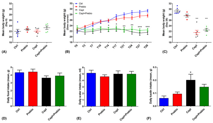
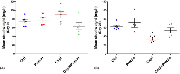
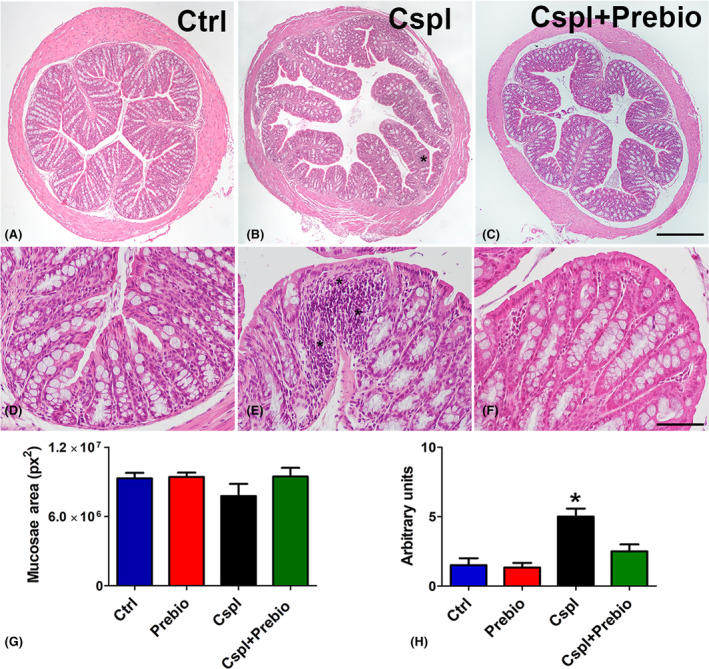
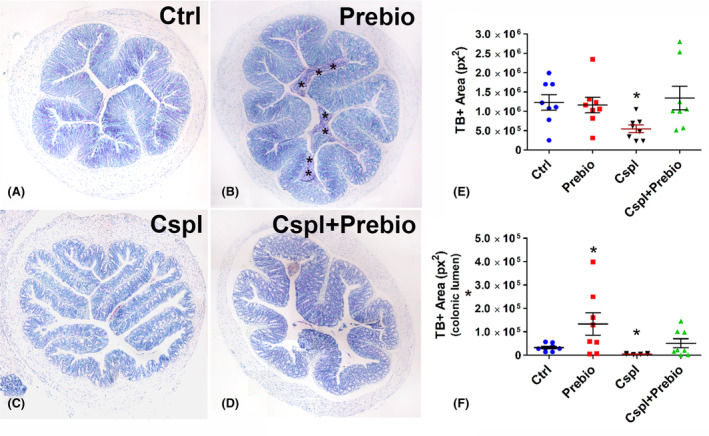
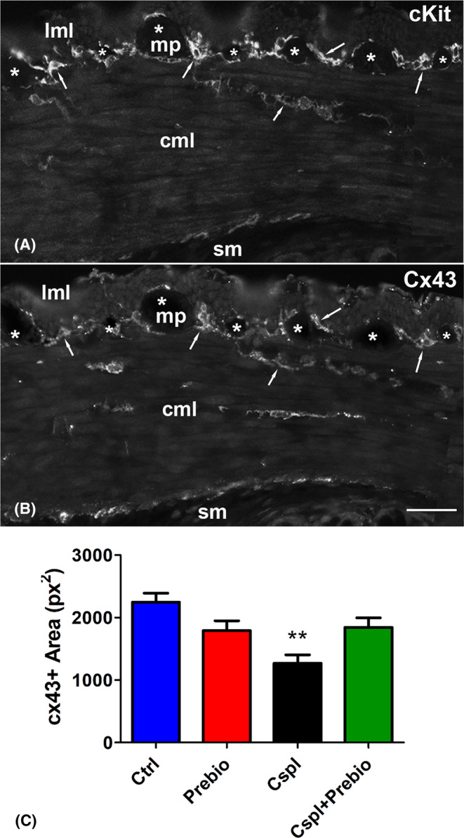
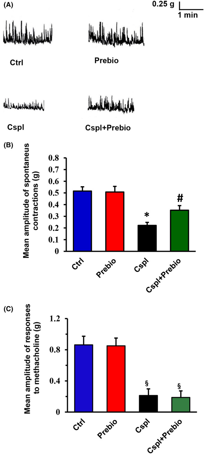
Cisplatin is an antimitotic drug able to cause acute and chronic gastrointestinal side effects. Acute side effects are attributable to mucositis while chronic ones are due to neuropathy. Cisplatin has also antibiotic properties inducing dysbiosis which enhances the inflammatory response, worsening local damage. Thus, a treatment aimed at protecting the microbiota could prevent or reduce the toxicity of chemotherapy. Furthermore, since a healthy microbiota enhances the effects of some chemotherapeutic drugs, prebiotics could also improve this drug effectiveness. We investigated whether chronic cisplatin administration determined morphological and functional alterations in mouse proximal colon and whether a diet enriched in prebiotics had protective effects. The results showed that cisplatin caused lack of weight gain, increase in kaolin intake, decrease in stool production and mucus secretion. Prebiotics prevented increases in kaolin intake, changes in stool production and mucus secretion, but had no effect on the lack of weight gain. Moreover, cisplatin determined a reduction in amplitude of spontaneous muscular contractions and of Connexin (Cx)43 expression in the interstitial cells of Cajal, changes that were partially prevented by prebiotics. In conclusion, the present study shows that daily administration of prebiotics, likely protecting the microbiota, prevents most of the colonic cisplatin-induced alterations.

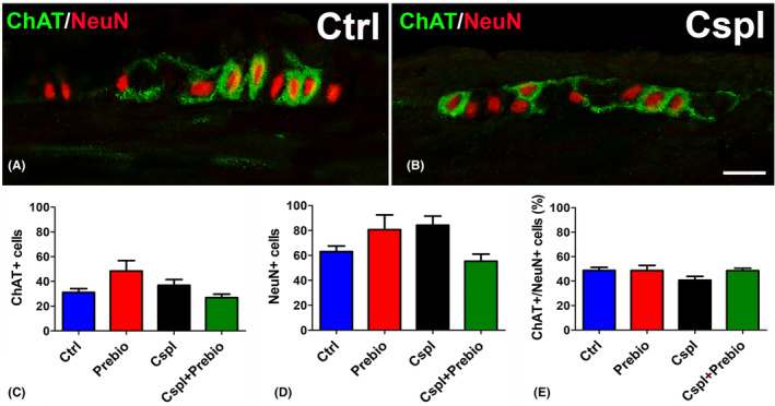
Keywords: Connexin 43; choline acetyl-transferase (ChAT); interstitial cells of Cajal; methacholine; mucus secretion, PGP9.5; spontaneous contractile activity.


Conflict of interest statement

The authors declare no conflict of interest.

Figures

FIGURE 1
FIGURE 1
(A) The body weight was measured from day T‐4 to T0. The results were similar among all the groups. (B) Time‐course of the body weight gain during the 28 days of treatment. With time the Cspl‐ and Cspl+Prebio‐treated mice did not show any weight gain contrary to the Ctrl and Prebio mouse groups. (C) The body weight before the sacrifice was significantly lower than in Cspl and Cspl+Prebio mice. Daily food (D) and water (E) intake were similar in all groups during the treatment. (F) The kaolin intake during the treatment was significantly higher for the Cspl mice compared to all the other groups. Data are expressed as mean ± SEM. B, C = *p < 0.05, **p < 0.005, ***p < 0.001 vs. Ctrl and Prebio mice. F = *p < 0.05 vs. all the other groups (ANOVA, post hoc Newman–Keuls test).
FIGURE 2
FIGURE 2
Stool production. (A) The faecal production the day before the beginning of the treatment was similar among the four groups of mice. (B) At the end of the treatment (28th day), the Cspl group showed a significant decrease in faecal production vs. all the other groups. Data are expressed as mean ± SEM. *p < 0.05 vs. all the other groups (ANOVA, post hoc Newman–Keuls test).
FIGURE 3
FIGURE 3
Haematoxylin and eosin staining. Ctrl (A–D), Cspl (B, E), Cspl+Prebio (C, F) mouse groups. Note the thinness of the villi and the dilatation of the crypts in Cspl‐treated mice compared with the other two groups of animals. Bar (A–C) = 200 μm; (D–F) = 50 μm. Asterisks indicate inflammatory cell infiltrates in the mucosa. Quantitation of the mucosae area (G) showed no difference among the groups of mice. Score analysis (H) was performed as described in Materials and Methods. Values are the median‐interquartile range. *p < 0.05 vs. all the other groups (ANOVA, Kruskal–Wallis, post hoc Newman–Keuls's test).
FIGURE 4
FIGURE 4
Toluidine blue staining. (A–D) The dye stained the goblet cells and the glandular cells in the crypts in all groups of mice. In Prebio group (B), the colonic lumen was filled with TB+ mucous secretion (asterisks). (E, F) Bar = 200 μm. Quantitation of TB+ material was significantly decreased in the epithelium (E) and in the lumen (F) of the Cspl group and significantly increased in the lumen of the Prebio group (F). *p < 0.05 vs. all the other groups (ANOVA, post hoc Newman–Keuls test).
FIGURE 5
FIGURE 5
c‐Kit‐ and Cx43‐immunoreactivity (IR). (A) c‐Kit‐IR was detected in the ICC at the myenteric plexus (MY‐ICC), in muscle layers (IM‐ICC) and at the border with the submucosa (SM‐ICC). (B) Cx43‐IR overlapped that of c‐Kit. (A, B) The arrows highlight some of the several sites of overlapping between the two markers. The asterisks indicate the myenteric ganglia. Bar = 50 μm. (C) Quantitation of the Cx43 labelling showed a significant decrease in the Cspl‐treated group. cml, circular muscle layer; lml, longitudinal muscle layer; mp, myenteric plexus; sm, submucosa. **p < 0.005 vs. all the other groups (ANOVA, post hoc Newman–Keuls test).
FIGURE 6
FIGURE 6
Choline Acetyl‐Transferasi (ChAT, green)‐ and Neuronal Nuclei (NeuN, red)‐IR. (A, B) NeuN‐IR was detected in the nuclei of several myenteric neurons. Some of them showed the ChAT labelling in the cytoplasm and processes. Bar = 20 μm. Quantitation of the total number of NeuN‐ and ChAT‐IR neurons (C, D) and of the percentage of the cholinergic ones with respect to the total myenteric neurons (E) showed no differences among the four groups.
FIGURE 7
FIGURE 7
(A) Typical tracings showing the spontaneous mechanical activity recorded in preparations from the different animal groups. In strips from Prebio‐treated animals (right‐hand upper trace), the amplitude of the spontaneous contractions was not different with respect to that obtained in the Ctrl mice (left‐hand upper trace). The amplitude of the spontaneous contractions was significantly reduced in preparations from Cspl‐treated animals (left‐hand lower trace). In strips from Cspl+Prebio‐treated mice (right‐hand lower trace), the amplitude of the spontaneous contractions was significantly increased with respect to Cspl mice. The motility pattern showed no differences among preparations from the four animal groups. (B) Bar chart showing the mean amplitude of the spontaneous contractions in preparations from the different animal groups. No statistical differences in the amplitude of the spontaneous contractions were present between preparations from Ctrl and Prebio mice. Note the great reduction in amplitude of the spontaneous contractions in strips from Cspl mice and its partial recovery in Cspl+Prebio‐treated mice. (C) Bar chart showing the mean amplitude of the direct muscle contractions elicited by methacholine in preparations from the different animal groups. No statistical differences were revealed between preparations from Ctrl‐ and Prebio‐treated mice. Note the great reduction in amplitude of the response to methacholine in strips from Cspl‐ and Cspl+Prebio‐treated mice. All values are means ± SEM of 5–8 preparations. *p < 0.05 vs. all the other groups; #p < 0.05 vs. Cspl mice § p < 0.05 vs. Ctrl and Prebio‐treated mice (ANOVA, post hoc Bonferroni's test).

Similar articles

Cited by

References

    1. Qi L, Luo Q, Zhang Y, Jia F, Zhao Y, Wang F. Advances in toxicological research of the anticancer drug cisplatin. Chem Res Toxicol. 2019;32:1469‐1486. doi:10.1021/acs.chemrestox.9b00204 - DOI - PubMed
    1. McQuade RM, Carbone SE, Stojanovska V, et al. Role of oxidative stress in oxaliplatin‐induced enteric neuropathy and colonic dysmotility in mice. Br J Pharmacol. 2016;173:3502‐3521. doi:10.1111/bph.13646 - DOI - PMC - PubMed
    1. Perše M. Cisplatin mouse models: treatment, toxicity and translatability. Biomedicines. 2021;9:1406. doi:10.3390/biomedicines9101406 - DOI - PMC - PubMed
    1. Stojanovska V, Sakkal S, Nurgali K. Platinum‐based chemotherapy: gastrointestinal immunomodulation and enteric nervous system toxicity. Am J Physiol Liver Physiol. 2015;308:G223‐G232. doi:10.1152/ajpgi.00212.2014 - DOI - PubMed
    1. Fuertes M, Castilla J, Alonso C, Pérez J. Cisplatin biochemical mechanism of action: from cytotoxicity to induction of cell death through interconnections between apoptotic and necrotic pathways. Curr Med Chem. 2003;10:257‐266. doi:10.2174/0929867033368484 - DOI - PubMed

Publication types

LinkOut - more resources