. 2024 Mar;28(6):e18203.
doi: 10.1111/jcmm.18203.

Supplementing probiotics during intermittent fasting proves more effective in restoring ileum and colon tissues in aged rats

Affiliations

Supplementing probiotics during intermittent fasting proves more effective in restoring ileum and colon tissues in aged rats

Hikmet Taner Teker et al. J Cell Mol Med. 2024 Mar.

Abstract

This study aimed to explore the impact of SCD Probiotics supplementation on biomolecule profiles and histopathology of ileum and colon tissues during a 30-day intermittent fasting (IF) program. Male Sprague-Dawley rats, aged 24 months, underwent 18-h daily fasting and received 3 mL (1 × 10⁸ CFU) of SCD Probiotics. The differences in biomolecule profiles were determined using FTIR Spectroscopy and two machine learning techniques, Linear Discriminant Analysis (LDA) and Support Vector Machine (SVM), which showed significant differences with high accuracy rates. Spectrochemical bands indicating alterations in lipid, protein and nucleic acid profiles in both tissues. The most notable changes were observed in the group subjected to both IF and SCD Probiotics, particularly in the colon. Both interventions, individually and in combination, decreased protein carbonylation levels. SCD Probiotics exerted a more substantial impact on membrane dynamics than IF alone. Additionally, both IF and SCD Probiotics were found to have protective effects on intestinal structure and stability by reducing mast cell density and levels of TNF-α and NF-κB expression in ileum and colon tissues, thus potentially mitigating age-related intestinal damage and inflammation. Furthermore, our results illustrated that while IF and SCD Probiotics individually instigate unique changes in ileum and colon tissues, their combined application yielded more substantial benefits. This study provides evidence for the synergistic potential of IF and SCD Probiotics in combating age-related intestinal alterations.

Keywords: NF-κB; SCD probiotics; TNF-α; aging; intermittent fasting; intestinal tissue.


Conflict of interest statement

The authors have declared that no competing interests exist.

Figures

FIGURE 1
FIGURE 1
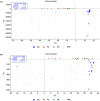
LDA discrimination plot for ileum samples in the full (4000–650 cm−1) spectral region (A). Control group and application groups significantly differed in overall biomolecule content, with 100% accuracy for whole content. LDA discrimination plot in the lipid (3000–2700 cm−1) spectral region (B). Control group and application groups clustered in distinct regions. Largest difference was seen in SCD Probiotics versus control, but similar to intermittent fasting effect. CIL (control), FIL (intermittent fasting), PIL (SCD Probiotics) and the FPIL applications (in which the intermittent fasting and SCD Probiotics were applied together).
FIGURE 2
FIGURE 2
Quantitative changes in ileum‐associated spectrochemical parameters include band area ratios for: (A) Acyl chain length of fatty acids (A2922/A2955): IF group saw an increase; SCD Probiotics and combined treatment groups showed a decrease, (B) Protein phosphorylation (A1239/A2955): Decreased in all groups, (C) protein conformation (A1653/A1545) and (A1080/A1545): (A1653/A1545) increased in IF and SCD Probiotics, but unchanged in combined treatment; (A1080/A1545) increased in IF and SCD Probiotics but unchanged in combined treatment, (D) protein carbonylation (A1740/A1545): Decreased in IF and combined treatment; no change in SCD Probiotics group. The data were analysed using One‐way anova and unpaired t‐test, and the significance levels were stated as *p < 0.05 and ****p ≤ 0.0001. Decreased in IF and combined treatment; no change in SCD Probiotics group. CIL (control), FIL (intermittent fasting), PIL (SCD Probiotics) and the FPIL applications (in which the intermittent fasting and SCD Probiotics were applied together).
FIGURE 3
FIGURE 3
LDA discrimination plot for colon samples in the full (4000–650 cm−1) spectral region (A). LDA analysis revealed significant differentiation between control and application groups, with 97.73% accuracy rate. LDA discrimination plot for colon samples in the lipid (3000–2700 cm−1) spectral region (B). Lipid profiles showed significant differentiation with 95.45% accuracy. CC (control), FC (intermittent fasting), PC (SCD Probiotics) and the FPC applications (in which the intermittent fasting and SCD Probiotics were applied together).
FIGURE 4
FIGURE 4
Quantitative changes in colon‐associated spectrochemical parameters include band area ratios for: (A) acyl chain length of fatty acids (A2922/A2955), (B) protein phosphorylation (A1239/A2955): Increased in IF and SCD Probiotics groups, (C) protein phosphorylation (A1080/A1545): Increased in all groups, (D) protein conformation (A1653/A1545): Increased only in SCD Probiotics group, (E) protein carbonylation (A1740/A1545): Decreased in all groups, with the greatest decrease in the combined treatment group. The data were analysed using one‐way anova and unpaired t‐test, and the significance levels were stated as *p < 0.05, **p ≤ 0.01 and ****p ≤ 0.0001. CC (control), FC (intermittent fasting), PC (SCD Probiotics) and the FPC applications (in which the intermittent fasting and SCD Probiotics were applied together).
FIGURE 5
FIGURE 5
Representative images of H&E staining and quantification of lymphatic infiltration area fraction (%) in all groups of ileum tissues (A). Red arrow shows lymphatic infiltrates. Representative images of H&E staining and quantification of Paneth cells intensity of area fraction (%) in all groups of ileum tissues (B). Yellow arrow shows Paneth cells. Areas in the H&E‐stained microphotographs of all groups were magnified in the photos to which they belonged to the area of interest. Values are expressed as mean ± SEM; n = 7 rats in each group. The significance levels were stated as *p < 0.05, **p ≤ 0.01 and ****p ≤ 0.0001. (One‐way anova and nonparametric Mann–Whitney U test). Scale bar = 50 μm and 100 μm. CIL (control), FIL (intermittent fasting), PIL (SCD Probiotics) and the FPIL applications (in which the intermittent fasting and SCD Probiotics were applied together).
FIGURE 6
FIGURE 6
Representative images of H&E staining and quantification of lymphatic infiltration area fraction (%) in all groups of rat colon tissues. Red arrow shows lymphatic infiltrates. Areas in the H&E‐stained microphotographs of all groups were magnified in the photos to which they belonged to the area of interest. Values are expressed as mean ± SEM; n = 7 rats in each group. The significance levels were stated as *p < 0.05 and ****p ≤ 0.0001. (One‐way anova and nonparametric Mann–Whitney U test). Scale bar = 100 μm. CC (control), FC (intermittent fasting), PC (SCD Probiotics) and the FPC applications (in which the intermittent fasting and SCD Probiotics were applied together).
FIGURE 7
FIGURE 7
A TNF‐α and NF‐κB staining intensity in ileum (A) and colon (B). TNF‐α: Tumour Necrosis Factor‐alpha, NF‐κB: Nuclear factor kappa‐light‐chain‐enhancer of activated B cells. IHC staining images of TNF‐α and NF‐κB expression in the rat ileum. Graphs of TNF‐α and NF‐κB staining intensity in the ileum and colon tissues as measured in ImageJ (FIJI). Values are expressed as mean ± SEM; n = 7 rats in each group. The significance levels were stated as *p < 0.05, **p ≤ 0.01 ***p ≤ 0.001 and ****p ≤ 0.0001. (One‐way anova and nonparametric Mann–Whitney U test). (IHC staining, Scale bar: 100 μm). CIL (control), FIL (intermittent fasting), PIL (SDC Probiotics) and the FPIL applications (in which the intermittent fasting and SCD Probiotics were applied together); CC (control), FC (intermittent fasting), PC (SCD Probiotics), and the FPC applications (in which the intermittent fasting and SCD Probiotics were applied together).

Similar articles

Cited by

References

    1. Castellano JM. Blood‐based therapies to combat aging. Gerontology. 2019;65(1):84‐89. - PMC - PubMed
    1. Lee JH, Verma N, Thakkar N, Yeung C, Sung HK. Intermittent fasting: physiological implications on outcomes in mice and men. Phys Ther. 2020;35(3):185‐195. - PubMed
    1. de Cabo R, Mattson MP. Effects of intermittent fasting on health, aging, and disease. N Engl J Med. 2019;381(26):2541‐2551. - PubMed
    1. Ceylani T, Taner H, Samgane G, Gurbanov R. Intermittent fasting‐induced biomolecular modifications in rat tissues detected by ATR‐FTIR spectroscopy and machine learning algorithms. Anal Biochem. 2022;654:114825. doi:10.1016/j.ab.2022.114825 - DOI - PubMed
    1. Bagherniya M, Butler AE, Barreto GE, Sahebkar A. The effect of fasting or calorie restriction on autophagy induction: a review of the literature. Ageing Res Rev. 2018;47:183‐197. - PubMed

LinkOut - more resources