. 2024 Apr;20(4):2843-2860.
doi: 10.1002/alz.13737. Epub 2024 Mar 6.

Nanoscale imaging of pT217-tau in aged rhesus macaque entorhinal and dorsolateral prefrontal cortex: Evidence of interneuronal trafficking and early-stage neurodegeneration

Affiliations

Nanoscale imaging of pT217-tau in aged rhesus macaque entorhinal and dorsolateral prefrontal cortex: Evidence of interneuronal trafficking and early-stage neurodegeneration

Dibyadeep Datta et al. Alzheimers Dement. 2024 Apr.

Abstract

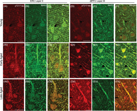
Introduction: Tau phosphorylated at threonine-217 (pT217-tau) is a novel fluid-based biomarker that predicts onset of Alzheimer's disease (AD) symptoms, but little is known about how pT217-tau arises in the brain, as soluble pT217-tau is dephosphorylated post mortem in humans.

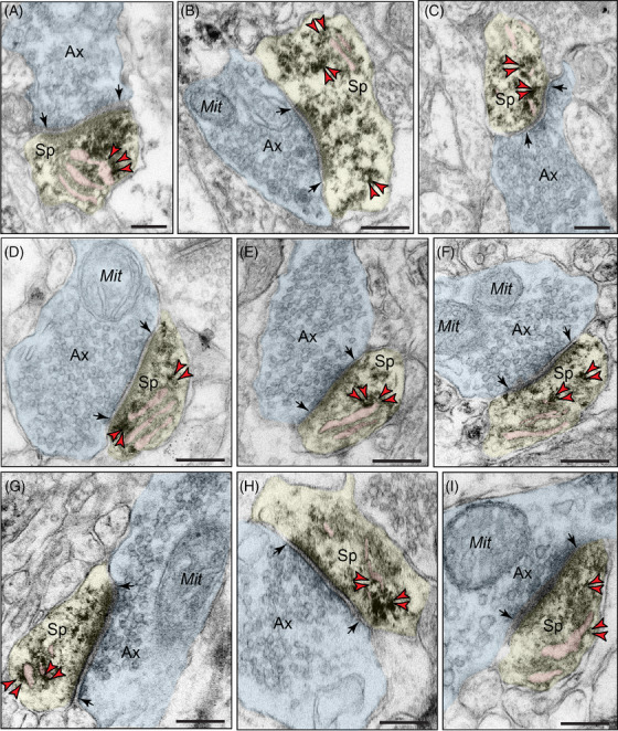
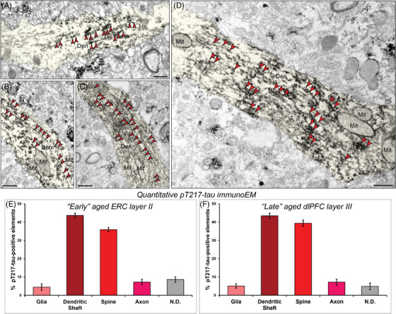
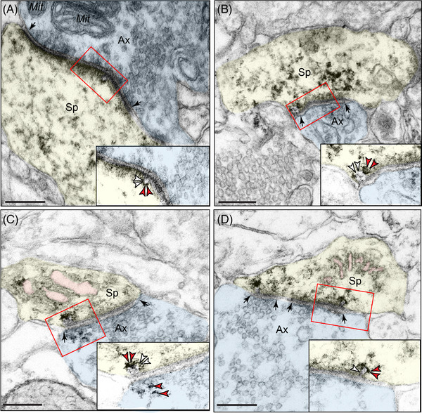
Methods: We used multilabel immunofluorescence and immunoelectron microscopy to examine the subcellular localization of early-stage pT217-tau in entorhinal and prefrontal cortices of aged macaques with naturally occurring tau pathology and assayed pT217-tau levels in plasma.

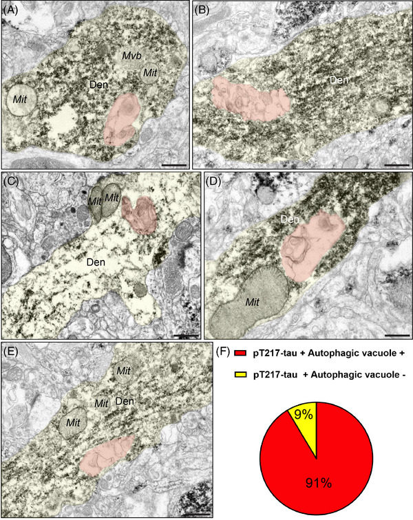
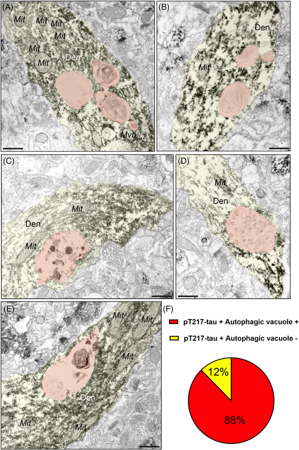
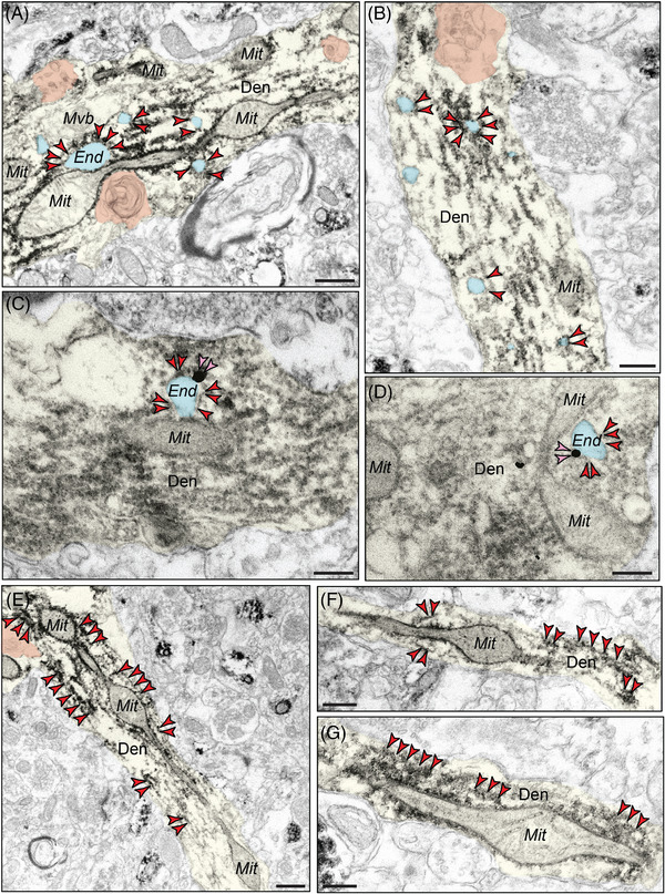
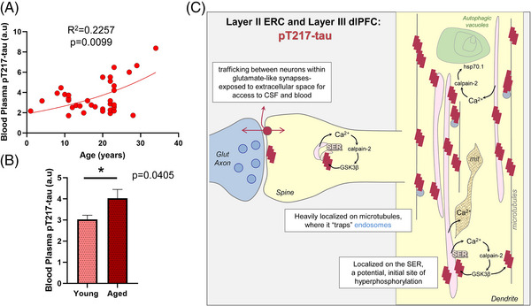
Results: pT217-tau was aggregated on microtubules within dendrites exhibiting early signs of degeneration, including autophagic vacuoles. It was also seen trafficking between excitatory neurons within synapses on spines, where it was exposed to the extracellular space, and thus accessible to cerebrospinal fluid (CSF)/blood. Plasma pT217-tau levels increased across the age span and thus can serve as a biomarker in macaques.

Discussion: These data help to explain why pT217-tau predicts degeneration in AD and how it gains access to CSF and plasma to serve as a fluid biomarker.

Keywords: biomarker; dorsolateral prefrontal cortex; entorhinal cortex; plasma; tau phosphorylated at threonine‐217; trafficking.


Conflict of interest statement

Dr. Zhongcong Xie provided consulting service to Baxter pharmaceutical company; Shanghai 9th and 10th hospitals; NanoMosaic, Inc.; and Anesthesiology and Perioperative Science in the past 36 months. All other co‐authors have nothing to disclose. Author disclosures are available in the supporting information.

Figures

FIGURE 1
FIGURE 1
Spatial and temporal localization pattern of pT217‐tau across vulnerable cortical regions. Multiple‐label immunofluorescence showing pT217‐tau labeling (red) co‐localized in excitatory neurons (MAP2, green) in ERC layer II and dlPFC layer III across age span in rhesus macaques at high magnification. In adult (8–10 years) rhesus macaques, pT217‐tau immunolabeling was observed within the perisomatic compartment and the nucleus in a subset of excitatory cells (Ai‐Ci, Di‐Fi), including delicate expression in proximal apical dendrites in ERC layer II cell islands and dlPFC layer III microcircuits. With advancing age, immunofluorescence revealed pT217‐tau accumulating in proximal and more distal apical and basal dendrites in ERC layer II excitatory cells and dlPFC deep layer III pyramidal cells, both in “early” aged monkeys (18–19 years; Aii‐Cii, Dii‐Fii) and “late” aged monkeys (28–34 years; Aiii‐Ciii, Diii‐Fiii). Scale bars: 24 μm. dlPFC, dorsolateral prefrontal cortex; ERC, entorhinal cortex; pT217‐tau, tau phosphorylated at threonine‐217
FIGURE 2
FIGURE 2
Postsynaptic localization of pT217‐tau in dendritic spines in rhesus macaque ERC and dlPFC. Immunoperoxidase labeling revealed that pT217‐tau immunolabeling was concentrated in dendritic spines near asymmetric, glutamate‐like synapses in rhesus macaque in “early” aged (18–24 years) macaque ERC layer II (A‐C) and “late” aged (26–31 years) macaque dlPFC layer III (D‐I). pT217‐tau was observed directly in association with the smooth endoplasmic reticulum (SER) spine apparatus (pseudocolored pink). pT217‐tau immunolabeling is also observed subjacent to the postsynaptic density, near axospinous, asymmetric glutamatergic synapses (A‐C, D, F, G, I). Synapses are between arrows. Red arrowheads point to pT217‐tau immunoreactivity. Profiles are pseudocolored for clarity. Scale bars, 200 nm. Ax, axon; dlPFC, dorsolateral prefrontal cortex; ERC, entorhinal cortex; Mit, mitochondria; pT217‐tau, tau phosphorylated at threonine‐217; Sp, dendritic spine
FIGURE 3
FIGURE 3
Aggregation of pT217‐tau along microtubules in dendritic shafts. ImmunoEM demonstrates aggregation of pT217‐tau along dendritic microtubules in putative excitatory cells in “early” aged (18–24 years) macaque ERC layer II (A‐B) and pyramidal cells in “late” aged (26–31 years) macaque dlPFC layer III (C‐D). Ultrastructural examination revealed dense accumulation of pT217‐tau occurring in parallel microtubule bundles in horizontal dendrites. Red arrowheads point to pT217‐tau immunoreactivity. Profiles are pseudocolored for clarity. Scale bars, 200 nm. Quantitative analysis of pT217‐tau immunoEM localization in rhesus macaque “early” aged (18–24 years) ERC layer II circuits (E). The prevalence of pT217‐tau in various cellular subcompartments in layer II of the ERC neuropil, expressed as percentage of pT217‐tau profile (e.g., dendrite) per total pT217‐tau profiles. We analyzed a total of 1205 pT217‐tau immunopositive profiles in “early” aged (18–24 years) ERC layer II neuropil. pT217‐tau is primarily expressed in postsynaptic subcompartments in “early” aged (18–24 years) ERC layer II microcircuits, with foremost expression in dendritic shafts, and significant expression within dendritic spines. Quantitative analysis of pT217‐tau immunoEM localization in rhesus macaque in “late” aged (26–31 years) macaque dlPFC layer III (F). The prevalence of pT217‐tau in various cellular subcompartments in layer III of the dlPFC neuropil, expressed as percentage of pT217‐tau profile (e.g., dendritic shaft) per total pT217‐tau profiles. We analyzed a total of 1254 pT217‐tau immunopositive profiles in “late” aged (26–31 years) macaque dlPFC layer III. pT217‐tau is primarily expressed in postsynaptic subcompartments in dlPFC layer III microcircuits, with greatest expression in dendritic shafts, and significant expression within dendritic spines. For additional details regarding quantitative assessment and profile identification see Materials and Methods section. Nondetermined (N.D) are profiles that could not be unequivocally categorized. Den, dendrite; dlPFC, dorsolateral prefrontal cortex; ERC, entorhinal cortex; Mit, mitochondria; pT217‐tau, tau phosphorylated at threonine‐217
FIGURE 4
FIGURE 4
Trans‐synaptic trafficking of pT217‐tau in aging rhesus macaques. Early‐stage, soluble pT217‐tau propagates between excitatory neurons within glutamate‐like synapses in “early” aged (18–24 years) macaque ERC layer II (A‐B) and in “late” aged (26–31 years) macaque dlPFC layer III (C‐D). Trans‐synaptic propagation of pT217‐tau occurs via omega‐shaped endosome‐like vesicular profiles on the plasma membrane within dendritic spines, specifically within the synapse (insets). All dendritic spines receive axospinous Type I asymmetric glutamatergic‐like synapses. Red arrowheads point to pT217‐tau immunoreactivity. Black arrows point to the synapse; white arrowheads indicate an omega‐shaped profile on the plasma membrane. The SER is pseudocolored in pink. Profiles are pseudocolored for clarity. Scale bars, 200 nm. Ax, axon; dlPFC, dorsolateral prefrontal cortex; ERC, entorhinal cortex; Mit, mitochondria; pT217‐tau, tau phosphorylated at threonine‐217; SER, smooth endoplasmic reticulum; Sp, dendritic spine
FIGURE 5
FIGURE 5
Concomitant autophagic degeneration in pT217‐tau immunopositive dendrites in ERC. Association of pT217‐tau immunolabeling in degenerating dendrites containing autophagic vacuoles in ERC layer II (A‐E). Autophagic vacuoles with multilamellar bodies (pseudocolored in orange) were observed in principal dendrites immunopositive for pT217‐tau in “early” aged (18–24 years) rhesus macaque ERC layer II. Systematic quantification (N = 116 profiles) in ERC layer II revealed robust concordance between signatures of autophagic degeneration and immunopositivity for pT217‐tau (N = 106; 91%) within dendrites (F). The percentage of pT217‐tau‐immunopositive dendrites showing signatures of autophagic degeneration is shown using a pie chart (F). Profiles are pseudocolored for clarity. Scale bars, 200 nm. Den, dendrite; dlPFC, dorsolateral prefrontal cortex; ERC, entorhinal cortex; Mit, mitochondria; pT217‐tau, tau phosphorylated at threonine‐217
FIGURE 6
FIGURE 6
Concomitant autophagic degeneration in pT217‐tau immunopositive dendrites in dlPFC. Association of pT217‐tau immunolabeling in degenerating dendrites containing autophagic vacuoles in dlPFC layer III (A‐E). Autophagic vacuoles with multilamellar bodies (pseudocolored in orange) were observed in principal dendrites immunopositive for pT217‐tau in pyramidal cells in “late” aged (26–31 years) macaque dlPFC layer III. Systematic quantification (N = 82 profiles) in dlPFC layer III revealed robust concordance between signatures of autophagic degeneration and immunopositivity for pT217‐tau (N = 71; 88%) within dendrites (F). The percentage of pT217‐tau‐immunopositive dendrites showing signatures of autophagic degeneration is shown using a pie chart (F). Profiles are pseudocolored for clarity. Scale bars, 200 nm. Den, dendrite; dlPFC, dorsolateral prefrontal cortex; ERC, entorhinal cortex; Mit, mitochondria; pT217‐tau, tau phosphorylated at threonine‐217
FIGURE 7
FIGURE 7
Evidence of pT217‐tau surrounding endosomes in dendritic shafts, oftentimes near dysmorphic mitochondrial profiles. pT217‐tau (indicated by red arrowheads) aggregating on microtubules in dendrites where it surrounds enlarged endosomes (pseudocolored in cyan) in “early” aged (18–24 years) macaque ERC layer II and in “late” aged (26–31 years) macaque dlPFC layer III (A‐D). Using dual‐label immunoEM, the current data show evidence of pT217‐tau surrounding endosomes containing Aβ42 (indicated by pink arrowheads) in dendrites in aged macaque dlPFC, suggesting that pT217‐tau may be involved in the etiology of amyloid pathology (C‐D). Aggregations of pT217‐tau are often seen near dysmorphic mitochondria, characterized by MOAS morphological phenotypes, indicative of impaired mitochondrial fission and fusion (A, E‐G). MOAS‐like morphological disturbances have been described previously in aging rhesus macaques, consistent with local calcium dysregulation. Autophagic vacuolar degeneration (pseudocolored in orange) is also observed in several dendritic shafts, consistent with signatures of neurite dystrophy (A, B, E). Profiles are pseudocolored for clarity. Scale bars, 200 nm. Aβ, amyloid beta; Den, dendrite; dlPFC, dorsolateral prefrontal cortex; End, endosome; ERC, entorhinal cortex; Mit, mitochondria; MOAS, mitochondria on a string; pT217‐tau, tau phosphorylated at threonine‐217
FIGURE 8
FIGURE 8
Increased pT217‐tau in blood plasma in aging rhesus macaques, and a working model of pT217‐tau etiology and toxic actions. Age‐related elevation in pT217‐tau in blood plasma in aging rhesus macaques (A). Results from a regression analysis performed in all animals (N = 36) indicated statistically significant increases in pT217‐tau signal with advancing age (R 2 = 0.2257, P = 0.0099). Quantification of pT217‐tau in blood plasma grouped by age (B). Young animals (N = 15) are compared to aged animals (N = 21) via a two‐tailed Welch t test (*P = 0.0405). SEM is plotted for each group. Summary schematic of pT217‐tau expression patterns in “early” aged (18–24 years) macaque ERC layer II and in “late” aged (26–31 years) macaque dlPFC layer III microcircuits (C). A schematic illustration of pT217‐tau localization in dendrites, and its potential etiology and toxic actions. pT217‐tau is primarily located in glutamatergic dendrites and dendritic spines, consistent with the known origins of tau pathology in dendrites in humans. The current study found evidence of pT217‐tau trafficking between neurons at glutamate‐like synapses, interfacing with the extracellular space to become accessible in CSF and plasma. This may contribute to tau “seeding” pathology through an interconnected network of glutamatergic neurons. Aggregations of pT217‐tau were prominently expressed on microtubules, as well as on the calcium‐storing SER, where increased calcium release may drive GSK3β hyperphosphorylation of tau at T217. These dendrites often showed signs of pathology: autophagic vacuoles that are an early sign of autophagic degeneration and abnormal mitochondria (mit), known as MOAS. The aggregations of pT217‐tau on microtubules could be seen to “trap” endosomes, interfering with intracellular trafficking needed for healthy dendrites. These “endosomal traffic jams” may also increase the production of Aβ by increasing the time APP spends in endosomes with exposure to β‐secretase, a hypothesis to be tested in future research. Aβ, amyloid beta; APP, amyloid precursor protein; CSF, cerebrospinal fluid; dlPFC, dorsolateral prefrontal cortex; End, endosome; ERC, entorhinal cortex; Mit, mitochondria; MOAS, mitochondria on a string; pT217‐tau, tau phosphorylated at threonine‐217; SEM, standard error of the mean; SER, smooth endoplasmic reticulum

Update of

Similar articles

Cited by

References

    1. Palmqvist S, Janelidze S, Quiroz YT, et al. Discriminative accuracy of plasma phospho‐tau217 for Alzheimer disease vs other neurodegenerative disorders. JAMA. 2020. 324:772‐781. - PMC - PubMed
    1. Barthelemy NR, Li Y, Joseph‐Mathurin N, et al. A soluble phosphorylated tau signature links tau, amyloid and the evolution of stages of dominantly inherited Alzheimer's disease. Nat Med. 2020. 26:398‐407. - PMC - PubMed
    1. Mattsson‐Carlgren N, Salvado G, et al. Prediction of longitudinal cognitive decline in preclinical Alzheimer disease using plasma biomarkers. JAMA Neurol. 2023. 80:360‐369. - PMC - PubMed
    1. Salvado G, Ossenkoppele R, Ashton NJ, et al. Specific associations between plasma biomarkers and postmortem amyloid plaque and tau tangle loads. EMBO Mol Med. 2023. 15:e17123. - PMC - PubMed
    1. Hansson O, Blennow K, Zetterberg H, Dage J. Blood biomarkers for Alzheimer's disease in clinical practice and trials. Nat Aging. 2023. 3:506‐519. - PMC - PubMed

MeSH terms