. 2024 Apr 10;15(4):e0137823.
doi: 10.1128/mbio.01378-23. Epub 2024 Mar 6.

Regulation of phase separation and antiviral activity of Cactin by glycolytic enzyme PGK via phosphorylation in Drosophila

Affiliations

Regulation of phase separation and antiviral activity of Cactin by glycolytic enzyme PGK via phosphorylation in Drosophila

Dongchao Chen et al. mBio. .

Abstract

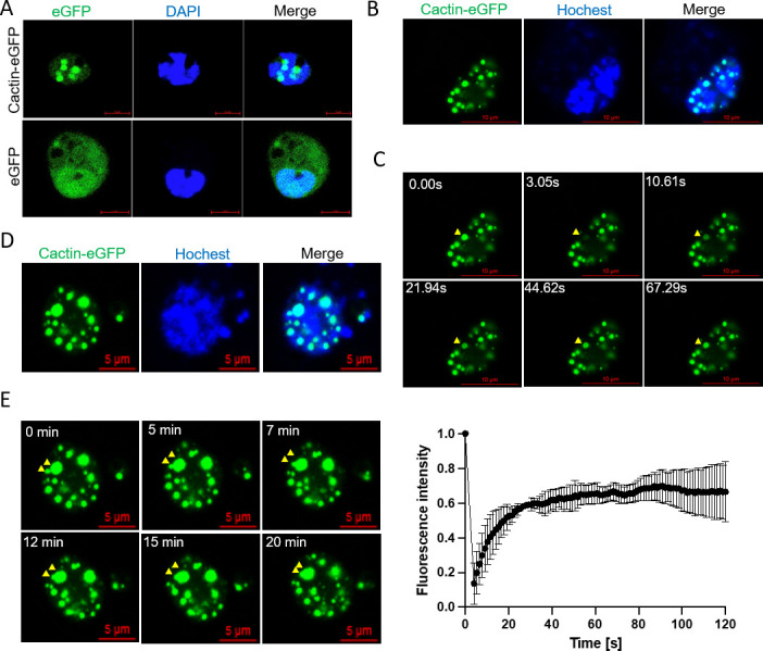
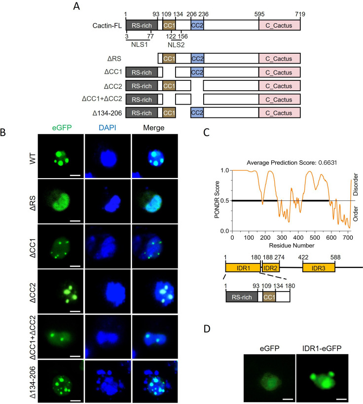
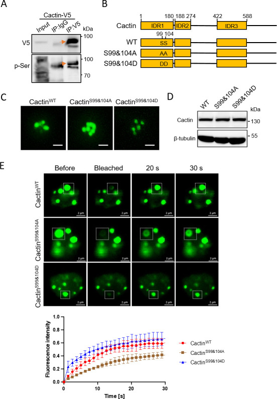
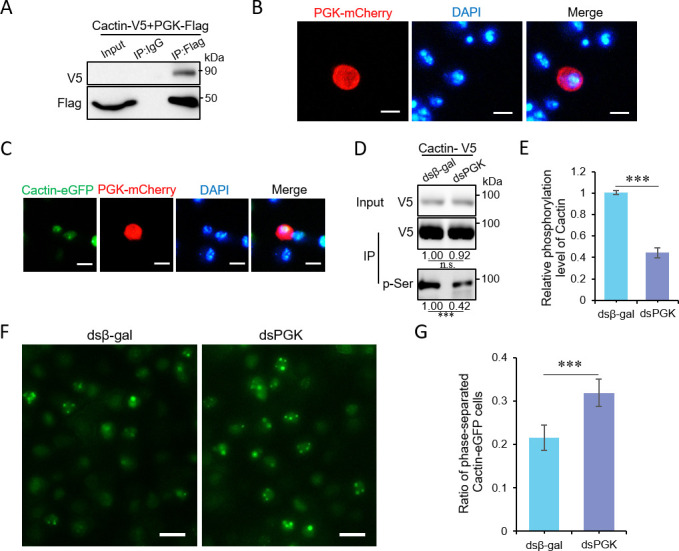
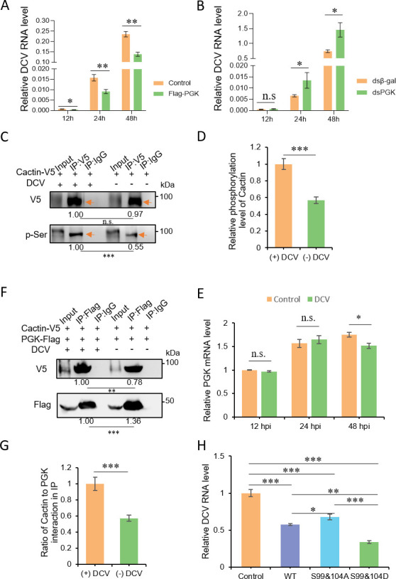
Liquid-liquid phase separation (LLPS) plays a crucial role in various biological processes in eukaryotic organisms, including immune responses in mammals. However, the specific function of LLPS in immune responses in Drosophila melanogaster remains poorly understood. Cactin, a highly conserved protein in eukaryotes, is involved in a non-canonical signaling pathway associated with Nuclear factor-κB (NF-κB)-related pathways in Drosophila. In this study, we investigated the role of Cactin in LLPS and its implications for immune response modulation. We discovered that Cactin undergoes LLPS, forming droplet-like particles, primarily mediated by its intrinsically disordered region (IDR). Utilizing immunoprecipitation and mass spectrometry analysis, we identified two phosphorylation sites at serine residues 99 and 104 within the IDR1 domain of Cactin. Co-immunoprecipitation and mass spectrometry further revealed phosphoglycerate kinase (PGK) as a Cactin-interacting protein responsible for regulating its phosphorylation. Phosphorylation of Cactin by PGK induced a transition from stable aggregates to dynamic liquid droplets, enhancing its ability to interact with other components in the cellular environment. Overexpression of PGK inhibited Drosophila C virus (DCV) replication, while PGK knockdown increased replication. DCV infection also increased Cactin phosphorylation. We also found that phosphorylation enhances the antiviral ability of Cactin by promoting liquid-phase droplet formation. These findings demonstrate the role of Cactin-phase separation in regulating DCV replication and highlight the modulation of its antiviral function through phosphorylation, providing insights into the interplay between LLPS and antiviral defense mechanisms.

Importance: Liquid-liquid phase separation (LLPS) plays an integral role in various biological processes in eukaryotic organisms. Although several studies have highlighted its crucial role in modulating immune responses in mammals, its function in immune responses in Drosophila melanogaster remains poorly understood. Our study investigated the role of Cactin in LLPS and its implications for immune response modulation. We identified that phosphoglycerate kinase (PGK), an essential enzyme in the glycolytic pathway, phosphorylates Cactin, facilitating its transition from a relatively stable aggregated state to a more dynamic liquid droplet phase during the phase separation process. This transformation allows Cactin to rapidly interact with other cellular components, enhancing its antiviral properties and ultimately inhibiting virus replication. These findings expand our understanding of the role of LLPS in the antiviral defense mechanism, shedding light on the intricate mechanisms underlying immune responses in D. melanogaster.

Keywords: Cactin; Drosophila C virus; Drosophila melanogaster; antiviral immunity; liquid–liquid phase separation; phosphoglycerate kinase.


Conflict of interest statement

The authors declare no conflict of interest.

Figures

Fig 1
Fig 1
Cactin forms droplet-like particles through LLPS in Drosophila S2 cells (A) Drosophila S2 cells transfected with pMT-Cactin-eGFP or pMT-eGFP plasmids were stained with DAPI to visualize the nucleus. Confocal images depict the formation of Cactin-eGFP droplet-like structures, while free eGFP fails to form droplets (scale bar, 5 µm). (B) Drosophila S2 cells transfected with the pMT-Cactin-eGFP plasmid were stained with Hoechst 33342 to label the nucleus in living cells (scale bar, 10 µm). (C) FRAP analysis of Cactin droplet-like structures in S2 cells (same cell as panel B). Montages on the top illustrate the FRAP process of a droplet (scale bar, 10 µm). The position is indicated by a yellow arrow. The graph on the bottom displays the recovery of fluorescence intensity over time. Data correspond to mean ± SD with n = 3. (D) Drosophila S2 cells transfected with the pMT-Cactin-eGFP plasmid were stained with Hochest 33342 to label the nucleus in living cells (scale bar, 5 µm). (E) Representative images demonstrate the time-dependent fusion of two Cactin-eGFP droplets in S2 cells (same cell as panel D). Montages depict the phase fusion process of a droplet (scale bar, 5 µm). The position is indicated by a yellow arrow.
Fig 2
Fig 2
The IDR1 domain of Cactin is crucial for phase separation. (A) Domain map of Cactin, featuring an N-terminal RS-rich domain, two coiled coils (CC1 and CC2), and the Cactus-interacting domain at the C-terminus. Schematic representation of the two NLS domains of Cactin, with positions of the five deletion variants indicated. (B) Confocal images of S2 cells transfected with Cactin-eGFP WT or variants, treated with DAPI for nuclear visualization (scale bar, 5 µm). (C) Disorder confidence score (top), diagrammatic representation of identified Cactin IDR domains (middle), and the structural domain encompassed by IDR1 (bottom). IDRs were predicted using the online tool PONDR. (D) Confocal images illustrating the condensed state of eGFP and IDR1-eGFP in S2 cells (scale bars, 5 µm).
Fig 3
Fig 3
Phosphorylation of the IDR1 domain in Cactin regulates phase separation. (A) Phosphorylation of Cactin-V5 in S2 cells, detected using an anti-V5 antibody for Cactin-V5 enrichment and an anti-pSer antibody for detecting Cactin-V5 phosphorylation. The orange arrow indicates the phosphorylated form. Representative results from triplicate experiments are shown. (B) Schematic representation of Cactin phosphorylation mutants, highlighting the positions of two point mutations within Cactin variants (CactinS99&104A and CactinS99&104D). (C) Confocal images showing the condensed state of CactinWT-eGFP, CactinS99&104A-eGFP, and CactinS99&104D-eGFP in S2 cells (scale bars, 5 µm). (D) Western blotting analyses of CactinWT-eGFP, CactinS99&104A-eGFP, and CactinS99&104D-eGFP in panel C. Representative results from triplicate experiments are shown. (E) FRAP measurement of CactinWT-eGFP, CactinS99&104A-eGFP, and CactinS99&104D-eGFP in S2 cells (scale bar, 2 µm). Montages on the top show the processes of FRAP of droplets. The graph on the bottom shows the recovery of fluorescence intensity over time. Data correspond to the mean ± SD with n = 3.
Fig 4
Fig 4
PGK-mediated phosphorylation regulates the phase separation of Cactin. (A) Immunoprecipitation performed in S2 cells transfected with pMT-PGK-Flag and pMT-Cactin-V5 plasmids. Representative results from triplicate experiments are shown. (B) Drosophila S2 cells transfected with pMT-PGK-mCherry plasmids, stained with DAPI to locate the nucleus. Confocal images illustrating the location of PGK-mCherry in S2 cells (scale bar, 5 µm). (C) Drosophila S2 cells transfected with pMT-Cactin-eGFP and pMT-PGK-mCherry plasmids, stained with DAPI to locate the nucleus. Confocal images showing the colocalization of Cactin-eGFP and PGK-mCherry in S2 cells (scale bar, 5 µm). (D–E) Cells transfected with pMT-Cactin-V5 plasmids 24 h earlier, treated with dsRNAs against control (β-gal) or PGK, followed by 48 h for immunoprecipitation. Anti-V5 antibody was used to detect Cactin-V5 enrichment, and phosphorylation of Cactin-V5 was detected with an anti-pSer antibody (D). Phosphorylated Cactin band density was normalized to total Cactin band density and compared to the control (E). Error bars represent the SD of three replicates. Representative results from triplicate experiments are shown in D. Data correspond to the mean ± SD with n = 3. ∗∗∗P < 0.001 (Student’s t-test). (F–G) Confocal images showing phase-separated cells in stable Cactin-eGFP-expressing cells pretreated 48 h earlier with dsRNAs against control (β-gal) or PGK, followed by a 12-h period of induced expression (F) (scale bar, 20 µm). The ratio of Cactin-eGFP cells undergoing phase separation was calculated (G). A total of eight fields were quantified, with an average of about 45 cells in each field. Data correspond to the mean ± SD with n = 8. ∗∗∗P < 0.001 (Student’s t-test).
Fig 5
Fig 5
Cactin phase separation regulates DCV replication. (A) S2 cells were transfected with pMT-PGK-Flag or pMT-eGFP (control) plasmids for 48 h and then infected with DCV, and reverse transcription quantitative PCR (RT-qPCR) analysis of DCV RNA levels at 12, 24, and 48 hpi, shown values relative to those of rp49. Data correspond to mean ± SD with n = 3. ∗P < 0.05, ∗∗P < 0.01 (Student’s t-test). (B) S2 cells were pretreated with dsRNAs against a control (β-gal) or PGK for 48 h and then infected with DCV, and RT-qPCR analysis of DCV RNA levels at 12, 24, and 48 hpi, shown values relative to those of rp49. Data correspond to mean ± SD with n = 3. ∗P < 0.05, n.s., not significant (Student’s t-test). (C–D) Immunoprecipitation in S2 cells transfected with pMT-Cactin-V5 plasmids, uninfected or infected, with DCV (infection initiated 48 h after transfection). Anti-V5 antibody detected Cactin-V5 enrichment, and phosphorylation of Cactin-V5 was detected with an anti-pSer antibody (C). Phosphorylated Cactin band density was normalized to the total Cactin band density and compared to the control (D). Representatives from triplicate experiments are shown (C). Data correspond to mean ± SD with n = 3. ∗∗∗P < 0.001 (Student’s t-test). (E) Reverse transcription quantitative PCR (RT-qPCR) analysis of PGK gene expression levels at 12, 24, and 48 h in DCV-infected S2 cells, shown values relative to those of rp49. Data correspond to mean ± SD with n = 3. ∗P < 0.05; n.s., not significant (Student’s t-test). (F–G) Immunoprecipitation in S2 cells transfected with pMT-Cactin-V5 or pMT-PGK-Flag plasmids, uninfected or infected, with DCV (infection initiated 48 h after transfection) (F). Quantification of Cactin and PGK interactions under conditions of DCV infection and non-infection (G). Representatives from triplicate experiments are shown in F. Data correspond to mean ± SD with n = 3. ∗∗∗P < 0.001 (Student’s t-test). (H) S2 cells were transfected with pMT-CactinWT-eGFP, pMT-CactinS99&104A-eGFP, pMT-CactinS99&104D-eGFP, and pMT-eGFP plasmids (negative control) for 48 h and then infected with DCV, and viral RNA quantification with RT-qPCR at 48 hpi, shown values relative to those of rp49. Data correspond to mean ± SD with n = 3. ∗P < 0.05; ∗∗P < 0.01; ∗∗∗P < 0.001 (Student’s t-test).

Similar articles

Cited by

References

    1. Balagopal V, Parker R. 2009. Polysomes, P bodies and stress granules: states and fates of eukaryotic mRNAs. Curr Opin Cell Biol 21:403–408. doi:10.1016/j.ceb.2009.03.005 - DOI - PMC - PubMed
    1. Banani SF, Lee HO, Hyman AA, Rosen MK. 2017. Biomolecular condensates: organizers of cellular biochemistry. Nat Rev Mol Cell Biol 18:285–298. doi:10.1038/nrm.2017.7 - DOI - PMC - PubMed
    1. Boeynaems S, Alberti S, Fawzi NL, Mittag T, Polymenidou M, Rousseau F, Schymkowitz J, Shorter J, Wolozin B, Van Den Bosch L, Tompa P, Fuxreiter M. 2018. Protein phase separation: a new phase in cell biology. Trends Cell Biol 28:420–435. doi:10.1016/j.tcb.2018.02.004 - DOI - PMC - PubMed
    1. Wang Z, Zhang H. 2019. Phase separation, transition, and autophagic degradation of proteins in development and pathogenesis. Trends Cell Biol 29:417–427. doi:10.1016/j.tcb.2019.01.008 - DOI - PubMed
    1. Shin Y, Brangwynne CP. 2017. Liquid phase condensation in cell physiology and disease. Science 357:eaaf4382. doi:10.1126/science.aaf4382 - DOI - PubMed

LinkOut - more resources