. 2024 Mar 6;14(1):25.
doi: 10.1186/s13550-024-01085-7.

Glial reactivity in a mouse model of beta-amyloid deposition assessed by PET imaging of P2X7 receptor and TSPO using [11C]SMW139 and [18F]F-DPA

Affiliations

Glial reactivity in a mouse model of beta-amyloid deposition assessed by PET imaging of P2X7 receptor and TSPO using [11C]SMW139 and [18F]F-DPA

Obada M Alzghool et al. EJNMMI Res. .

Erratum in

Abstract

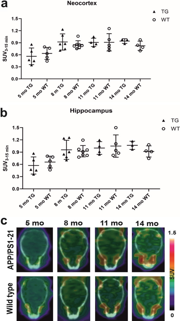
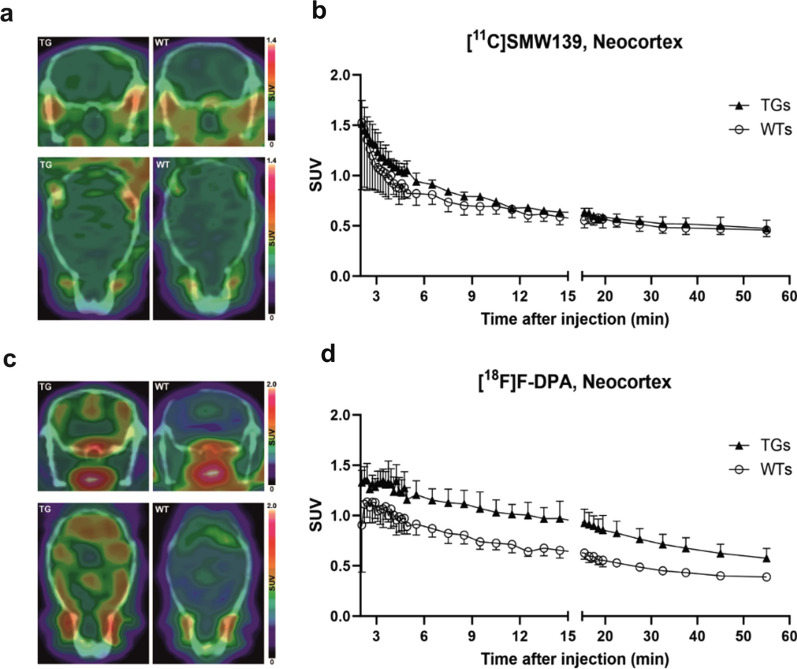
Background: P2X7 receptor has emerged as a potentially superior PET imaging marker to TSPO, the gold standard for imaging glial reactivity. [11C]SMW139 is the most recently developed radiotracer to image P2X7 receptor. The aim of this study was to image reactive glia in the APP/PS1-21 transgenic (TG) mouse model of Aβ deposition longitudinally using [11C]SMW139 targeting P2X7 receptor and to compare tracer uptake to that of [18F]F-DPA targeting TSPO at the final imaging time point. TG and wild type (WT) mice underwent longitudinal in vivo PET imaging using [11C]SMW139 at 5, 8, 11, and 14 months, followed by [18F]F-DPA PET scan only at 14 months. In vivo imaging results were verified by ex vivo brain autoradiography, immunohistochemical staining, and analysis of [11C]SMW139 unmetabolized fraction in TG and WT mice.

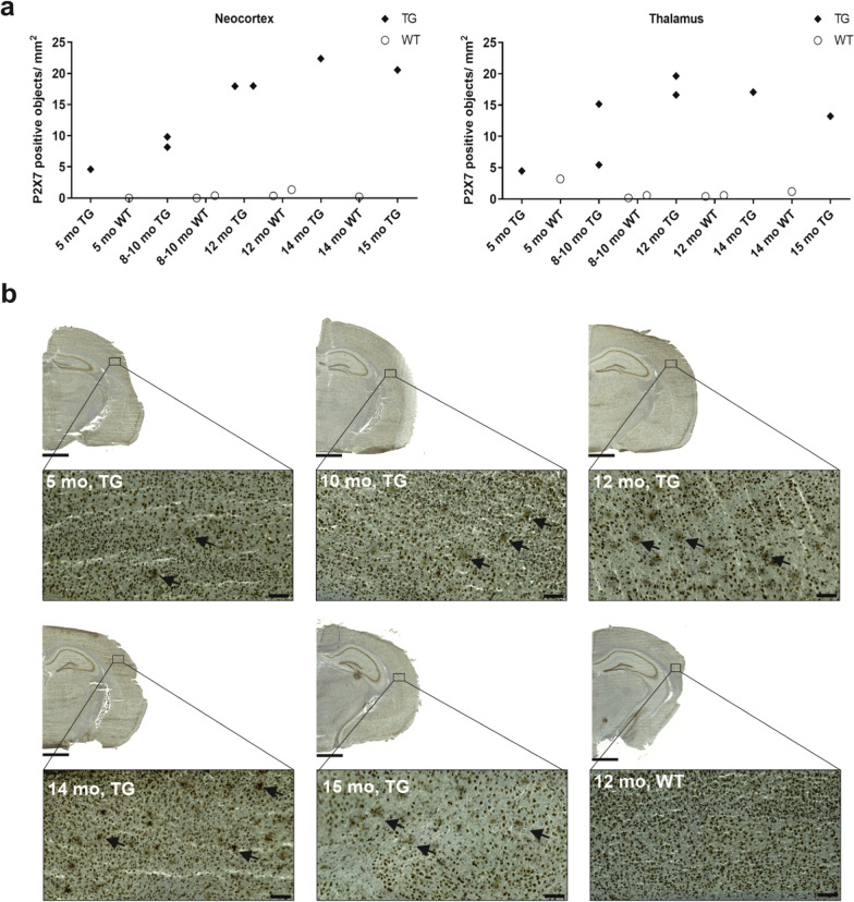
Results: Longitudinal change in [11C]SMW139 standardized uptake values (SUVs) showed no statistically significant increase in the neocortex and hippocampus of TG or WT mice, which was consistent with findings from ex vivo brain autoradiography. Significantly higher [18F]F-DPA SUVs were observed in brain regions of TG compared to WT mice. Quantified P2X7-positive staining in the cortex and thalamus of TG mice showed a minor increase in receptor expression with ageing, while TSPO-positive staining in the same regions showed a more robust increase in expression in TG mice as they aged. [11C]SMW139 was rapidly metabolized in mice, with 33% of unmetabolized fraction in plasma and 29% in brain homogenates 30 min after injection.

Conclusions: [11C]SMW139, which has a lower affinity for the rodent P2X7 receptor than the human version of the receptor, was unable to image the low expression of P2X7 receptor in the APP/PS1-21 mouse model. Additionally, the rapid metabolism of [11C]SMW139 in mice and the presence of several brain-penetrating radiometabolites significantly impacted the analysis of in vivo PET signal of the tracer. Finally, [18F]F-DPA targeting TSPO was more suitable for imaging reactive glia and neuroinflammatory processes in the APP/PS1-21 mouse model, based on the findings presented in this study and previous studies with this mouse model.

Keywords: APP/PS1-21; Alzheimer’s disease; Microglia; P2X7; P2Y12; Positron emission tomography; TSPO; [11C]SMW139; [18F]F-DPA.


Conflict of interest statement

The authors declare that they have no competing interests.

Figures

Fig. 1
Fig. 1
In vivo PET imaging study design and mice counts at each imaging time point. The green box illustrates that at 14 months, mice were imaged with both radiotracers [11C]SMW139 and [18F]F-DPA. TG transgenic, WT wild type
Fig. 2
Fig. 2
[11C]SMW139 longitudinal brain uptake in APP/PS1-21 transgenic (TG) and wild type (WT) mice. a and b Longitudinal [11C]SMW139 standardized uptake values (SUVs) in the neocortex and hippocampus of TG and WT mice at 5, 8, 11 and 14 months of age. Statistical analysis: linear mixed model with compound symmetry covariance structure. c Representative axial brain PET/CT images of [11C]SMW139 longitudinal SUVs in the same TG and WT mice at 5, 8, 11 and 14 months of age
Fig. 3
Fig. 3
In vivo PET imaging using [11C]SMW139 and [18F]F-DPA in the same APP/PS1-21 transgenic (TG) and age-matched wild type (WT) mice at 14 months. a and c Representative coronal and axial brain PET/CT images of TG (n = 1) and WT (n = 1) mice at 14 months imaged with [11C]SMW139 (a) and [18F]F-DPA (c). [11C]SMW139 images were summed over 3–15 min and adjusted to the same color scale. [18F]F-DPA were summed over 25–50 min and scaled to the same color scale. b and d Averaged [11C]SMW139 (b) and [18F]F-DPA (d) time-activity curves in the neocortex of the same TG (n = 3) and WT (n = 4) mice at 14 months. Error bars indicate standard deviation
Fig. 4
Fig. 4
[11C]SMW139 and [18F]F-DPA standardized uptake value (SUV) in the same APP/PS1-21 transgenic (TG) and wild type (WT) mice at 14 months. a and c [11C]SMW139 averaged SUV in the neocortex (a) and hippocampus (c) of TG (n = 3) and WT (n = 4) mice. b and d [18F]F-DPA averaged SUV in the neocortex (b) and hippocampus (d) of the same TG and WT mice. Statistical test: t-test with Welch’s correction. SUV values are presented with mean and standard deviation. *p < 0.05
Fig. 5
Fig. 5
Ex vivo brain autoradiography imaging using [11C]SMW139 in APP/PS1-21 transgenic (TG) and wild type (WT) mice. a Representative autoradiography images of TG (n = 1) and WT (n = 1) mice at 8–10 months. Images are adjusted to the same color bar. b and c [11C]SMW139 standardized uptake value (SUV) in cortex (b) and thalamus (c) of TG (n = 4, 5, 2, 2) and WT (n = 4, 7, 1, 5) mice at 5, 8–10, 12 and 14 months
Fig. 6
Fig. 6
Immunohistochemical staining of P2X7 receptor in brain cryosections of APP/PS1-21 transgenic (TG) and wild type (WT) mice. a Quantification of P2X7-positive staining as object counts/mm2. Staining was evaluated in the cortex and thalamus of TG and WT mice at 5, 8–10, 12, 14, and 15 months. b Representative images of P2X7-positive staining in TG and WT mice at the investigated time points. Arrows point at the P2X7-positive staining detected by the artificial intelligence object detection algorithm. Scale bar = 1000 µm for the half-hemisphere brain section image, 100 µm for the cortex view image. Magnification = 1.5× for the half-hemisphere brain section image, 15.0 × for the cortex view image
Fig. 7
Fig. 7
Immunohistochemical staining of TSPO in brain cryosections of APP/PS1-21 transgenic (TG) and wild type (WT) mice. a Quantification of TSPO-positive staining as object counts/mm2. Staining was evaluated in the cortex and thalamus of TG and WT mice at 5, 8–10, 12, 14, and 15 months. Error bars indicate standard deviation. b Representative images of TSPO-positive staining in TG and WT mice at the investigated time points. Scale bar = 1000 µm for the half-hemisphere brain section image, 100 µm for the cortex view image. Magnification = 1.5× for the half-hemisphere brain section image, 15.0 × for the cortex view image
Fig. 8
Fig. 8
Fractions of unmetabolised [11C]SMW139 in APP/PS1-21 transgenic (TG) and wild type (WT) mice plasma and brain homogenates at 10, 30, and 45 min. a and b Percentage of unmetabolised [11C]SMW139 over all radioactivity in male and female mice in plasma (a) and brain homogenates (b). Error bars indicate standard deviation

References

    1. Gao HM, Hong JS. Why neurodegenerative diseases are progressive: uncontrolled inflammation drives disease progression. Trends Immunol. 2008;29(8):357–365. doi: 10.1016/j.it.2008.05.002. - DOI - PMC - PubMed
    1. Block ML, Zecca L, Hong JS. Microglia-mediated neurotoxicity: uncovering the molecular mechanisms. Nat Rev Neurosci. 2007;8:57–69. doi: 10.1038/nrn2038. - DOI - PubMed
    1. Wyss-Coray T, Mucke L. Inflammation in neurodegenerative disease—a double-edged sword. Neuron. 2002;35:419–432. doi: 10.1016/S0896-6273(02)00794-8. - DOI - PubMed
    1. Savio LEB, Mello PA, da Silva CG, Coutinho-Silva R. The P2X7 receptor in inflammatory diseases: Angel or demon? Front Pharmacol. 2018;9:52. doi: 10.3389/fphar.2018.00052. - DOI - PMC - PubMed
    1. Yu Y, Ugawa S, Ueda T, Ishida Y, Inoue K, Kyaw Nyunt A, et al. Cellular localization of P2X7 receptor mRNA in the rat brain. Brain Res. 2008;15(1194):45–55. doi: 10.1016/j.brainres.2007.11.064. - DOI - PubMed

LinkOut - more resources