. 2024 Aug;46(4):3889-3909.
doi: 10.1007/s11357-024-01111-5. Epub 2024 Mar 6.

Ergothioneine promotes longevity and healthy aging in male mice

Affiliations

Ergothioneine promotes longevity and healthy aging in male mice

Makoto Katsube et al. Geroscience. 2024 Aug.

Abstract

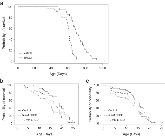
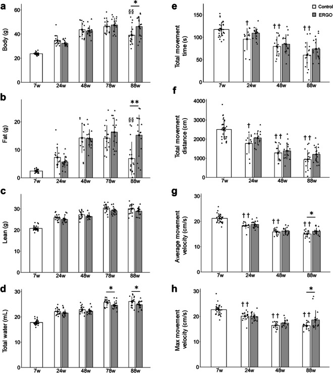
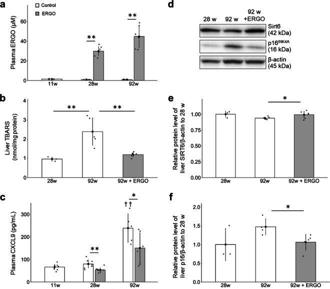
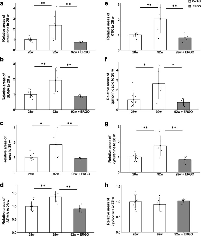
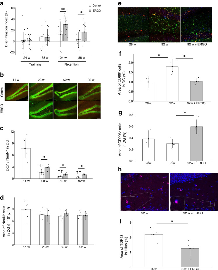
Healthy aging has emerged as a crucial issue with the increase in the geriatric population worldwide. Food-derived sulfur-containing amino acid ergothioneine (ERGO) is a potential dietary supplement, which exhibits various beneficial effects in experimental animals although the preventive effects of ERGO on aging and/or age-related impairments such as frailty and cognitive impairment are unclear. We investigated the effects of daily oral supplementation of ERGO dissolved in drinking water on lifespan, frailty, and cognitive impairment in male mice from 7 weeks of age to the end of their lives. Ingestion of 4 ~ 5 mg/kg/day of ERGO remarkably extended the lifespan of male mice. The longevity effect of ERGO was further supported by increase in life and non-frailty spans of Caenorhabditis elegans in the presence of ERGO. Compared with the control group, the ERGO group showed significantly lower age-related declines in weight, fat mass, and average and maximum movement velocities at 88 weeks of age. This was compatible with dramatical suppression by ERGO of the age-related increments in plasma biomarkers (BMs) such as the chemokine ligand 9, creatinine, symmetric dimethylarginine, urea, asymmetric dimethylarginine, quinolinic acid, and kynurenine. The oral intake of ERGO also rescued age-related impairments in learning and memory ability, which might be associated with suppression of the age-related decline in hippocampal neurogenesis and TDP43 protein aggregation and promotion of microglial shift to the M2 phenotype by ERGO ingestion. Ingestion of ERGO may promote longevity and healthy aging in male mice, possibly through multiple biological mechanisms.

Keywords: Age-related impairments; Anti-aging; Ergothioneine; Frailty; Healthy aging; Hippocampal neurogenesis; Lifespan; Longevity.


Conflict of interest statement

The authors declare no competing interests.

Figures

Fig. 1
Fig. 1
Oral intake of ERGO promoted lifespan and non-frailty span. a Survival curves for mice with daily intake of water containing 0.055 mg/mL ERGO (solid line, n = 36) or water alone (dashed line, n = 35). b Lifespan and c non-frailty span curves for C. elegans cultured on agar with 5 mM (dashed line, n = 46) and 10 mM ERGO (solid line, n = 36) or without ERGO (dotted line, n = 40). Significant differences between groups were determined by using the log-rank test. C. elegans, Caenorhabditis elegans
Fig. 2
Fig. 2
ERGO prevented frailty in mice. Age-dependent changes in a body weight in mice with daily intake of water containing 0.055 mg/mL ERGO (gray columns, n = 16) or water alone (white columns, n = 32 and 16 for 7 weeks and others, respectively). b Fat mass, c lean mass, and d total water content were measured by EchoMRI. To evaluate frailty, e total movement time, f total movement distance, g average movement velocity, and h maximum movement velocity were also evaluated by OFT. Data represent mean ± SEM. **P < 0.01, *P < 0.05 versus corresponding control (Welch t-test); §§P < 0.01 versus control at ages 78 weeks (Tukey’s test). ††P < 0.01, P < 0.05 versus control at 7 weeks of age (Tukey’s test). OFT, open field test
Fig. 3
Fig. 3
ERGO suppressed cellular senescence in mice. a The plasma ERGO levels in mice with daily intake of water alone (white columns, n = 8, 8, and 6 for 11, 28, and 92 weeks, respectively) or water containing 0.055 mg/mL ERGO (gray columns, n = 8 and 6 for 28 and 92 weeks, respectively) were quantified using UHPLC-MS/MS. **P < 0.01 versus corresponding control (Welch t-test). b Hepatic concentration of TBARS was measured in control (white columns, n = 3 and 6 for 28 and 92 weeks, respectively) and ERGO groups (gray column, n = 6). **P < 0.01 versus control group at 92 weeks of age (Dunnett’s test). c Plasma CXCL9 concentration was measured in control (white columns, n = 8, 8, and 6 for 11, 28, and 92 weeks, respectively) and ERGO groups (gray columns, n = 8 and 6 for 28 and 92 weeks, respectively). **P < 0.01, *P < 0.05 versus corresponding control (Welch t-test); ††P < 0.01 versus control at 11 weeks of age (Tukey’s test). The protein levels of d, e SIRT6 and d, f p16 in the liver lysates were quantified by western blotting analysis in control (white columns, n = 3 and 6 for 28 and 92 weeks, respectively) and ERGO groups (gray columns, n = 6). The intensity of each band was normalized by that of β-actin. *P < 0.05 versus control group at 92 weeks of age (Dunnett’s test). The bar graph data represent mean ± SEM. CXCL9, chemokine (CXC motif) ligand 9; SIRT6, NAD+-dependent protein deacetylase sirtuin-6; TBARS, thiobarbituric acid-reactive substances
Fig. 4
Fig. 4
ERGO suppressed age-dependent changes in several plasma biomarkers. a The plasma levels of creatinine, b SDMA, c urea, d ADMA, e KTR, f quinolinic acid, g kynurenine, and h tryptophan were measured by CE-TOF–MS. The white and gray columns represent the mice with daily intake of water alone (n = 10 and 5 for 28 and 92 weeks, respectively) and water containing 0.055 mg/mL ERGO (n = 4), respectively. Data represent mean ± SEM. **P < 0.01, *P < 0.05 versus control group at 92 weeks of age (Dunnett’s test). ADMA, asymmetrical dimethylarginine; KTR, kynurenine/tryptophan ratio; SDMA, symmetric dimethylarginine
Fig. 5
Fig. 5
ERGO improved age-related hippocampal impairment. a DI values for the training and retention trials were measured at 24 and 88 weeks of age. The white and gray columns represent the mice with daily intake of water alone and water containing 0.055 mg/mL ERGO, respectively (n = 16 for each group); **P < 0.01, *P < 0.05 versus corresponding control (Welch t-test). b Immunohistochemical detection of the newborn neuron marker Dcx (red) and neuronal nuclei marker NeuN (green) in the DG. Scale bar, 100 µm. c Dcx+/NeuN+ ratio, which was determined by dividing the area of Dcx+ cells by the area of NeuN+ cells and d area of NeuN+ cells in the DG. *P < 0.05 versus corresponding control (Welch t-test); ††P < 0.01 versus control at 11 weeks of age (Tukey’s test). The white and gray columns represent the control (n = 6 and 4 for 11 weeks and others, respectively) and ERGO groups (n = 4 for each group), respectively. e Immunohistochemical staining of the M1 microglial marker CD86 (green), M2 microglial marker CD206 (blue), and microglial marker Iba1 (red) in the DG. Scale bar, 50 µm. f Area of CD86+ cells and g that of CD206+ cells in the DG. The white and gray columns represent the control (n = 3 and 4 for 28 and 92 weeks, respectively) and ERGO groups (n = 4), respectively. *P < 0.05 versus control group at 92 weeks of age (Dunnett’s test). h Immunohistochemical staining of brain aggregate protein marker TDP43 (pink) and nuclear marker DAPI (blue) in the hilus. Scale bar, 20 µm. i Area of TDP43+ cells in the hilus at 92 weeks of age. The white and gray columns represent the control (n = 4) and ERGO groups (n = 4), respectively. *P < 0.05 versus control (Welch t-test). The bar graph data represent mean ± SEM. Dcx, doublecortin; DG, dentate gyrus; DI, discrimination index; NORT, novel object recognition test; TDP43, TAR DNA-binding protein of 43 kDa
Fig. 6
Fig. 6
ERGO inhibited the histamine-metabolizing enzyme and promoted polarization of anti-inflammatory M2 microglia. a Dose-dependent inhibitory effect of ERGO (0, 10, 30, 60, 100, and 500 µM) on histamine metabolism in mouse brain cytosol (20 µg/mL) and b the effect of ERGO (0, 10, 30, 100, and 500 µM) on rhHNMT (10 nM). c Lineweaver–Burk plots on the data of the inhibition of rhHNMT by ERGO. Data represent mean ± SEM (n = 3). d Immunohistochemical detection of HNMT (green) and the microglial marker Iba1 (red) in mouse hippocampus. Scale bar, 5 µm. e Immunocytochemical detection of HNMT (green) and Iba1 (red) in mouse PMG. Scale bar, 50 µm. f qRT–PCR determination of the relative levels of the M1 microglial marker CD86 and g M2 microglial marker CD206 mRNA in mouse PMG. Data are the mean ratio ± SEM of transcript levels normalized to gapdh (n = 4–6). *P < 0.05 versus control (Dunnett’s test). PMG, primary cultured microglia; rhHNMT, recombinant human histamine N-methyltransferase; qRT–PCR, quantitative reverse transcription-polymerase chain reaction

References

    1. Sayed N, HuangY, Nguyen K, Krejciova-Rajaniemi Z, Grawe AP, Gao T, Tibshirani R, Hastie T, Alpert A, Cui L, Kuznetsova T, Rosenberg-Hasson Y, Ostan R, Monti D, Lehallier B, Shen-Orr SS, Maecker HT, Dekker CL, Wyss-Coray T, … Furman D. An inflammatory aging clock (iAge) based on deep learning tracks multimorbidity immunosenescence frailty and cardiovascular aging. Nat Aging 2021 1:598–615. 10.1038/s43587-021-00082-y - PMC - PubMed
    1. Ma Y, Matsuwaki T, Yamanouchi K, Nishihara M. Involvement of progranulin in modulating neuroinflammatory responses but not neurogenesis in the hippocampus of aged mice. Exp Gerontol. 2017;95:1–8. doi: 10.1016/j.exger.2017.05.003. - DOI - PubMed
    1. Wilson AC, Dugger BN, Dickson DW, Wang D-S. TDP-43 in aging and Alzheimer’s disease—a review. Int J Clin Exp Pathol. 2011;4(2):147–155. - PMC - PubMed
    1. Heneka MT, Carson MJ, Khoury JEL Landreth GE, Brosseron F, Feinstein DL, Jacobs AH, Wyss-Coray T, Vitorica J, Ransohoff RM, Herrup K, Frautschy SA, Finsen B, Brown GC, Verkhratsky A, Yamanaka K, Koistinaho J, Latz E, Halle A, … Kummer MP. Neuroinflammation in Alzheimer’s disease. Lancet Neurol. 2015,14(4):388–405.10.1016/S1474-4422(15)70016-5. - PMC - PubMed
    1. Ritzel RM, Doran SJ, Glaser EP, Meadows VE, Faden AI, Stoica BA, Loane DJ. Old age increases microglial senescence exacerbates secondary neuroinflammation and worsens neurological outcomes after acute traumatic brain injury in mice. Neurobiol Aging. 2019;77:194–206. doi: 10.1016/j.neurobiolaging.2019.02.010. - DOI - PMC - PubMed

Publication types

LinkOut - more resources