. 2024 Apr 1;35(4):ar59.
doi: 10.1091/mbc.E24-01-0012. Epub 2024 Mar 6.

Loss of Grp170 results in catastrophic disruption of endoplasmic reticulum function

Affiliations

Loss of Grp170 results in catastrophic disruption of endoplasmic reticulum function

Melissa J Mann et al. Mol Biol Cell. .

Abstract

GRP170 (Hyou1) is required for mouse embryonic development, and its ablation in kidney nephrons leads to renal failure. Unlike most chaperones, GRP170 is the lone member of its chaperone family in the ER lumen. However, the cellular requirement for GRP170, which both binds nonnative proteins and acts as nucleotide exchange factor for BiP, is poorly understood. Here, we report on the isolation of mouse embryonic fibroblasts obtained from mice in which LoxP sites were engineered in the Hyou1 loci (Hyou1LoxP/LoxP). A doxycycline-regulated Cre recombinase was stably introduced into these cells. Induction of Cre resulted in depletion of Grp170 protein which culminated in cell death. As Grp170 levels fell we observed a portion of BiP fractionating with insoluble material, increased binding of BiP to a client with a concomitant reduction in its turnover, and reduced solubility of an aggregation-prone BiP substrate. Consistent with disrupted BiP functions, we observed reactivation of BiP and induction of the unfolded protein response (UPR) in futile attempts to provide compensatory increases in ER chaperones and folding enzymes. Together, these results provide insights into the cellular consequences of controlled Grp170 loss and provide hypotheses as to why mutations in the Hyou1 locus are linked to human disease.


Figures

FIGURE 1:
FIGURE 1:
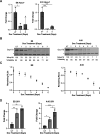
Hyou1fl/fl MEF cells expressing Cre recombinase are rapidly out-competed by cells that do not. (A) GFP and mCherry expression were measured by FACS as described in the text at the indicated days after incubation with doxycycline as well as UT (untreated). (B) The indicated transcripts were quantified by RT-PCR after doxycline treatment to induce Cre synthesis. (C) Corresponding protein expression of the transcripts measured in (B) were determined by western blotting and quantified. The signals were quantified and graphically displayed as a percent of their value in untreated cells (n = 3). Data presented represent the mean ± SD.
FIGURE 2:
FIGURE 2:
Inducible expression of Cre recombinase in single cell clones of Hyou1fl/fl MEFs leads to loss of Grp170, which decreases cell viability. (A) The expression of Hyou1 transcripts were measured in the E8 and A10 clones after Cre induction (n = 3). (B) The resulting effect on Grp170 protein expression was assessed by western blot with levels relative to untreated (UT) cells displayed below each time point. A nonspecific band is indicated (*). (C) Cell viability was determined each day after Cre induction using a Cell Titer-Glo assay. Signals are expressed relative to that in untreated cells (UT) (n = 3). (D) Transcripts for Sil1, the other ER NEF, were quantified by RT-PCR after loss of Grp170 in each clone. Data represent the mean ± SD; *p < 0.05, **p < 0.01, ***p < 0.001.
FIGURE 3:
FIGURE 3:
As Grp170 levels fall, AMPylated pools of BiP are reactivated to increase the pool of functional BiP. Cells from the (A) A10 and (B) E8 clone were incubated with doxycycline for the indicated days, and cell lysates were prepared for western blotting. The top panel portrays a membrane blotted with a combination of anti-Grp170 and anti-BiP, whereas the bottom panel is from a membrane blotted with a monoclonal antibody specific for AMPylated BiP. An aliquot of recombinant AMPylated BiP was loaded on the right of the gel, and the parental clone (P) was loaded to the left (n = 3). A band migrating between Grp170 and BiP (*) was detected with the anti-Grp170 rabbit polyclonal serum, although its identity is unknown.
FIGURE 4:
FIGURE 4:
Loss of Grp170 increases steady-state BiP binding to an unfolded client and slows its degradation. (A) Cells from the E8 clones were transfected with an expression vector encoding the NS1 LC 4 d after incubation with doxycycline (+), and lysates were prepared the following day. Untreated cells (–) served as a control. The mouse κ LC was immunoprecipitated from lysates, resolved on reducing SDS-gels, transferred, and blotted with anti-BiP or anti-κ, n = 3. (B) E8 cells that had been treated with doxycycline or left untreated were transfected with an expression vector encoding the NHK variant of α1AT. The next day, the cells were pulsed with [35S]-labeled methionine and cysteine for 30 min before chasing for the indicated times. Lysates were prepared, and 90% of the lysate from a p60 dish was immunoprecipitated with anti-α1AT antibody for each time-point and electrophoresed on reducing SDS-polyacrylamide gels. Signals were determined on a phosphorimager and represented as a percentage of that observed at the start of the chase (t = 0). Data represent the mean ± SD (n = 2).
FIGURE 5:
FIGURE 5:
The relative percent of an aggregation-prone protein and BiP in the insoluble fraction increases as Grp170 is depleted. (A) Cre recombinase expression was induced in E8 cells for the indicated days. Cells were lysed with NP40 buffer and centrifuged at 20,000 g to separate NP40 soluble and insoluble material. The insoluble material was extracted by heating in Laemmli buffer and sonication. The samples loaded on the NP-40 insoluble gel (bottom) are six times that loaded on the NP40 soluble gel (top). After electrophoresis and transfer, membranes were blotted for BiP and Grp170 (n = 3). A nonspecific band is indicated (*) (B) Cre recombinase expression was induced in E8 cells which were transfected with an expression plasmid for A1PiZ. Cells were harvested at 5 d postdox induction (48 h posttransfection). Cells were lysed and lysate was centrifuged at 100,000 g for 1 h to separate soluble and insoluble material. Samples loaded on the gel correspond to 20% of the supernatant and the entire pellet fraction. After electrophoresis and transfer, membranes were probed with antibodies against A1PiZ and Grp170. Data represent the mean ± SD; *p < 0.05, **p < 0.01 (n = 4).
FIGURE 6:
FIGURE 6:
The UPR is activated in the absence of Grp170. (A) The expression of UPR target transcripts was measured in the E8 clone after Cre induction for the indicated days by qPCR (n = 3). Data represent the mean ± SD; *p < 0.05, **p < 0.01, ***p < 0.001, ****p < 0.0001. (B) Western blot analysis was also performed for UPR targets (B), Gadd34 (C), an apoptotic marker (D), and loading controls (actin or tubulin). For (D) cells were also treated with 1 μm staurosporine (STS) as a positive control for apoptosis. Representative images are shown.
FIGURE 7:
FIGURE 7:
ER morphology is altered as Grp170 is depleted. (A) E8 cells grown on coverslips were treated for the indicated times to induce Cre-lox editing of Grp170. Cells were stained for KDEL followed by anti-Rabbit 647 secondary antibody to mark the ER. Scale bar in the bottom right panel is 10 µm (Bottom). (B) Whole cell area was quantified using Nikon NIS-Elements software. Violin plots depicting the individual data points as well as the median, and first and third quartiles are plotted, ***p < 0.0001 (n = 21–37). (C) Protein lysates from the E8 clone treated with doxycycline for the indicated days were subject to western blot and probed with anti-Climp63 antibody. Data represent the mean ± SD; *p < 0.05, **p < 0.01 (n = 3).

Update of

References

    1. Adams BM, Oster ME, Hebert DN (2019). Protein quality control in the endoplasmic reticulum. Protein J 38, 317–329. - PMC - PubMed
    1. Amin-Wetzel N, Saunders RA, Kamphuis MJ, Rato C, Preissler S, Harding HP, Ron D (2017). A J-Protein cochaperone recruits BiP to monomerize IRE1 and repress the unfolded protein response. Cell 171, 1625–1637. - PMC - PubMed
    1. Anttonen AK, Mahjneh I, Hämäläinen RH, Lagier-Tourenne C, Kopra O, Waris L, Anttonen M, Joensuu T, Kalimo H, Paetau A, et al. (2005). The gene disrupted in Marinesco-Sjögren syndrome encodes SIL1, an HSPA5 cochaperone. Nat Genet 37, 1309–1311. - PubMed
    1. Appenzeller-Herzog C, Ellgaard L (2008). The human PDI family: versatility packed into a single fold. Biochim Biophys Acta 1783, 535–548. - PubMed
    1. Balchin D, Hayer-Hartl M, Hartl FU (2016). In vivo aspects of protein folding and quality control. Science 353, aac4354. - PubMed

Substances