Review
. 2024 Mar 6;10(1):114.
doi: 10.1038/s41420-024-01868-w.

The potential role of hydrogen sulfide in cancer cell apoptosis

Affiliations
Review

The potential role of hydrogen sulfide in cancer cell apoptosis

Wei Gao et al. Cell Death Discov. .

Abstract

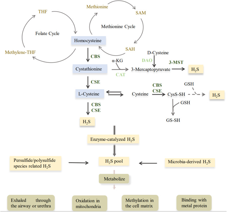
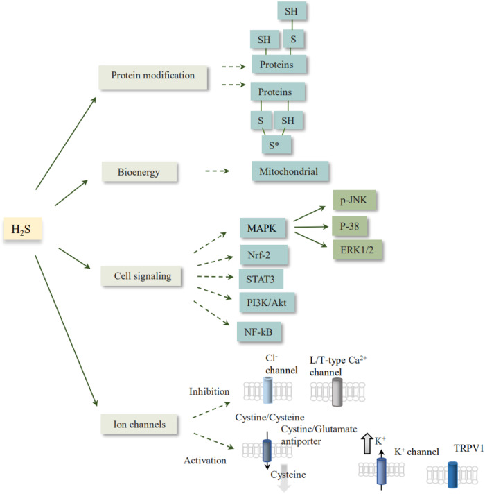
For a long time, hydrogen sulfide (H2S) has been considered a toxic compound, but recent studies have found that H2S is the third gaseous signaling molecule which plays a vital role in physiological and pathological conditions. Currently, a large number of studies have shown that H2S mediates apoptosis through multiple signaling pathways to participate in cancer occurrence and development, for example, PI3K/Akt/mTOR and MAPK signaling pathways. Therefore, the regulation of the production and metabolism of H2S to mediate the apoptotic process of cancer cells may improve the effectiveness of cancer treatment. In this review, the role and mechanism of H2S in cancer cell apoptosis in mammals are summarized.


Conflict of interest statement

The authors declare no competing interests.

Figures

Fig. 1
Fig. 1. The production and metabolism of H2S.
In mammals, H2S is produced endogenously from cysteine, serine, homocysteine and other substrates primarily through the actions of three major enzymes. Non-enzymatic pathways: gut microbes as well as polysulfide-derived H2S. Metabolism of H2S: H2S can be oxidized in the mitochondria or metabolized by methylation in the cytoplasm. H2S is first oxidized by SQR in the mitochondria to form a persulfide. This persulfide is further oxidized by ETHE1 to produce SO32−, which is then converted to SO42− and S2O32− by sulfite oxidase and rhodan oxidase and excreted in the urine or by respiration. H2S can also be removed by binding to metalloproteins to form sulfheme. α-KG α-ketoglutaric acid, 3-MST 3-mercaptopyruvate sulfurtransferase, CAT cysteine aminotransferase, CBS cystathionine beta-synthase, CSE cystathionine gamma-lyase, CysS-SH cysteine persulfide, DAO D-amino acid oxidase, GSH glutathione, SAH S-adenosylhomocysteine, SAM S-adenosyl methionine, THF tetrahydrofolic acid.
Fig. 2
Fig. 2. The mechanisms of H2S function in the organism.
Promotion or inhibition of signaling pathways, post-translational modification of proteins, activation or shutdown of ion channels, and participation in mitochondrial metabolism. Akt protein kinase B, ERK extracellular signal-regulated kinase, JNK C-Jun N-terminal kinase, MAPK mitogen-activated protein kinase, NF-κB nuclear factor-kappa B, Nrf2 nuclear factor erythroid-2 related factor 2, PI3K phosphoinositide 3-kinase, STAT3 signal transducer and activator of transcription 3, TRPV1 transient receptor potential vanilloid 1.
Fig. 3
Fig. 3. Several common H2S donors.
DATS diallyl trisulfide, DADS diallyl disulfide, GYY4137 morpholin-4-ium 4-methoxyphenyl(morpholino) phosphinodithioate), SPRC S-propargyl-cysteine.
Fig. 4
Fig. 4. The ways of DATS and DADS in the generation of H2S.
DATS and DADS are two main active ingredients in garlic. DATS reacts rapidly with GSH to release H2S via thiol−disulfide exchange followed by allyl perthiol reduction by GSH. DADS only releases a minute amount of H2S via a sluggish reaction with GSH through an α-carbon nucleophilic substitution pathway. DATS diallyl trisulfide, DADS diallyl disulfide, GSH glutathione.
Fig. 5
Fig. 5. H2S regulates three pathways of apoptosis through multiple signaling pathways.
Cyt C cytochrome C, Fas fas cell surface death receptor, Fasl fas ligand, Smac second mitochondria-derived activator of caspases, JNK C-Jun N-terminal kinase, MAPK mitogen activated protein kinase, NF-κB nuclear factor-kappa B, PI3K phosphoinositide 3-kinase, Akt protein kinase B, ERs endoplasmic reticulum stress, ROS reactive oxygen species.

Similar articles

Cited by

References

    1. Ishigami M, Hiraki K, Umemura K, Ogasawara Y, Ishii K, Kimura H. A source of hydrogen sulfide and a mechanism of its release in the brain. Antioxid Redox Signal. 2009;11:205–14. doi: 10.1089/ars.2008.2132. - DOI - PubMed
    1. Kimura H. Hydrogen sulfide (H(2)S) and polysulfide (H(2)S(n)) signaling: the first 25 years. Biomolecules. 2021;11, 10.3390/biom11060896. - PMC - PubMed
    1. Shibuya N, Koike S, Tanaka M, Ishigami-Yuasa M, Kimura Y, Ogasawara Y, et al. A novel pathway for the production of hydrogen sulfide from D-cysteine in mammalian cells. Nat Commun. 2013;4:1366. doi: 10.1038/ncomms2371. - DOI - PubMed
    1. Kabil O, Banerjee R. Enzymology of H2S biogenesis, decay and signaling. Antioxid Redox Signal. 2014;20:770–82. doi: 10.1089/ars.2013.5339. - DOI - PMC - PubMed
    1. Nagy P. Mechanistic chemical perspective of hydrogen sulfide signaling. Methods Enzymol. 2015;554:3–29. doi: 10.1016/bs.mie.2014.11.036. - DOI - PubMed