. 2024 Mar 7;81(1):118.
doi: 10.1007/s00018-023-05081-3.

Tektin bundle interacting protein, TEKTIP1, functions to stabilize the tektin bundle and axoneme in mouse sperm flagella

Affiliations

Tektin bundle interacting protein, TEKTIP1, functions to stabilize the tektin bundle and axoneme in mouse sperm flagella

Xin-Yan Geng et al. Cell Mol Life Sci. .

Abstract

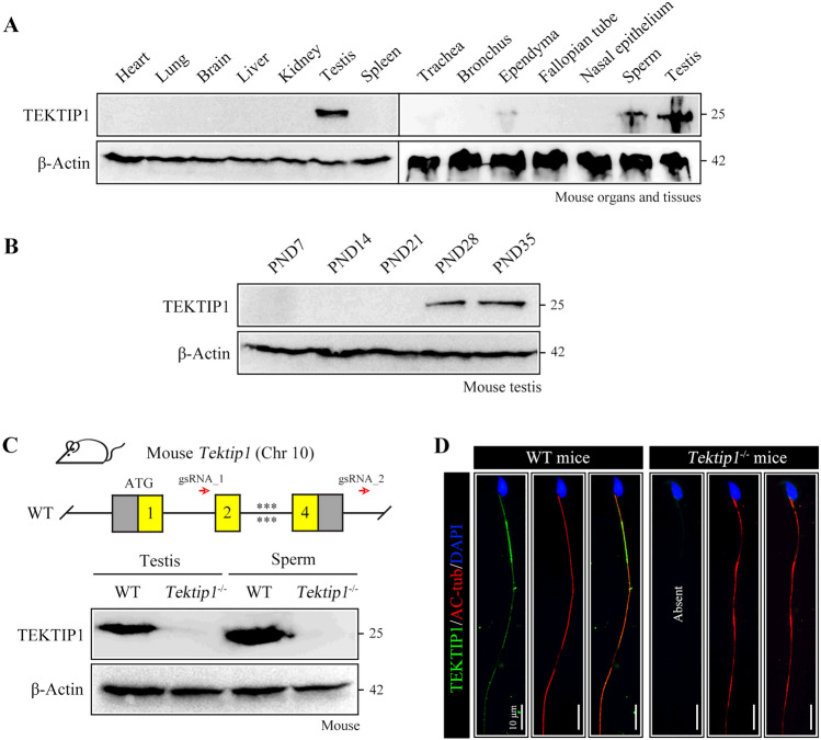
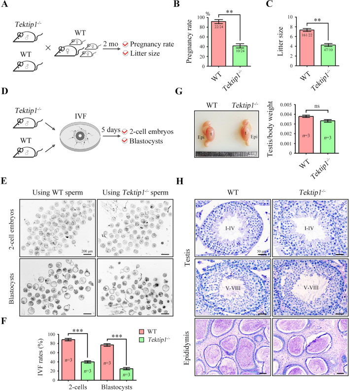
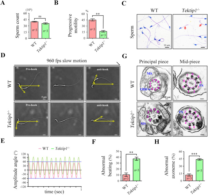
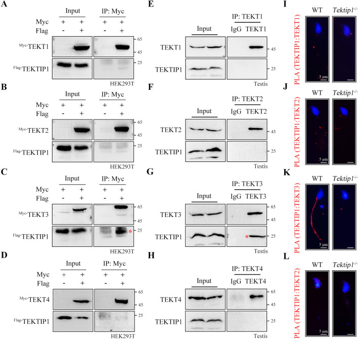
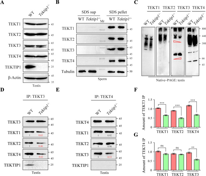
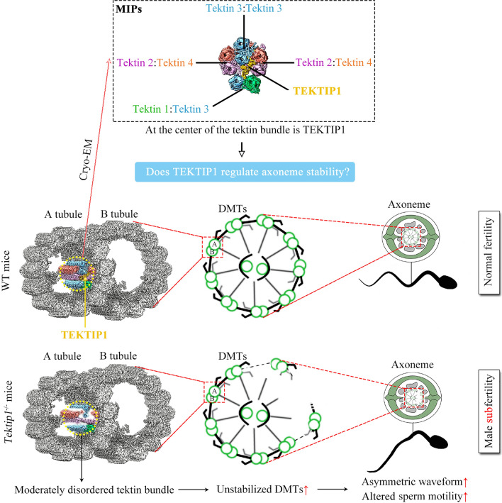
Tektins are microtubule inner proteins (MIPs) and localize at the inside lumen of doublet microtubules (DMTs) of cilia/flagella. TEKTIP1, a newly identified protein by cryo-electron microscopy (cryo-EM), is proposed to be localized at the center of the tektin bundle and hypothesized to recruit tektins or stabilize the bundle. However, the physiological role of TEKTIP1 is unknown. In this study, we generated Tektip1-knockout (Tektip1-/-) mice and showed that they were male subfertile primarily due to reduced sperm motility. A high percentage of sperm from Tektip1-/- mice showed moderately disorganized axoneme structures and abnormal flagellar waveforms. TEKTIP1 predominately interacted with TEKT3 among tektins. Loss of TEKTIP1 partially disturbed the organization of tektin bundle by mainly affecting the native status of TEKT3 and its interaction with other tektins. Collectively, our study reveals the physiological role and potential molecular mechanism of TEKTIP1 in axonemal structure and sperm motility, highlights the importance of MIPs in stabilizing DMTs, and suggests a potential relevance of TEKTIP1 deficiency to human asthenospermia. Tektip1-/- mice will be an excellent animal model to study the DMT organization of sperm flagella using cryo-EM in future.

Keywords: Knockout mice; Microtubule inner protein; Sperm motility; TEKTIP1; Tektins.


Conflict of interest statement

The authors declare no competing interests.

Figures

Fig. 1
Fig. 1
Expression of TEKTIP1 and establishment of Tektip1−/− mice. A Mouse TEKTIP1 expression in specified organs/tissues, detected via Western blotting and normalized to β-Actin. B Mouse TEKTIP1 expression in postnatal testes detected via Western blotting and normalized to β-Actin. PND, postnatal day. C Genomic features and knockout strategy of mouse Tektip1. Detailed information on the construction of Tektip1−/− mice is provided in Supplementary Fig. S3. TEKTIP1 protein expression in testis and sperm samples from Tektip1−/− mice and WT mice. β-Actin served as an internal control. D Immunofluorescence staining of sperm from WT mice and Tektip1−/− mice using anti-TEKTIP1 (green) and anti-AC-tub (acetylated-tubulin, red) antibodies. The nucleus was stained with DAPI (blue). Scale bar, 10 μm
Fig. 2
Fig. 2
Fertilizing ability and sperm parameters of Tektip1−/− mice. A Adult Tektip1−/− male mice and their littermate WT mice (n = 3 each group) were continuously coupled with adult WT female mice at a sex ratio of 1:2 for 2 months. B The pregnancy rate was calculated. Data are represented as the mean ± SD, Student’s t test (unpaired, two-tailed), **p < 0.01. C The litter size was calculated. Data are represented as the mean ± SD, Student’s t test (unpaired, two-tailed), **p < 0.01. D An in vitro fertilization (IVF) assay was performed using sperm from adult Tektip1−/− mice and WT mice (n = 3 for each group). E The images of two-cell embryos and blastocyst. F The percentage of two-cell embryos and blastocysts was calculated. Data are represented as the mean ± SD, Student’s t test (unpaired, two-tailed), ***p < 0.001. G Representative images of the testis and epididymis. Evaluation of the weight ratio of the testes/body from adult Tektip1−/− mice and WT mice (n = 3 for each group). ns not significant, t testis, Epi epididymis. Scale bar, 0.4 cm. H Histological evaluation of the testis and epididymis from adult Tektip1−/− and WT mice. Scale bar, 50 μm
Fig. 3
Fig. 3
Flagellar waveform and ultrastructural assessment of sperm from Tektip1−/− mice. A Sperm counts were determined using a fertility counting chamber under a light microscope. Data are represented as the mean ± SD (n = 3 each group), Student’s t test (unpaired, two-tailed), ns, not significant. B Progressive motility was assessed via computer-assisted semen analysis (CASA). Data are represented as the mean ± SD (n = 3 each group), Student’s t test (unpaired, two-tailed), **p < 0.01. C Light microscopy analysis of sperm from Tektip1−/− mice and WT mice using Papanicolaou staining. Red arrows indicated bent-tail sperm. Scale bar, 10 μm. D Flagellar waveforms of head-tethered sperm from Tektip1−/− mice and WT mice. Scale bar, 5 μm. E The amplitude angle of flagellar waveforms was recorded for 1 s. Flagellar waveforms of sperm from WT mice were symmetrical (± 60°). Flagellar waveforms of sperm from Tektip1−/− mice were asymmetrical (-30° ~ 90°). F Percentage of sperm showing abnormal beating patterns from Tektip1−/− mice and WT mice. One hundred spermatids in each group were counted. Data are represented as the mean ± SD (n = 3 each group), Student’s t test (unpaired, two-tailed), **p < 0.01. G Transmission electron microscope (TEM) analysis showing the principle piece and mid-piece of sperm flagella from Tektip1−/− mice and WT mice. ODF, outer dense fibre; MS, mitochondrial sheath; FS, fibrous sheath. Scale bar, 100 nm. H Percentage of sperm showing abnormal axoneme structures from Tektip1−/− mice and WT mice. Twenty-five spermatids in each group were counted. Data are represented as the mean ± SD (n = 3 each group), Student’s t test (unpaired, two-tailed), ***p < 0.001
Fig. 4
Fig. 4
TEKTIP1 interacts with TEKT3 but not TEKT1, TEKT2, or TEKT4. A–D Myc-tagged plasmids of TEKT1 ~ 4 and Flag-tagged plasmid of TEKTIP1 were constructed. Flag-tagged TEKTIP1 was immunoprecipitated with Myc-tagged TEKT3 but not Myc-tagged TEKT1, TEKT2, or TEKT4 in HEK293T cell lysates using co-IP assays. E–H An interaction between endogenous TEKTIP1 and TEKT3 was identified in mouse testis lysates using a co-IP assay. Endogenous TEKTIP1 was not immunoprecipitated with TEKT1, TEKT2, or TEKT4. Rabbit IgG served as the negative control. I-L Representative immunofluorescence images from a PLA performed on sperm from WT and Tektip1.−/− mice. Closely interaction was revealed by positive signals (red). Sperm heads were stained with DAPI (blue)
Fig. 5
Fig. 5
Expression and connections of tektins after the loss of TEKTIP1. A Expression of TEKT1 ~ 4 was analysed by Western blotting (SDS‒PAGE) in testis lysates of adult Tektip1−/− mice and WT mice. β-Actin served as a loading control. B Sperm samples were treated with SDS-EDTA solution. Supernatant and pellet samples were resolved by SDS‒PAGE. Expression of TEKT1 ~ 4 was analysed by Western blotting (SDS‒PAGE) in sperm lysates of adult Tektip1−/− mice and WT mice. C Expression of TEKT1 ~ 4 was analysed by Western blotting (Native‒PAGE) in testis lysates of adult Tektip1−/− mice and WT mice under native conditions. D Endogenous TEKT3 was immunoprecipitated from testis protein lysates of Tektip1−/− mice and WT mice. The amounts of TEKT1, TEKT2, or TEKT4 coimmunoprecipitated by TEKT3 were further detected by Western blotting. E The amounts of TEKT1, TEKT2, or TEKT3 coimmunoprecipitated by endogenous TEKT4 were detected in testis protein lysates of Tektip1−/− mice and WT mice. F Bar graph representing the band intensities of TEKT3-immunoprecipitated TEKT1, TEKT2, or TEKT4. Data represent the mean ± SD (n = 3 each group), Student’s t test (unpaired, two-tailed), ***p < 0.001. G Bar graph representing the band intensities of TEKT4-immunoprecipitated TEKT1, TEKT2, or TEKT3. Data represent the mean ± SD (n = 3 each group), Student’s t test (unpaired, two-tailed), **p < 0.01; ns, not significant
Fig. 6
Fig. 6
Schematic diagram of the physiological role of TEKTIP1 in tektin bundle, sperm motility and male fertility. Although TEKTIP1 does not regulate the expression of tektins and other MIP proteins, loss of TEKTIP1 causes moderately disordered tektins bundle at the level of protein–protein interaction. As a consequence, the percentage of sperm exhibiting unstabilized DMTs, asymmetric waveform, and altered sperm motility was increased, finally leading to male subfertility of Tektip1−/− mice. MIP microtubule inner protein, DMT doublet microtubule. Some cartoon elements were modified from reference [4, 28].

Similar articles

Cited by

References

    1. Satir P, Christensen ST. Overview of structure and function of mammalian cilia. Annu Rev Physiol. 2007;69:377–400. doi: 10.1146/annurev.physiol.69.040705.141236. - DOI - PubMed
    1. Inaba K. Sperm flagella: comparative and phylogenetic perspectives of protein components. Mol Hum Reprod. 2011;17:524–538. doi: 10.1093/molehr/gar034. - DOI - PubMed
    1. Ma MS, et al. Structure of the decorated ciliary doublet microtubule. Cell. 2019;179:909–922. doi: 10.1016/j.cell.2019.09.030. - DOI - PMC - PubMed
    1. Gui M, et al. De novo identification of mammalian ciliary motility proteins using cryo-EM. Cell. 2021;184:5791–5806. doi: 10.1016/j.cell.2021.10.007. - DOI - PMC - PubMed
    1. Ichikawa M, Bui KH. Microtubule inner proteins: a meshwork of luminal proteins stabilizing the doublet microtubule. BioEssays. 2018;40:1700209. doi: 10.1002/bies.201700209. - DOI - PubMed

LinkOut - more resources