. 2024 Mar 6;22(1):162.
doi: 10.1186/s12964-024-01539-4.

Interleukin-4 from curcumin-activated OECs emerges as a central modulator for increasing M2 polarization of microglia/macrophage in OEC anti-inflammatory activity for functional repair of spinal cord injury

Affiliations

Interleukin-4 from curcumin-activated OECs emerges as a central modulator for increasing M2 polarization of microglia/macrophage in OEC anti-inflammatory activity for functional repair of spinal cord injury

Jianbin Guo et al. Cell Commun Signal. .

Abstract

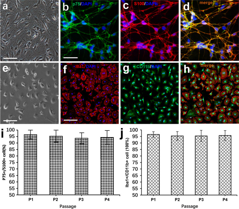
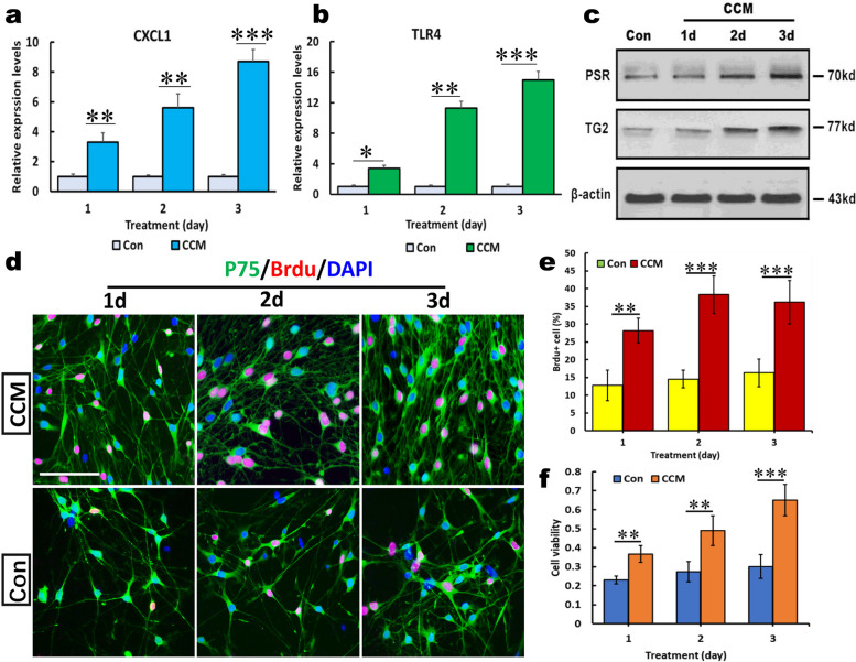
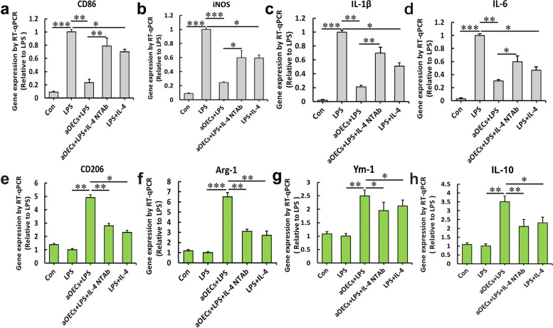
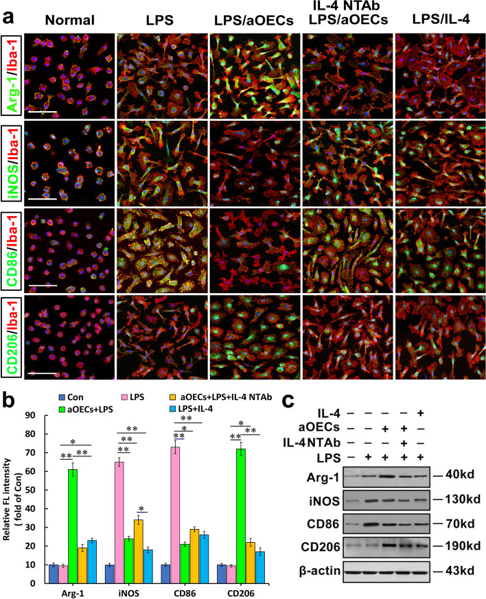
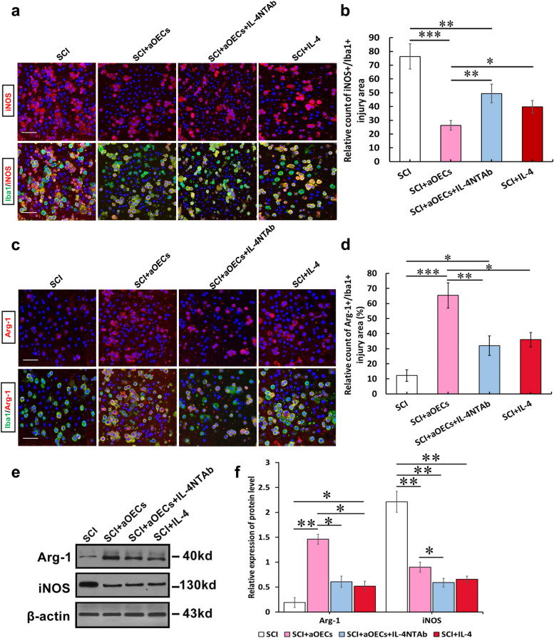
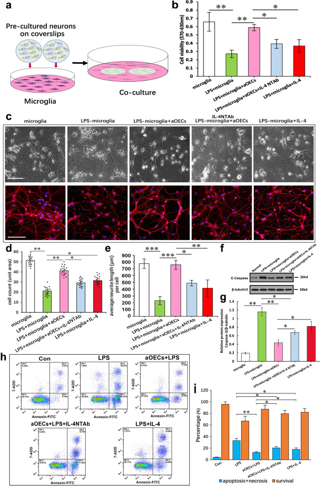
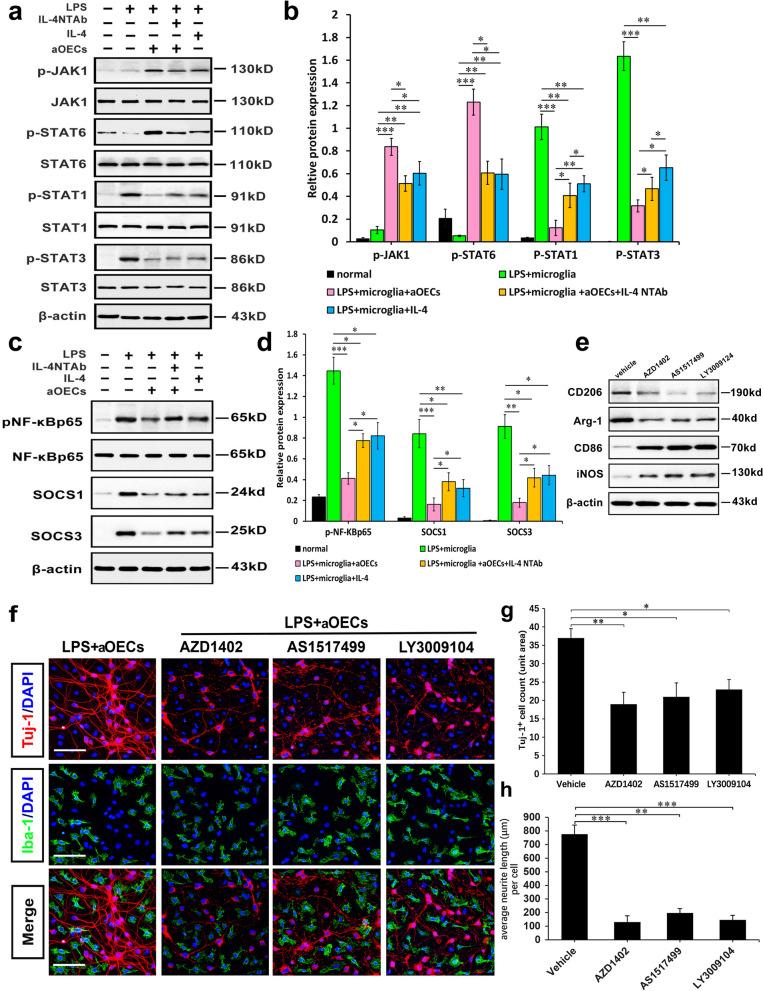
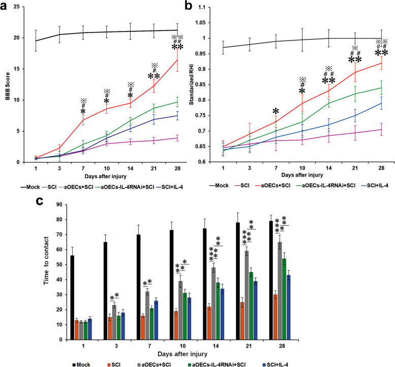
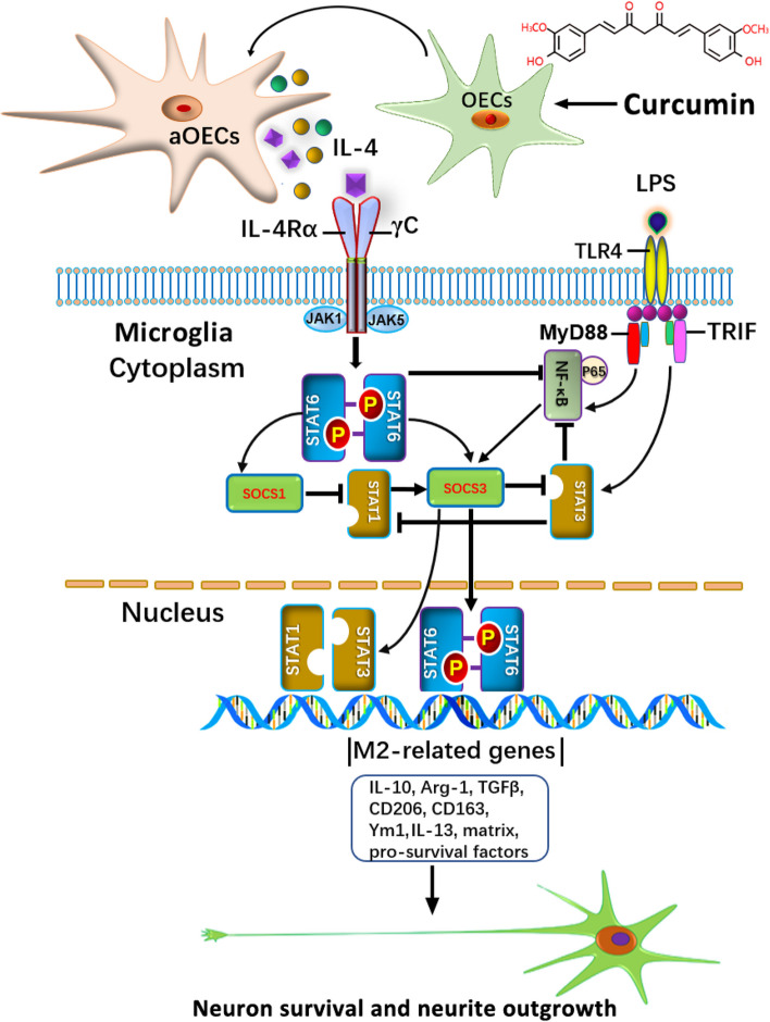
Microglia/macrophages are major contributors to neuroinflammation in the central nervous system (CNS) injury and exhibit either pro- or anti-inflammatory phenotypes in response to specific microenvironmental signals. Our latest in vivo and in vitro studies demonstrated that curcumin-treated olfactory ensheathing cells (aOECs) can effectively enhance neural survival and axonal outgrowth, and transplantation of aOECs improves the neurological outcome after spinal cord injury (SCI). The therapeutic effect is largely attributed to aOEC anti-inflammatory activity through the modulation of microglial polarization from the M1 to M2 phenotype. However, very little is known about what viable molecules from aOECs are actively responsible for the switch of M1 to M2 microglial phenotypes and the underlying mechanisms of microglial polarization. Herein, we show that Interleukin-4 (IL-4) plays a leading role in triggering the M1 to M2 microglial phenotype, appreciably decreasing the levels of M1 markers IL‑1β, IL‑6, tumour necrosis factor-alpha (TNF-α) and inducible nitric oxide synthase (iNOS) and elevating the levels of M2 markers Arg-1, TGF-β, IL-10, and CD206. Strikingly, blockade of IL-4 signaling by siRNA and a neutralizing antibody in aOEC medium reverses the transition of M1 to M2, and the activated microglia stimulated with the aOEC medium lacking IL-4 significantly decreases neuronal survival and neurite outgrowth. In addition, transplantation of aOECs improved the neurological function deficits after SCI in rats. More importantly, the crosstalk between JAK1/STAT1/3/6-targeted downstream signals and NF-κB/SOCS1/3 signaling predominantly orchestrates IL-4-modulated microglial polarization event. These results provide new insights into the molecular mechanisms of aOECs driving the M1-to-M2 shift of microglia and shed light on new therapies for SCI through the modulation of microglial polarization.

Keywords: Activated olfactory ensheathing cells; Interleukin-4; JAK1/STAT1/STAT3/STAT6; Microglia/macrophage polarization; Neuroinflammation.


Conflict of interest statement

The authors declare no competing interests.

Figures

Fig. 1
Fig. 1
Morphological and biochemical characteristics of primary olfactory ensheathing cells (OECs) and microglial cells. a Phase-contrast microscopy showing morphology of primary OECs after purification at 5 days in vitro. b, c and d Double immunostaining with p75 and S100 in purified OECs, respectively. c Primary microglial cells at 3 days in vitro. e, f and g Representative photomicrographs of immunofluorescence for Iba1 and CD11b in purified microglial cells. h Percentages of p75+/S100+ cells among purified cells after sub-culture from passages 1–4. i Percentages of IBa1+/CD11b+ cells among purified cells after sub-culture from passages 1–4. The error bars indicate the SD of triplicate values. All data are reported as the means ± SEM. Scale bars, 100 mm
Fig. 2
Fig. 2
The effect of curcumin (CCM) on the activation of olfactory ensheathing cells. a and b Real-time polymerase chain reaction showed that mRNA levels of CXCL1 and Toll-like receptor 4 (TLR4) in OECs stimulated with CCM for 1, 2 and 3 days, respectively. c Western blot analysis of TG2 and PSR expression in OECs under the indicated conditions for 1, 2, and 3 days. β-actin served as a loading control of total proteins. d Representative photomicrographs of BrdU incorporation into p75-positive cells to assess OEC proliferative capacity after treatment with CCMs for 1, 2, and 3 days. e Quantification analysis of BrdU-positive OECs. f Quantification of the proliferation rate in MTT assays revealed that CCM significantly promoted OEC proliferation. OEC proliferation was time-dependent. All data are reported means ± SEM, and representative of 3 independent experiments with similar findings. **p < 0.01 and ***p < 0.001 vs controls (Con). Scale bars, 100 mm
Fig. 3
Fig. 3
In vitro analysis of M1 and M2 polarization markers in microglial cells following exposure to proinflammatory stimuli and blockade of IL-4 signaling from aOECs. mRNA expression for M1 phenotypic genes CD86 (a), iNOS (b), IL-1β (c) and IL-6 (d) or M2 phenotypic genes CD206 (e), Arg-1 (f), YM-1 (g), IL-10 (h) using real time RT-PCR. Data are reported as means ± SEM of 3 independent experiments. *p < 0.05, **p < 0.01, and ***p < 0.001 compared with the corresponding controls
Fig. 4
Fig. 4
Immunohistochemical analysis of involvement of IL-4 from aOECs in microglial polarization from M1 to M2 switch in LPS-induced microglial cells. a Immunofluorescence revealed expression of M1 markers (iNOS and CD86) and M2 markers (Arg-1 and CD206), respectively, in Iba1-positive cells treated under the indicated conditions for 36 h. b Quantification of the cytosol intensity for the indicated M1 and M2 markers. c Western blot analysis of M1 and M2 marker expression in microglial cells treated with the indicated conditions. All data are shown as the mean ± SEM of 3 independent experiments and normalized to normal controls. *p < 0.05, **p < 0.01 vs corresponding controls (Con). Scale bars = 100 μm
Fig. 5
Fig. 5
IL-4 from aOECs increased microglial anti-inflammatory polarization and reduced the pro-inflammatory cells after SCI. a Representative images of immunostaining of iNOS and Iba1 in saline, OECs, IL-4, or IL-4-si OECs-treated rats at 14 dpi. Scale bar = 100 μm. b Quantification of iNOS-positive microglia in the bilateral areas rostral and caudal to the lesion site. c Immunostaining of Arg-1-positive cells in saline, OECs, IL-4, or IL-4-si OECs-treated rats at 14 dpi. Scale bar = 100 μm. d Quantification of Arg-1-positive microglia in the bilateral areas rostral and caudal to the lesion site. Note that aOEC treatment increased the numbers of Arg-1-positive microglia/macrophages, the effect was significantly weakened by IL-4-si and NTAb n = 5/group. e Western blot analysis of Arg-1 and iNOS expression in the injury area at 14 dpi under the indicated treatments. f Quantification of expression levels of Arg-1 and iNOS normalized to β-actin. n = 10, *p < 0.05, **p < 0.01 ***p < 0.001
Fig. 6
Fig. 6
aOECs counteract LPS-induced pro-inflammatory insult of microglia to spinal cord neurons. a Schematic diagram showing co-culture of neurons with microglia treated with aOECs and other stimuli, respectively. b The viability of neurons co-cultured with microglia undergoing the indicated treatments, respectively. c Representative photographs of neurons co-cultured with LPS-treated microglia in the presence or absence of aOECs plus/minus IL-4 NTAb, respectively. d Quantitative assessment of the number of β-tubulinIII positive cells. Note that aOECs significantly suppressed the decreased number of β-tubulinIII positive cells by pro-inflammatory insult of microglia. Reversely, Blockade of IL-4 signaling by IL-4 NTAb results in a decrease of β-tubulinIII positive cells. e Quantitative assessment of neurite length under indicated treatments. **p < 0.05, **p < 0.01, and ***p < 0.001 vs their respective control, analysis of variance post hoc test. f Western blot analysis of C-caspase-3 expression in neurons under the indicated treatments. g Quantification of expression levels of C-caspase-3 normalized to β-tubulinIII. h The analysis of the apoptotic and viable neurons co-cultured with microglial cells undergoing the indicated treatments via flow cytometry. Notably, four quadrants represent the percentage of live, apoptotic and necrotic cells undergoing the indicated treatments, respectively. i Quantification of the relative number of apoptotic and necritic neurons co-cultured with microglia undergoing the indicated treatments. n = 3, *p < 0.05 and **p < 0.01 vs their respective controls. Scale bar = 100 μm
Fig. 7
Fig. 7
aOECs reversed neuroinflammation through crosstalk of JAK1/STAT1/3/6 signaling pathways and NF-κB/SOCS1/SOCS3 cascades to orchestrate IL-4-mediated M2 polarization of microglia. a Representative western blot band showed the levels of total protein (JAK1, STAT1, STAT3, and STAT6) and phosphorylated protein for the above-mentioned molecules in microglial cells under indicated stimulation for 24 h. b Quantitative analysis of the levels of phosphorylated protein (JAK1, STAT1, STAT3, and STAT6) in a, which were quantified and normalized to their non- phosphorylation. (n = 3/group). c Representative western blot band showed the levels of NF-κB, SOCS1, SOCS3, and phosphorylated NF-κB expression in microglial cells under the indicated conditions. β-actin served as a loading control. d Quantitative analysis of the levels of phosphorylated NF-κB, SOCS1, and SOCS3 (n = 3/group). All data are presented as means ± SEM. *P < 0.05, **p < 0.01, and ***p < 0.001. e Western blot analysis of M1 and M2 marker expression in microglial cells treated with the indicated treatments (several selective antagonists AZD1402 for IL-4Rα, AS1517499 for STAT6, and LY3009104 for JAK1). f The representative photographs of neurons co-cultured with microglia in the indicated conditions. Note that microglia pre-treated with several selective antagonists significantly inhibited the neuronal survival and neurite outgrowth. Scale bar = 100 μm. g Quantitative assessment of the number of Tuj-1+ cells co-cultured with microglia treated with antagonist as indicated, respectively. h Quantitative assessment of average length of neurite under indicated treatments. *p < 0.05, **p < 0.01, and ***p < 0.001 vs the corresponding controls
Fig. 8
Fig. 8
Transplantation of aOECs improved the neurological functions in rat models of SCI. a BBB scores within the different observation periods in distinct treated rats after compression injury of spinal cord. b RHI values at different time points after SCI. c Contact time in mock, aOEC transplantation, OEC-IL-4-RNAi transplantation, and IL-4 delivery rats at the indicative time post-surgery. Note that BBB scores and RHI values in distinct treated rats n = 5/group. *P/#P/P < 0.05, **P/##P/※※P < 0.01, and ***P < 0.001 represent aOEC-transplanted rats compared with SCI (*), OEC-IL-4-RNAi transplantation + SCI (#), and IL-4 delivery + SCI (※) rats at the indicative time post-surgery, respectively
Fig. 9
Fig. 9
Schematic diagram showing the underlying molecular mechanism by which aOECs suppress LPS-induced neuroinflammation by regulating the switch of M1 to M2 microglial phenotypes mainly via IL-4 mediation

Similar articles

Cited by

References

    1. Streit WJ, Mrak RE, Griffin W. SMicroglia and neuroinflammation: a pathological perspective. J Neuroinflamm. 2004;1:14. doi: 10.1186/1742-2094-1-14. - DOI - PMC - PubMed
    1. Nichols MR, St-Pierre MK, Wendeln AC, Makoni NJ, Gouwens LK, Garrad EC, et al. Inflammatory mechanisms in neurodegeneration. J Neurochem. 2019;2019(149):562–581. doi: 10.1111/jnc.14674. - DOI - PMC - PubMed
    1. Block ML, Zecca L, Hong JS. Microglia-mediated neurotoxicity: uncovering the molecular mechanisms. Nat Rev Neurosci. 2007;8:57–69. doi: 10.1038/nrn2038. - DOI - PubMed
    1. Witcher KG, Bray CE, Chunchai T, Zhao F, O'Neil SM, Gordillo AJ, et al. Traumatic brain injury causes chronic cortical inflammation and neuronal dysfunction mediated by microglia. J Neurosci. 2021;41:1597–1616. doi: 10.1523/JNEUROSCI.2469-20.2020. - DOI - PMC - PubMed
    1. Kempuraj D, Ahmed ME, Selvakumar GP, Thangavel R, Dhaliwal AS, Dubova I, et al. Brain injury-mediated neuroinflammatory response and alzheimer's disease. Neuroscientist. 2020;26:134–155. doi: 10.1177/1073858419848293. - DOI - PMC - PubMed

Publication types