

Circulating cell-free DNA as a biomarker for diagnosis of Schistosomiasis japonica
- PMID: 38449022
- PMCID: PMC10918879
- DOI: 10.1186/s13071-024-06203-x
Circulating cell-free DNA as a biomarker for diagnosis of Schistosomiasis japonica
Abstract
Background: Schistosomiasis, a neglected tropical disease, remains an important public health problem. Although there are various methods for diagnosing schistosomiasis, many limitations still exist. Early diagnosis and treatment of schistosomiasis can significantly improve survival and prognosis of patients.
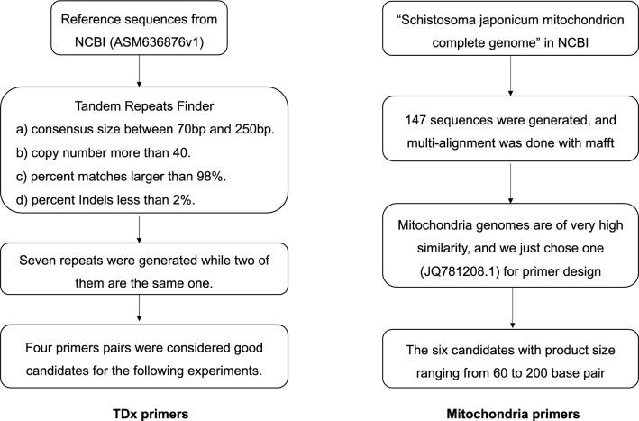
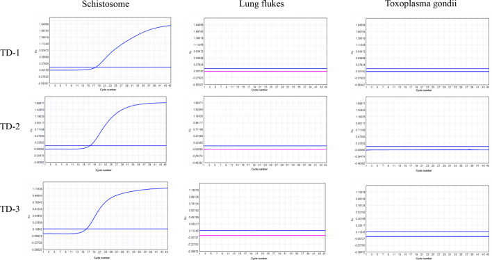
Methodology: Circulating cell-free (cf)DNA has been widely used in the diagnosis of various diseases. In our study, we evaluated the diagnostic value of circulating cfDNA for schistosomiasis caused by Schistosoma japonicum. We focused on the tandem sequences and mitochondrial genes of S. japonicum to identify highly sensitive and specific targets for diagnosis of Schistosomiasis japonica.
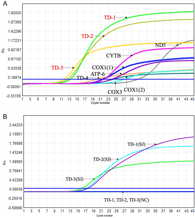
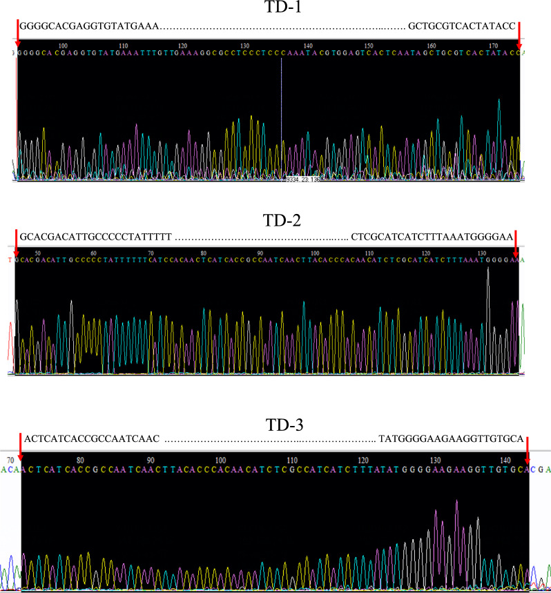
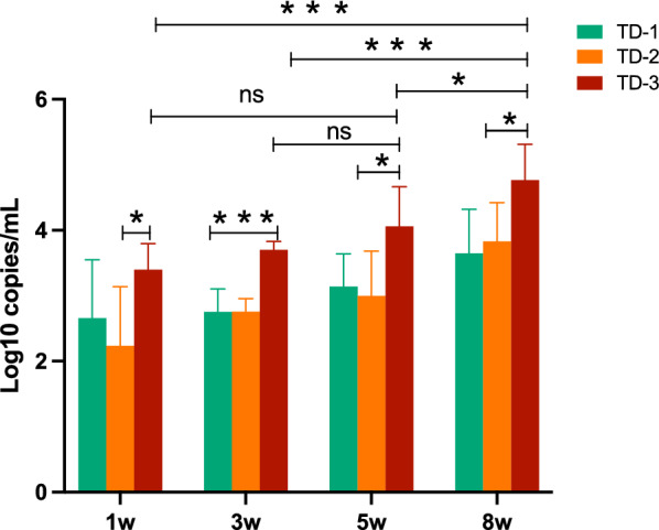
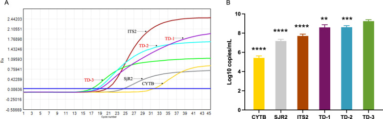
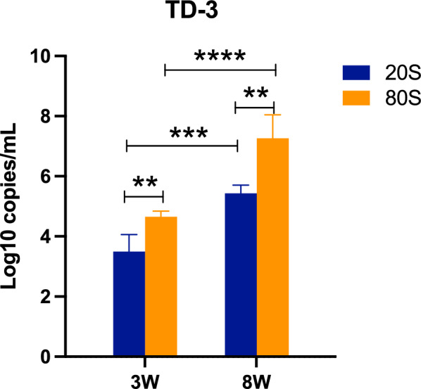
Results: Through data screening and analysis, we ultimately identified four specific tandem sequences (TD-1, TD-2, TD-3. and TD-4) and six mitochondrial genes (COX1(1), COX1(2), CYTB, ATP6, COX3, and ND5). We designed specific primers to detect the amount of circulating cfDNA in S. japonicum-infected mouse and chronic schistosomiasis patients. Our results showed that the number of tandem sequences was significantly higher than that of the mitochondrial genes. A S. japonicum infection model in mice suggested that infection of S. japonicum can be diagnosed by detecting circulating cfDNA as early as the first week. We measured the expression levels of circulating cfDNA (TD-1, TD-2, and TD-3) at different time points and found that TD-3 expression was significantly higher than that of TD-1 or TD-2. We also infected mice with different quantities of cercariae (20 s and 80 s). The level of cfDNA (TD-3) in the 80 s infection group was significantly higher than in the 20 s infection group. Additionally, cfDNA (TD-3) levels increased after egg deposition. Meanwhile, we tested 42 patients with chronic Schistosomiasis japonica and circulating cfDNA (TD-3) was detected in nine patients.
Conclusions: We have screened highly sensitive targets for the diagnosis of Schistosomiasis japonica, and the detection of circulating cfDNA is a rapid and effective method for the diagnosis of Schistosomiasis japonica. The levels of cfDNA is correlated with cercariae infection severity. Early detection and diagnosis of schistosomiasis is crucial for patient treatment and improving prognosis.
Keywords: Schistosomiasis japonica; Diagnosis; cfDNA.
© 2024. The Author(s).
Conflict of interest statement
The authors declare no conflict.
Figures
Similar articles
-
Circulating cell-free mitochondrial DNA fragment: A possible marker for early detection of Schistosoma japonicum.Infect Genet Evol. 2021 Mar;88:104683. doi: 10.1016/j.meegid.2020.104683. Epub 2020 Dec 24. Infect Genet Evol. 2021. PMID: 33348056
-
Detection of circulating cell-free DNA to diagnose Schistosoma japonicum infection.Acta Trop. 2020 Nov;211:105604. doi: 10.1016/j.actatropica.2020.105604. Epub 2020 Jun 26. Acta Trop. 2020. PMID: 32598919
-
Clinical diagnostic value of viable Schistosoma japonicum eggs detected in host tissues.BMC Infect Dis. 2017 Apr 4;17(1):244. doi: 10.1186/s12879-017-2362-4. BMC Infect Dis. 2017. PMID: 28376858 Free PMC article.
-
Reviews and advances in diagnostic research on Schistosoma japonicum.Acta Trop. 2021 Jan;213:105743. doi: 10.1016/j.actatropica.2020.105743. Epub 2020 Nov 4. Acta Trop. 2021. PMID: 33159894 Review.
-
Schistosoma japonicum infection in the pig as a model for human schistosomiasis japonica.Acta Trop. 2000 Sep 18;76(2):85-99. doi: 10.1016/s0001-706x(00)00103-0. Acta Trop. 2000. PMID: 10936567 Review.
References
-
- Degarege A, Degarege D, Veledar E, Erko B, Nacher M, Beck-Sague CM, Madhivanan P. Plasmodium falciparum infection status among children with schistosoma in Sub-Saharan Africa: a systematic review and meta-analysis. PLoS Negl Trop Dis. 2016;10:e0005193. doi: 10.1371/journal.pntd.0005193. - DOI - PMC - PubMed
-
- Wu C, Chen Q, Fang Y, Wu J, Han Y, Wang Y, Yang Y, Chu M, Feng Y, Tan L, Guo X, Hu W, Wang Z. Schistosoma japonicum egg specific protein SjE16.7 recruits neutrophils and induces inflammatory hepatic granuloma initiation. PLoS Negl Trop Dis. 2014;8:e2703. doi: 10.1371/journal.pntd.0002703. - DOI - PMC - PubMed
-
- López-Abán J, Vicente B, Kabbas-Piñango E, Hernández-Goenaga J, Sánchez-Montejo J, Aguiriano M, Del Olmo E, Vanegas M, Patarroyo MA, Muro A. T cell peptides derived from invasive stages of schistosoma mansoni as potential schistosomiasis vaccine. J Clin Med. 2021;10:445. doi: 10.3390/jcm10030445. - DOI - PMC - PubMed
MeSH terms
Substances
Grants and funding
LinkOut - more resources
Full Text Sources
