. 2024 Mar 6;29(1):155.
doi: 10.1186/s40001-024-01752-4.

Tibial cortex transverse transport promotes ischemic diabetic foot ulcer healing via enhanced angiogenesis and inflammation modulation in a novel rat model

Affiliations

Tibial cortex transverse transport promotes ischemic diabetic foot ulcer healing via enhanced angiogenesis and inflammation modulation in a novel rat model

Wencong Qin et al. Eur J Med Res. .

Abstract

Background: Tibial Cortex Transverse Transport (TTT) represents an innovative surgical method for treating lower extremity diabetic foot ulcers (DFUs), yet its underlying mechanisms remain elusive. Establishing an animal model that closely mirrors clinical scenarios is both critical and novel for elucidating the mechanisms of TTT.

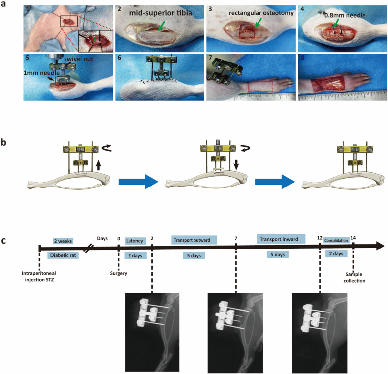
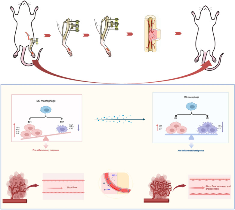
Methods: We established a diabetic rat model with induced hindlimb ischemia to mimic the clinical manifestation of DFUs. TTT was applied using an external fixator for regulated bone movement. Treatment efficacy was evaluated through wound healing assessments, histological analyses, and immunohistochemical techniques to elucidate biological processes.

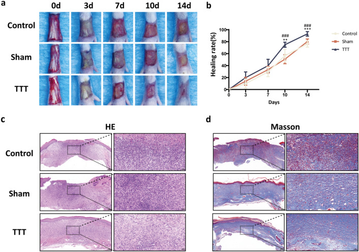
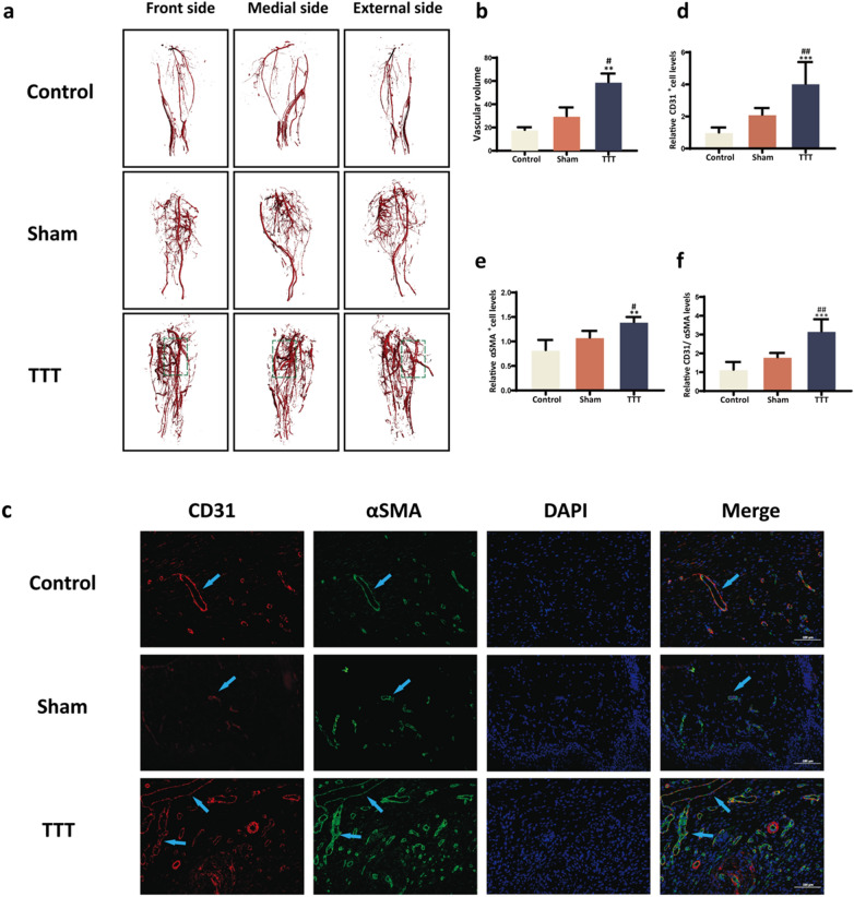
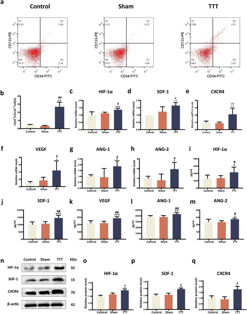
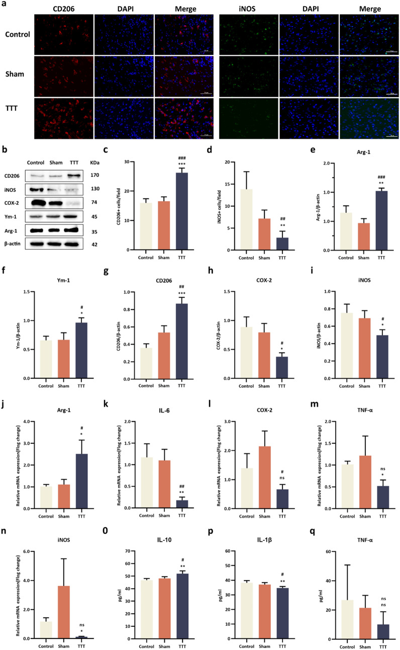
Results: The TTT group demonstrated expedited wound healing, improved skin tissue regeneration, and diminished inflammation relative to controls. Marked neovascularization and upregulation of angiogenic factors were observed, with the HIF-1α/SDF-1/CXCR4 pathway and an increase in EPCs being pivotal in these processes. A transition toward anti-inflammatory M2 macrophages indicated TTT's immunomodulatory capacity.

Conclusion: Our innovative rat model effectively demonstrates the therapeutic potential of TTT in treating DFUs. We identified TTT's roles in promoting angiogenesis and modulating the immune system. This paves the way for further in-depth research and potential clinical applications to improve DFU management strategies.


Conflict of interest statement

The authors declare that they have no competing interests.

Figures

Fig. 1
Fig. 1
Surgical Procedure of TTT. a Creation of an animal model and detailed surgical steps. b Illustration of bone transport adjustment using an external fixator. c Workflow for the TTT operation and post-operative X-ray imaging is utilized to verify the alignment and status of the cortical bone fragments
Fig. 2
Fig. 2
TTT promotes diabetic wound healing and improves the quality of newly generated skin tissue. a Representative images displaying wound healing progression and b respective wound healing rates across the three groups on days 0, 3, 7, 10 and 14 days post-surgery (n = 10). c-d H&E and Masson trichrome staining of skin sections from rat feet on day 14 post-surgery. Scale bar = 200 μm (left) and 50 μm (right). *P < 0.05, **P < 0.01, ***P < 0.001, TTT vs. Control; #P < 0.05, ##P < 0.01, ###P < 0.001, TTT vs. Sham
Fig. 3
Fig. 3
Promotion of Angiogenesis in Diabetic Rats' Hindlimbs and foot ulcers by TTT. a-b Representative images alongside quantitative vessel volume analyses from micro-CT scans, post vascular perfusion, in the three study groups (n = 4). c Dual immunofluorescence staining of wound sections highlighting CD31 (red) and αSMA (green), with nuclear DAPI (blue). Scale bar represents 100 μm. d-f Semi-quantitative assessment of CD31, αSMA, and combined CD31/αSMA expressions reveals enhanced angiogenesis in the TTT-treated wounds. Comparative data are normalized to the control group (n = 6). Statistical significance indicated as *P < 0.05, **P < 0.01, ***P < 0.001 for TTT versus control; #P < 0.05, ##P < 0.01, ###P < 0.001 for TTT versus sham
Fig. 4
Fig. 4
TTT activates the HIF-1α/SDF-1/CXCR4 pathway and stimulates epcs mobilization in diabetic rats. a Flow cytometry analysis of VEGR2, CD133, CD34-labeled EPCs in the peripheral blood of diabetic rats (n = 4). b Proportion of CD34+/CD133+cells. c–h RT-qPCR analysis of mRNA expression levels for genes associated with angiogenesis in wound tissues, including HIF1α, SDF-1, CXCR4, VEGF, ANG-1, and ANG-2 (n = 6). i–m ELISA results showing the levels of HIF-1α, SDF-1, VEGF, ANG-1, and ANG-2 in the serum of diabetic rats (n = 6).n–q Western blot analysis and quantification of HIF-1α, SDF-1, and CXCR4 protein expression in wound tissues (n = 6). All protein levels were standardized against β-actin and then normalized to the Control group. *P < 0.05, **P < 0.01, ***P < 0.001 for TTT vs. Control; #P < 0.05, ##P < 0.01, ###P < 0.001 for TTT vs. Sham
Fig. 5
Fig. 5
TTT enhances M2 macrophage polarization and modulates inflammatory response in diabetic foot ulcers. a Immunofluorescence Analysis: Wound sections were probed with CD206 (red) and iNOS (green) antibodies, and cell nuclei were counterstained with DAPI (blue). Semi-quantitative analysis indicated a marked reduction in M1 macrophages and an augmentation in M2 macrophages within the TTT-treated group. b–i Western Blotting and Quantitative Analysis: Investigations were conducted on the expression levels of proteins CD206, iNOS, COX2, Ym1, and Arg1 in wound tissues (sample size: n = 6). Protein quantifications were normalized against β-actin. j–n Quantitative RT-PCR: mRNA levels of cytokines and enzymes, including IL-6, TNF-α, iNOS, COX2, and Arg-1, were quantified in wound tissues (n = 6). o–q ELISA Measurements: Concentrations of cytokines IL-10, IL-1β, and TNF-α were measured in the serum of diabetic rats (n = 6). Statistical significance is denoted as follows: *P < 0.05, **P < 0.01, ***P < 0.001 for comparisons of TTT versus Control; #P < 0.05, ##P < 0.01, ###P < 0.001 for TTT versus Sham
Fig. 6
Fig. 6
Mechanisms by which TTT promotes healing of ischemic diabetic ulcers. The local tibial cortex of the pulled tibia promotes the expression of angiogenic factors during slow movement and simultaneously regulates the local immune microenvironment of the wound, accelerating wound healing

Similar articles

Cited by

References

    1. Zhang P, Lu J, Jing Y, Tang S, Zhu D, Bi Y. Global epidemiology of diabetic foot ulceration: a systematic review and meta-analysis. Ann Med. 2017;49:106–116. doi: 10.1080/07853890.2016.1231932. - DOI - PubMed
    1. Lavery LA, Hunt NA, Ndip A, Lavery DC, Van Houtum W, Boulton AJM. Impact of chronic kidney disease on survival after amputation in individuals with diabetes. Diabetes Care. 2010;33:2365–2369. doi: 10.2337/dc10-1213. - DOI - PMC - PubMed
    1. Ilizarov GA. The tension-stress effect on the genesis and growth of tissues: Part I. The influence of stability of fixation and soft-tissue preservation. Clin Orthop Relat Res. 2014;238:249–81. doi: 10.1007/978-1-4471-5451-8_136. - DOI - PubMed
    1. Ilizarov GA. The tension-stress effect on the genesis and growth of tissues: Part II. The influence of the rate and frequency of distraction. Clin Orthop Relat Res. 1989;238:263–85. doi: 10.1007/978-1-4471-5451-8_137. - DOI - PubMed
    1. Chen Y, Kuang X, Zhou J, Zhen P, Zeng Z, Lin Z, et al. Proximal tibial cortex transverse distraction facilitating healing and limb salvage in severe and recalcitrant diabetic foot ulcers. Clin Orthop Relat Res. 2020;478(4):836. doi: 10.1097/CORR.0000000000001075. - DOI - PMC - PubMed