

Morphological analysis of the jugular foramen in dry human skulls in northeastern Brazil
- PMID: 38449076
- PMCID: PMC11184423
- DOI: 10.5115/acb.23.218
Morphological analysis of the jugular foramen in dry human skulls in northeastern Brazil
Abstract
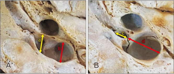
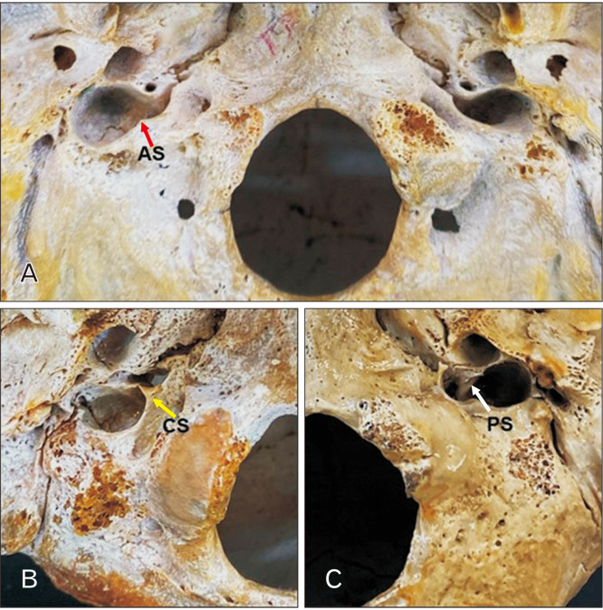
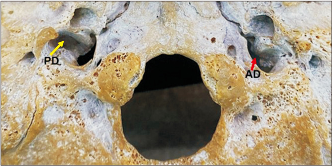
The jugular foramen (JF) is located between the temporal and occipital bones. The JF is a primary pathway for venous outflow from the skull and passage of nerves. Variations are common in this region and may have clinical and surgical implications. To analyze the sexual dimorphism and JF morphology in skulls from Northeastern Brazil. 128 human skulls from the Anatomy Laboratory of the Federal University of Paraíba, 64 male and 64 female, were selected and the JFs analyzed for bone septation and the presence of a dome. Data analysis considered P<0.05 as significant. On at least one side, complete septation was observed in 26 skulls (20.3%), incomplete septation in 93 skulls (72.6%) and 61 skulls (47.6%) did not present septation. In 114 skulls (89%), 47.6% female and 41.4% male, have a unilateral presence of the dome and 71 (55.4%) have it bilaterally. Posterolateral compartment diameters and JF area had higher values on the right side in the total sample and separated by sex (P<0.05). Most morphometric variables of the anteromedial compartment were higher in male than in female (P<0.05), fact that was not observed in the posterolateral compartment (P>0.05). This study showed a higher prevalence of complete septation in males compared to females. Morphometric analysis presented a peculiar morphology of the JF in this study. These results suggests that the surgical approach to diseases that affect the JF may be peculiar to the studied population, confirming the importance of morphological analysis of the skull base.
Keywords: Anatomy; Brazil; Jugular foramen; Skull.
Conflict of interest statement
No potential conflict of interest relevant to this article was reported.
Figures
References
-
- Pereira GAM, Lopes PTC, Santos AMPV, Krebs WD. Morphometric aspects of the jugular foramen in dry skulls of adult individuals in Southern Brazil. J Morphol Sci. 2010;27:3–5.
-
- Standring S. Gray's anatomy: the anatomical basis of clinical practice. 40th ed. Churchill Livingstone; 2008. pp. 423–34.
-
- Caldeira JVC, Godas AG de L, Carvalho GB de A, Silva KRT da, Machado AR da SR, Almeida PF de, Silva AV da. [Measurement of the jugular foramen in dry skulls from the midwest region of Brazil]. Braz J Health Rev. 2020;3:14614–28. doi: 10.34119/bjhrv3n5-256. Portuguese. - DOI
LinkOut - more resources
Full Text Sources