. 2024 Feb 21:18:1356703.
doi: 10.3389/fnins.2024.1356703. eCollection 2024.

Mito-metformin protects against mitochondrial dysfunction and dopaminergic neuronal degeneration by activating upstream PKD1 signaling in cell culture and MitoPark animal models of Parkinson's disease

Affiliations

Mito-metformin protects against mitochondrial dysfunction and dopaminergic neuronal degeneration by activating upstream PKD1 signaling in cell culture and MitoPark animal models of Parkinson's disease

Muhammet Ay et al. Front Neurosci. .

Abstract

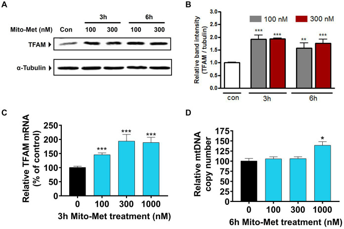
Impaired mitochondrial function and biogenesis have strongly been implicated in the pathogenesis of Parkinson's disease (PD). Thus, identifying the key signaling mechanisms regulating mitochondrial biogenesis is crucial to developing new treatment strategies for PD. We previously reported that protein kinase D1 (PKD1) activation protects against neuronal cell death in PD models by regulating mitochondrial biogenesis. To further harness the translational drug discovery potential of targeting PKD1-mediated neuroprotective signaling, we synthesized mito-metformin (Mito-Met), a mitochondria-targeted analog derived from conjugating the anti-diabetic drug metformin with a triphenylphosphonium functional group, and then evaluated the preclinical efficacy of Mito-Met in cell culture and MitoPark animal models of PD. Mito-Met (100-300 nM) significantly activated PKD1 phosphorylation, as well as downstream Akt and AMPKα phosphorylation, more potently than metformin, in N27 dopaminergic neuronal cells. Furthermore, treatment with Mito-Met upregulated the mRNA and protein expression of mitochondrial transcription factor A (TFAM) implying that Mito-Met can promote mitochondrial biogenesis. Interestingly, Mito-Met significantly increased mitochondrial bioenergetics capacity in N27 dopaminergic cells. Mito-Met also reduced mitochondrial fragmentation induced by the Parkinsonian neurotoxicant MPP+ in N27 cells and protected against MPP+-induced TH-positive neurite loss in primary neurons. More importantly, Mito-Met treatment (10 mg/kg, oral gavage for 8 week) significantly improved motor deficits and reduced striatal dopamine depletion in MitoPark mice. Taken together, our results demonstrate that Mito-Met possesses profound neuroprotective effects in both in vitro and in vivo models of PD, suggesting that pharmacological activation of PKD1 signaling could be a novel neuroprotective translational strategy in PD and other related neurocognitive diseases.

Keywords: MitoPark; PKD1; Parkinson’s disease; metformin; mitochondria; mitochondrial biogenesis; neuroprotection.


Conflict of interest statement

AnK and VA have an equity interest in PK Biosciences Corporation and Probiome Therapeutics, located at the University of Georgia, Athens, GA. The terms of this arrangement have been reviewed and approved by the University of Georgia in accordance with their conflict-of-interest policies. The remaining authors declare that the research was conducted in the absence of any commercial or financial relationships that could be construed as a potential conflict of interest.

Figures

Figure 1
Figure 1
Activation of PKD1 by Mito-Met in N27 cells. (A) N27 dopaminergic neuronal cells were treated with metformin (100 and 1,000 μM) for 3 h. (B) N27 cells were treated with Mito-Met (100 and 300 nM) for 3 h. Cell lysates were prepared and subjected to Western blot analysis. Representative immunoblots of PKD1 S744/748 phosphorylation are shown. (C) N27 cells were treated with 100 and 300 nM Mito-Met for 3 and 6 h. Cell lysates were prepared and subjected to Western blot analysis. Representative immunoblots of total PKD1, PKD1 S744/748, and S916 phosphorylation are shown. (D) The graph represents the densitometric analysis of phospho-PKD1 S744/748 levels in (C). Results are the mean ± SEM of at least three independent experiments (*p ≤ 0.05; ***p < 0.001).
Figure 2
Figure 2
Activation of Akt and AMPK by Mito-Met in N27 cells. (A) N27 cells were treated with 100 and 300 nM Mito-Met for 3 and 6 h. Cell lysates were prepared and phospho-Akt (S473) and phospho-AMPKα (Thr172) levels were determined by Western blot analysis. (B) N27 cells were pretreated with 50 μM PKD1 inhibitor CID755673 for 1 h and then cotreated with 100 nM Mito-Met for 3 h. Cell lysates were prepared and subjected to Western blot analyses of phospho-PKD1 (S916), phospho-Akt (S473), and phospho-AMPKα (Thr172).
Figure 3
Figure 3
Mito-Met increases TFAM expression in N27 cells. (A) N27 cells were treated with 100 and 300 nM Mito-Met for 3 and 6 h. Cell lysates were prepared and TFAM levels were determined by Western blot analysis. (B) The graph represents the densitometric analysis of TFAM protein levels normalized to α-tubulin. (C) N27 cells were treated with Mito-Met (100–1,000 nM) for 3 h. Real-time RT-PCR analysis of TFAM mRNA level was performed. 18S rRNA served as internal control. (D) N27 cells were treated with Mito-Met (100–1,000 nM) for 6 h. Genomic DNA was isolated and mtDNA content was determined by quantitative PCR with SYBR green. 10 ng nuclear DNA and 1 ng mitochondrial DNA were amplified using the primers for mitochondrial ND1 and nuclear β-actin genes. Results are the mean ± SEM of three independent experiments (*p ≤ 0.05; **p < 0.01; ***p < 0.001; between the control and Mito-Met-treated samples).
Figure 4
Figure 4
Mito-Met increases mitochondrial bioenergetics capacity. (A–C) N27 dopaminergic neuronal cells were treated with 100 and 300 nM Mito-Met for 3 h. Rotenone (1 μM) was included as a positive control. N27 cell-containing culture plates were loaded into the Seahorse XF96 analyzer for the OCR measurement. Mitochondrial dynamics were measured using the sequential injection of oligomycin A (1 μg/mL), FCCP (1 μM), and antimycin A (10 μM) (A). Basal OCR (B) and ATP-linked respiration (C) were calculated from the output OCR values. Values represent the means ± SEM of four replicates (**p < 0.01; ***p < 0.001; between the control and Mito-Met-treated samples).
Figure 5
Figure 5
Mito-Met reduces MPP+-induced mitochondrial fragmentation. (A–C) N27 cells were pretreated with 300 nM Mito-Met for 1 h, and then cotreated with 300 μM MPP+ for 16 h. Cells were stained with the MitoTracker red dye. Images were taken at a magnification of 60X (A). Mitochondrial length (B) and degree of circularity (C) were quantified using ImageJ. Values represent the means ± SEM of two independent experiments performed in sextuplicate (*p ≤ 0.05; between the Mito-Met-pretreated and MPP+ (alone)-treated groups).
Figure 6
Figure 6
Mito-Met protects against MPP+-induced neurotoxicity in primary neurons. (A,B) The primary mesencephalic neurons were treated with 10 μM MPP+ in the presence or absence of Mito-Met (100 nM) for 24 h and immunostained with an TH antibody. Images were taken at 60X magnification using the Leica confocal microscope and 3D image reconstruction was performed using IMARIS software (A). The length of TH-positive neuronal processes in primary dopaminergic neurons from each coverslip was measured using MetaMorph software (B). The experiments were performed in sextuplicate (*p ≤ 0.05; ***p < 0.001).
Figure 7
Figure 7
Mito-Met improves motor deficits in MitoPark mice. (A) Treatment schedule of MitoPark mice with Mito-Met. 12-week-old MitoPark mice were treated with either saline or Mito-Met (10 mg/kg) via oral gavage three times per week for 8 weeks. The locomotor activities were measured using a VersaMax system 1 day prior to sacrifice. Moving track of mice (B), horizontal activity (C), total distance traveled (D), number of movements (E), and stereotypy counts (F). Values represent the means ± SEM of six mice per group (*p ≤ 0.05; **p < 0.01).
Figure 8
Figure 8
Mito-Met attenuates striatal dopamine depletion in MitoPark mice. (A,B) 12-week-old MitoPark mice were treated with either saline or Mito-Met (10 mg/kg) via oral gavage three times per week for 8 week. Mice were sacrificed 1 day after the last Mito-Met treatment and dopamine (A) and DOPAC (B) levels were measured from striatal tissues by HPLC analysis. Results are the means ± SEM of five mice per group (*p ≤ 0.05; **p < 0.01).

Similar articles

Cited by

References

    1. Andrzejewski S., Gravel S. P., Pollak M., St-Pierre J. (2014). Metformin directly acts on mitochondria to alter cellular bioenergetics. Cancer Metab 2:12. doi: 10.1186/2049-3002-2-12, PMID: - DOI - PMC - PubMed
    1. Armstrong M. J., Okun M. S. (2020). Diagnosis and treatment of Parkinson disease: a review. JAMA 323, 548–560. doi: 10.1001/jama.2019.22360 - DOI - PubMed
    1. Asaithambi A., Ay M., Jin H., Gosh A., Anantharam V., Kanthasamy A., et al. . (2014). Protein kinase D1 (PKD1) phosphorylation promotes dopaminergic neuronal survival during 6-OHDA-induced oxidative stress. PLoS One 9:e96947. doi: 10.1371/journal.pone.0096947, PMID: - DOI - PMC - PubMed
    1. Asaithambi A., Kanthasamy A., Saminathan H., Anantharam V., Kanthasamy A. G. (2011). Protein kinase D1 (PKD1) activation mediates a compensatory protective response during early stages of oxidative stress-induced neuronal degeneration. Mol. Neurodegener. 6:43. doi: 10.1186/1750-1326-6-43, PMID: - DOI - PMC - PubMed
    1. Ay M., Jin H., Harischandra D. S., Asaithambi A., Kanthasamy A., Anantharam V., et al. . (2015). Molecular cloning, epigenetic regulation, and functional characterization of Prkd1 gene promoter in dopaminergic cell culture models of Parkinson's disease. J. Neurochem. 135, 402–415. doi: 10.1111/jnc.13261, PMID: - DOI - PMC - PubMed

LinkOut - more resources