Review
. 2024 Feb 21:5:1363052.
doi: 10.3389/froh.2024.1363052. eCollection 2024.

New insights into the role of the oral leukoplakia microenvironment in malignant transformation

Affiliations
Review

New insights into the role of the oral leukoplakia microenvironment in malignant transformation

Wilfredo Alejandro González-Arriagada et al. Front Oral Health. .

Abstract

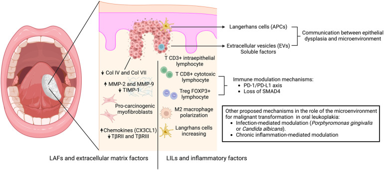
Oral leukoplakia is the most frequent and potentially malignant lesion of the oral cavity. Although dysplasia grading remains the main factor for risk assessment, challenges persist in determining the exact risk of transformation, and the literature has focused on studying alternative biomarkers. The interaction between dysplastic epithelial cells and the microenvironment starts early, and the communication is mainly mediated by lymphocytes, inflammatory factors, fibroblasts, and the extracellular matrix, leading to dysplastic progression. Leukoplakia-infiltrating leukocytes (LILs) and leukoplakia-associated fibroblasts (LAFs) play crucial roles in the dysplastic microenvironment. The immune response is related to intraepithelial T lymphocyte infiltration, mechanisms of immunosuppression coordinated by regulatory T cells, M2 macrophage polarization, and increased numbers of Langerhans cells; in contrast, fibroblastic and extracellular matrix factors are associated with increased numbers of pro-tumorigenic myofibroblasts, increased expression of metalloproteinases vs. decreased expression of TIMPs, and increased expression of chemokines and other inflammatory mediators. The microenvironment offers insights into the progression of leukoplakia to carcinoma, and understanding the complexity of the oral microenvironment in potentially malignant diseases aids in determining the risk of malignant transformation and proposing new therapeutic alternatives.

Keywords: dysplasia; fibroblast; lymphocyte; malignant transformation; microenvironment; oral cancer; oral leukoplakia; premalignant.


Conflict of interest statement

The authors declare that the research was conducted in the absence of any commercial or financial relationships that could be construed as a potential conflict of interest. The authors declared that they were editorial board members of Frontiers at the time of submission. This had no impact on the peer review process and the final decision.

Figures

Figure 1
Figure 1
Mechanisms involved in the interaction between oral dysplastic epithelial cells and the microenvironment leading to malignant transformation. The proposed mechanism of early communication involves antigen-presenting cells (Langerhans cells), extracellular vesicles (EVs) and soluble factors, which promote the stromal response. This response is mediated by: (A) leukoplakia-infiltrating lymphocytes (LILs) and inflammatory factors related to intraepithelial T lymphocyte infiltration, mechanisms of immunosuppression coordinated by regulatory T cells through the PD1 -PDL1 axis, Treg-related proteins (FOXP3, TGF-β, IL-6, and IL-10), and loss of SMAD4 associated with M2 macrophage polarization and increased Langerhans cell numbers; and (B) leukoplakia-associated fibroblasts and extracellular matrix factors related to increased pro-tumorigenic myofibroblasts, decreased collagen expression in the basement membrane, increased expression of metalloproteinases vs. decreased TIMPs, and increased expression of chemokines vs. decreased receptors of TGF-β. Other mechanisms have been suggested to induce malignant transformation, including infection-mediated modulation (Porphyromonas gingivalis or Candida albicans) and chronic inflammation.

Similar articles

Cited by

References

    1. Saldivia-Siracusa C, González-Arriagada WA. Difficulties in the prognostic study of oral leukoplakia: standardisation proposal of follow-up parameters. Front Oral Health. (2021) 2. 10.3389/froh.2021.614045 - DOI - PMC - PubMed
    1. Aguirre-Urizar JM, Lafuente-Ibáñez de Mendoza I, Warnakulasuriya S. Malignant transformation of oral leukoplakia: systematic review and meta-analysis of the last 5 years. Oral Dis. (2021) 27(8):1881–95. 10.1111/odi.13810 - DOI - PubMed
    1. Warnakulasuriya S, Ariyawardana A. Malignant transformation of oral leukoplakia: a systematic review of observational studies. J Oral Pathol Med. (2015) 45(3):155–66. 10.1111/jop.12339 - DOI - PubMed
    1. Jeong WJ, Paik JH, Cho SW, Sung MW, Kim KH, Ahn SH. Excisional biopsy for management of lateral tongue leukoplakia. J Oral Pathol Med. (2011) 41(5):384–8. 10.1111/j.1600-0714.2011.01106.x - DOI - PubMed
    1. Kuribayashi Y, Tsushima F, Morita K-I, Matsumoto K, Sakurai J, Uesugi A, et al. Long-term outcome of non-surgical treatment in patients with oral leukoplakia. Oral Oncol. (2015) 51(11):1020–5. 10.1016/j.oraloncology.2015.09.004 - DOI - PubMed

LinkOut - more resources