. 2024 Mar 4;13(2):tfae026.
doi: 10.1093/toxres/tfae026. eCollection 2024 Apr.

Evaluation of the toxic potential of Bisphenol-A glycidylmethacrylate (BisGMA) on the third instar larvae of transgenic Drosophila

Affiliations

Evaluation of the toxic potential of Bisphenol-A glycidylmethacrylate (BisGMA) on the third instar larvae of transgenic Drosophila

Nabeela Ibrahim et al. Toxicol Res (Camb). .

Abstract

Introduction: In the present study the cytotoxic and genotoxic effects of Bisphenol-A glycidyl methacrylate (BisGMA) was studied on the third instar larvae of transgenic Drosophila melanogaster (hsp70-lacZ)Bg9.

Materials and methods: The concentration of BisGMA i.e. 0.005, 0.010, 0.015 and 0.020 M were established in diet and the larvae were allowed to feed on it for 24 h.

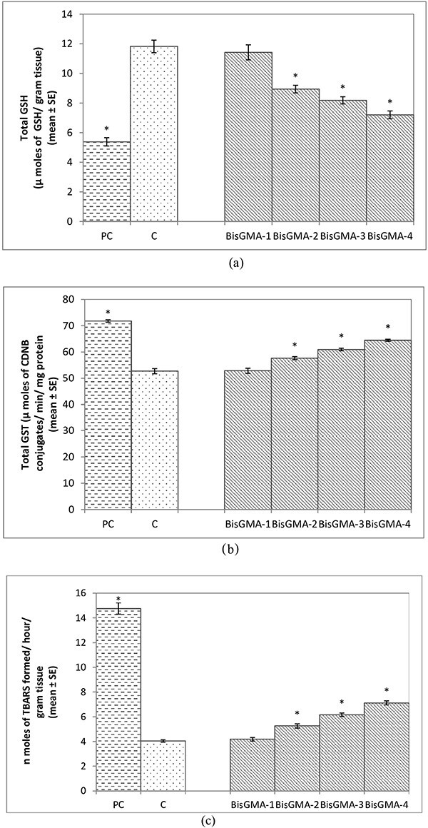
Results: A dose dependent significant increase in the activity of β-galactosidase was observed compared to control. A significant dose dependent tissue damage was observed in the larvae exposed to 0.010, 0.015 and 0.020 M of BisGMA compared to control. A dose dependent significant increase in the Oxidative stress markers was observed compared to control. BisGMA also exhibit significant DNA damaged in the third instar larvae of transgenic D. melanogaster (hsp70-lacZ)Bg9 at the doses of 0.010, 0.015 and 0.020 M compared to control.

Conclusion: BisGMA at 0.010, 0.015 and 0.020 M was found to be cytotoxic for the third instar larvae of transgenic D. melanogaster (hsp70-lacZ) Bg9.

Keywords: Drosophila; bisphenol-A-glycidylmethacrylate; cytotoxicity; genotoxicity; oxidative stress.


Conflict of interest statement

None declared.

Figures

Fig. 1
Fig. 1
Quantification of β- galactosidase activity in the third instar larvae of transgenic Drosophila melanogaster (hsp70-lac Z)Bg9 after the exposure to various doses of Bisphenol A- glycidyl methacrylate (Bis-GMA) for 24 h. [BisGMA-1 = 0.005 M; BisGMA-2 = 0.010 M; BisGMA-3 = 0.015 M; BisGMA-4 = 0.020 M; positive control (methyl methane sulphonate = 80 μM); C = control;*significant at P < 0.05 compared to control].
Fig. 2
Fig. 2
X-gal staining performed on the third instar larvae of transgenic Drosophila melanogaster (hsp70-lac Z)Bg9 after the exposure to various doses of Bisphenol A-glycidyl methacrylate(Bis-GMA) for 24 h [positive control (methyl methane sulphonate = 80 μM (a); control (b); BisGMA-1 = 0.005 M (c); BisGMA-2 = 0.010 M (d); BisGMA-3 = 0.015 M (e); BisGMA-4 = 0.020 M (f); BG-brain ganglia, SG-salivary gland, PV-Proventriculus, FG-foregut, MG-Midgut, HG-hindgut, MT-Malpighian tubule, GC-gastric caeca].
Fig. 3
Fig. 3
Trypan staining performed on the third instar larvae of transgenic Drosophila melanogaster (hsp70-lac Z)Bg9 after the exposure to various doses of Bisphenol A- glycidyl methacrylate (Bis-GMA) for 24 h [positive control (methyl methane sulphonate = 80 μM) (a); control (b); BisGMA-1 = 0.005 M (c); BisGMA-2 = 0.010 M (d); BisGMA-3 = 0.015 M (e); BisGMA-4 = 0.020 M (f); BG-brain ganglia, SG-salivary gland, PV-Proventriculus, FG-foregut, MG-Midgut, HG-hindgut, MT-Malpighian tubule, GC-gastric caeca].
Fig. 4
Fig. 4
Quantification of tissue damage after performing trypan staining on the third instar larvae of transgenic Drosophila melanogaster (hsp70-lac Z)Bg9 exposed to various doses of Bisphenol A- glycidyl methacrylate (Bis-GMA) for 24 h [BisGMA-1 = 0.005 M; BisGMA-2 = 0.010 M; BisGMA-3 = 0.015 M; BisGMA-4 = 0.020 M; positive control (methyl methane sulphonate = 80 μM); C = control; *significant at P < 0.05 compared to control].
Fig. 5
Fig. 5
a) Quantification of glutathione content in the third instar larvae of transgenic Drosophila melanogaster (hsp70-lac Z)Bg9 exposed to various doses of Bisphenol A- glycidyl methacrylate(Bis-GMA) for 24 h [BisGMA-1 = 0.005 M; BisGMA-2 = 0.010 M; BisGMA-3 = 0.015 M; BisGMA-4 = 0.020 M; positive control (methyl methane sulphonate = 80 μM); C = control; *significant at P < 0.05 compared to control]. b) Quantification of glutathione-S-transferase activity in the third instar larvae of transgenic D. melanogaster (hsp70-lac Z)Bg9 exposed to various doses of Bisphenol A- glycidyl methacrylate(Bis-GMA) for 24 h [BisGMA-1 = 0.005 M; BisGMA-2 = 0.010 M; BisGMA-3 = 0.015 M; BisGMA-4 = 0.020 M; positive control (methyl methane sulphonate = 80 μM); C = control; *significant at P < 0.05 compared to control]. c) Quantification of lipid peroxidation in the third instar larvae of transgenic D. melanogaster (hsp70-lac Z)Bg9 exposed to various doses of Bisphenol A- glycidyl methacrylate (Bis-GMA) for 24 h [BisGMA-1 = 0.005 M; BisGMA-2 = 0.010 M; BisGMA-3 = 0.015 M; BisGMA-4 = 0.020 M; positive control (methyl methane sulphonate =80 μM); C = control; *significant at P < 0.05 compared to control].
Fig. 6
Fig. 6
a) Quantification of apoptosis after performing acridine orange (AO)/Ethidium bromide (Et-Br) stanning on the midgut cells of third instar larvae of transgenic Drosophila melanogaster (hsp70-lac Z)Bg9 exposed to various doses of Bisphenol A- glycidyl methacrylate (Bis-GMA) for 24 h [BisGMA-1 = 0.005 M; BisGMA-2 = 0.010 M; BisGMA-3 = 0.015 M; BisGMA-4 = 0.020 M; positive control (methyl methane sulphonate = 80 μM); C = control; *significant at P < 0.05 compared to control]. b) Quantification of Caspase-3 activityin the mid gut cells of the third instar larvae of transgenic D. melanogaster (hsp70-lac Z)Bg9 exposed to various doses of Bisphenol A- glycidyl methacrylate (Bis-GMA) for 24 h [BisGMA-1 = 0.005 M; BisGMA-2 = 0.010 M; BisGMA-3 = 0.015 M; BisGMA-4 = 0.020 M; positive control (methyl methane sulphonate = 80 μM); C = control; *significant at P < 0.05 compared to control]. c) Quantification of Caspase-9 activityin the mid gut cells of the third instar larvae of transgenic D. melanogaster (hsp70-lac Z)Bg9 exposed to various doses of Bisphenol A- glycidyl methacrylate (Bis-GMA) for 24 h [BisGMA-1 = 0.005 M; BisGMA-2 = 0.010 M; BisGMA-3 = 0.015 M; BisGMA-4 = 0.020 M; positive control (methyl methane sulphonate = 80 μM); C = control; *significant at P < 0.05 compared to control].
Fig. 7
Fig. 7
a) Comet assay performed on the mid gut cells of the third instar larvae of transgenic Drosophila melanogaster (hsp70-lac Z)Bg9 exposed to various doses of Bisphenol A- glycidyl methacrylate (Bis-GMA) for 24 h [control (a); BisGMA-1 = 0.005 M (b); BisGMA-2 = 0.010 M (c); BisGMA-3 = 0.015 M (d); BisGMA-4 = 0.020 M (e); positive control (methyl methane sulphonate = 80 μM) (f)]. b) Quantification of DNA damage after performing comet assay on the mid gut cells of the third instar larvae of transgenic D. melanogaster (hsp70-lac Z)Bg9 exposed to various doses of Bisphenol A- glycidyl methacrylate(Bis-GMA) for 24 h [BisGMA-1 = 0.005 M; BisGMA-2 = 0.010 M; BisGMA-3 = 0.015 M; BisGMA-4 = 0.020 M; positive control (methyl methane sulphonate = 80 μM); C = control; *significant at P < 0.05 compared to control].

Similar articles

References

    1. Alkadhimi A, Motamedi F. Orthodontic adhesives for fixed appliances: a review of available systems. Dent Update. 2019:46(8):742–758.
    1. Gange P. The evolution of bonding in orthodontics. Am J Orthod Dentofacial Orthop. 2015:147(4):S56–S63. - PubMed
    1. Papakonstantinou AE, Eliades T, Cellesi F, Watts DC, Silikas N. Evaluation of UDMA's potential as a substitute for Bis-GMA in orthodontic adhesives. Dent Mater. 2013:29(8):898–905. - PubMed
    1. Ahrari F, Tavakkol Afshari J, Poosti M, Brook A. Cytotoxicity of orthodontic bonding adhesive resins on human oral fibroblasts. Eur J Orthod. 2010:32(6):688–692. - PubMed
    1. Angiero F, Farronato G, Dessy E, Magistro S, Seramondi R, Farronato D, Benedicenti S, Tetè S. Evaluation of the cytotoxic and genotoxic effects of orthodontic bonding adhesives upon human gingival papillae through immunohistochemical expression of p53, p63 and p16. Anticancer Res. 2009:29(10):3983–3987. - PubMed

LinkOut - more resources