

Diagnosis of benign and malignant nodules with a radiomics model integrating features from nodules and mammary regions on DCE-MRI
- PMID: 38450180
- PMCID: PMC10915177
- DOI: 10.3389/fonc.2024.1307907
Diagnosis of benign and malignant nodules with a radiomics model integrating features from nodules and mammary regions on DCE-MRI
Abstract
Objectives: To establish a radiomics model for distinguishing between the benign and malignant mammary gland nodules via combining the features from nodule and mammary regions on DCE-MRI.
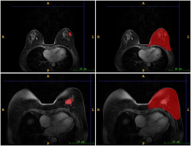
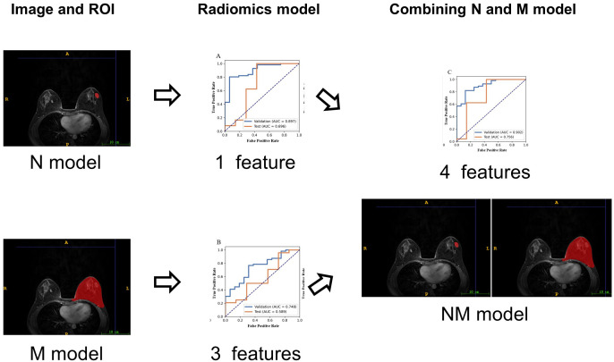
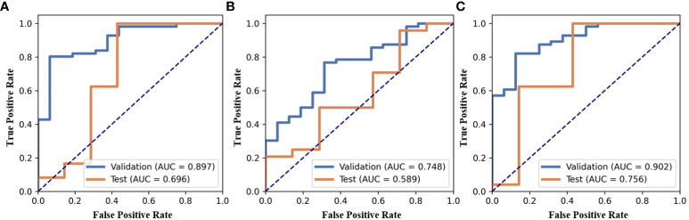
Methods: In this retrospective study, a total of 103 cases with mammary gland nodules (malignant/benign = 80/23) underwent DCE-MRI, and was confirmed by biopsy pathology. Features were extracted from both nodule region and mammary region on DCE-MRI. Three SVM classifiers were built for diagnosis of benign and malignant nodules as follows: the model with the features only from nodule region (N model), with the features only from mammary region (M model) and the model combining the features from nodule region and mammary region (NM model). The performance of models was evaluated with the area under the curve of receiver operating characteristic (AUC).
Results: One radiomic features is selected from nodule region and 3 radiomic features is selected from mammary region. Compared with N or M model, NM model exhibited the best performance with an AUC of 0.756.
Conclusions: Compared with the model only using the features from nodule or mammary region, the radiomics-based model combining the features from nodule and mammary region outperformed in the diagnosis of benign and malignant nodules.
Keywords: DCE-MRI; breast cancer; mammary region; nodule region; radiomics.
Copyright © 2024 Fan, Sun, Xu, Pan and Man.
Conflict of interest statement
The authors declare that the research was conducted in the absence of any commercial or financial relationships that could be construed as a potential conflict of interest.
Figures
References
LinkOut - more resources
Full Text Sources