Type III intermediate filaments in redox interplay: key role of the conserved cysteine residue
- PMID: 38451193
- PMCID: PMC11088922
- DOI: 10.1042/BST20231059
Type III intermediate filaments in redox interplay: key role of the conserved cysteine residue
Abstract
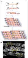
Intermediate filaments (IFs) are cytoskeletal elements involved in mechanotransduction and in the integration of cellular responses. They are versatile structures and their assembly and organization are finely tuned by posttranslational modifications. Among them, type III IFs, mainly vimentin, have been identified as targets of multiple oxidative and electrophilic modifications. A characteristic of most type III IF proteins is the presence in their sequence of a single, conserved cysteine residue (C328 in vimentin), that is a hot spot for these modifications and appears to play a key role in the ability of the filament network to respond to oxidative stress. Current structural models and experimental evidence indicate that this cysteine residue may occupy a strategic position in the filaments in such a way that perturbations at this site, due to chemical modification or mutation, impact filament assembly or organization in a structure-dependent manner. Cysteine-dependent regulation of vimentin can be modulated by interaction with divalent cations, such as zinc, and by pH. Importantly, vimentin remodeling induced by C328 modification may affect its interaction with cellular organelles, as well as the cross-talk between cytoskeletal networks, as seems to be the case for the reorganization of actin filaments in response to oxidants and electrophiles. In summary, the evidence herein reviewed delineates a complex interplay in which type III IFs emerge both as targets and modulators of redox signaling.
Keywords: Alexander disease; cysteine modification; intermediate filaments; lipoxidation; oxidative stress; vimentin.
© 2024 The Author(s).
Conflict of interest statement
The authors declare that there are no competing interests associated with the manuscript.
Figures


Similar articles
-
Vimentin single cysteine residue acts as a tunable sensor for network organization and as a key for actin remodeling in response to oxidants and electrophiles.Redox Biol. 2023 Aug;64:102756. doi: 10.1016/j.redox.2023.102756. Epub 2023 May 31. Redox Biol. 2023. PMID: 37285743 Free PMC article.
-
Vimentin disruption by lipoxidation and electrophiles: Role of the cysteine residue and filament dynamics.Redox Biol. 2019 May;23:101098. doi: 10.1016/j.redox.2019.101098. Epub 2019 Jan 8. Redox Biol. 2019. PMID: 30658903 Free PMC article.
-
Type III intermediate filaments as targets and effectors of electrophiles and oxidants.Redox Biol. 2020 Sep;36:101582. doi: 10.1016/j.redox.2020.101582. Epub 2020 Jul 17. Redox Biol. 2020. PMID: 32711378 Free PMC article. Review.
-
The redox-responsive roles of intermediate filaments in cellular stress detection, integration and mitigation.Curr Opin Cell Biol. 2024 Feb;86:102283. doi: 10.1016/j.ceb.2023.102283. Epub 2023 Nov 20. Curr Opin Cell Biol. 2024. PMID: 37989035 Review.
-
Oxidative stress elicits the remodeling of vimentin filaments into biomolecular condensates.Redox Biol. 2024 Sep;75:103282. doi: 10.1016/j.redox.2024.103282. Epub 2024 Jul 23. Redox Biol. 2024. PMID: 39079387 Free PMC article.
References
Publication types
MeSH terms
Substances
LinkOut - more resources
Full Text Sources
Miscellaneous

