Review
. 2024 Jan;45(4-6):533-547.
doi: 10.1007/s00281-024-01003-y. Epub 2024 Mar 7.

Physiological and immunological barriers in the lung

Affiliations
Review

Physiological and immunological barriers in the lung

Takahiro Kageyama et al. Semin Immunopathol. 2024 Jan.

Abstract

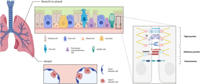
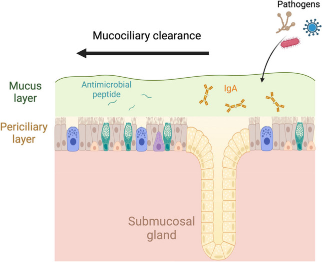
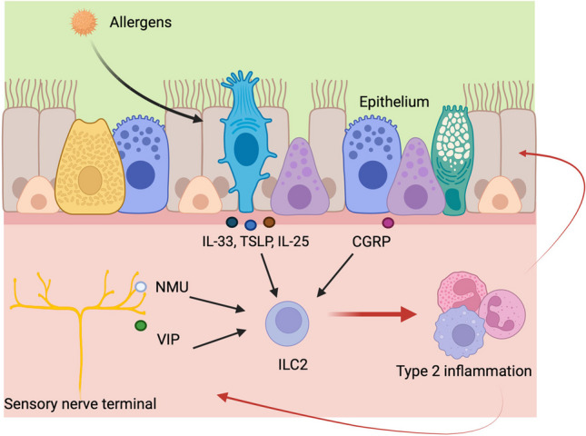
The lungs serve as the primary organ for respiration, facilitating the vital exchange of gases with the bloodstream. Given their perpetual exposure to external particulates and pathogens, they possess intricate protective barriers. Cellular adhesion in the lungs is robustly maintained through tight junctions, adherens junctions, and desmosomes. Furthermore, the pulmonary system features a mucociliary clearance mechanism that synthesizes mucus and transports it to the outside. This mucus is enriched with chemical barriers like antimicrobial proteins and immunoglobulin A (IgA). Additionally, a complex immunological network comprising epithelial cells, neural cells, and immune cells plays a pivotal role in pulmonary defense. A comprehensive understanding of these protective systems offers valuable insights into potential pathologies and their therapeutic interventions.

Keywords: Asthma; Immunological barriers; Lung; Physiological barriers.


Conflict of interest statement

The authors declare no competing interests.

Figures

Fig. 1
Fig. 1
Bronchial and alveolar epithelial cell types and intercellular adhesion in the respiratory epithelium. The bronchial epithelium is composed of basal cells, ciliated cells, club cells, goblet cells, pulmonary endocrine cells (PNECs), tuft cells, and ionocytes. Basal cells can differentiate into other AECs as well as self-renew. Ciliated cells play a major role in mucociliary clearance (MCC) by moving mucus. Club cells secrete anti-inflammatory proteins such as Scgb1a1. Goblet cells are critical for mucus production. Ionocytes express high levels of CFTR, which is thought to play a role in maintaining the hydration and pH of the airway. Alveolar epithelium is composed of AT1 cells and AT2 cells. AT1 cells are essential for gas exchange and barrier function, while AT2 cells produce surfactant and GM-CSF. AT2 cells also function as progenitor cells. Cell–cell adhesion complexes mainly consist of tight junctions (TJs), adherens junctions (AJs), and desmosomes. TJs are composed of occludin, claudin, and junctional adhesion molecules (JAMs), which adhere cell to cell, and each binds to zonula occludens (ZO) proteins intracellularly and then connects to actin fiber. TJs primarily regulate paracellular permeability. AJs are cadherin-catenin complexes located below TJs, linking to the actin cytoskeleton. AJs control cell morphology and kinetics. Desmosomes bind intermediate filaments intracellularly to consolidate mechanical stability
Fig. 2
Fig. 2
Mucus-mediated defense mechanisms. Mucus is mainly produced by secretory cells such as goblet cells and club cells. Submucosal glands also contribute substantially to the production of mucus. Its sticky nature prevents pathogens from invading the airway epithelium. Then, ciliated cells move mucus to extrude the captured foreign substances out of the body. This coordinated mechanism of mucus and epithelium is mucociliary clearance (MCC). In addition, mucus contains antimicrobial peptides and IgA and functions as a chemical barrier
Fig. 3
Fig. 3
The induction of type 2 inflammation through the interactions between epithelial cells, neurons, and immune cells. When allergens invade the respiratory epithelium, tuft cells release cytokines such as IL-25, and pulmonary neuroendocrine cells (PNECs) produce calcitonin gene–related peptide (CGRP), which can activate ILC2s. Epithelial cells release IL-33 and TSLP upon stimulation and/or damage, activating ILC2s. In addition, neuromedin U (NMU) and vasoactive intestinal peptide (VIP) from sensory neurons stimulate ILC2s to release cytokines, leading to type 2 inflammation. The induction of type 2 inflammation mobilizes various immune cells, which also act on the nervous system and epithelium to enhance inflammation

Similar articles

Cited by

References

    1. Davis JD, Wypych TP. Cellular and functional heterogeneity of the airway epithelium. Mucosal Immunol. 2021;14(5):978–990. doi: 10.1038/s41385-020-00370-7. - DOI - PMC - PubMed
    1. Hallstrand TS, Hackett TL, Altemeier WA, Matute-Bello G, Hansbro PM, Knight DA. Airway epithelial regulation of pulmonary immune homeostasis and inflammation. Clin Immunol. 2014;151(1):1–15. doi: 10.1016/j.clim.2013.12.003. - DOI - PubMed
    1. Saku A, Hirose K, Ito T, Iwata A, Sato T, et al. Fucosyltransferase 2 induces lung epithelial fucosylation and exacerbates house dust mite-induced airway inflammation. J Allergy Clin Immunol. 2019;144(3):698–709.e9. doi: 10.1016/j.jaci.2019.05.010. - DOI - PubMed
    1. Nishimura N, Yokota M, Kurihara S, Iwata A, Kageyama T, et al. Airway epithelial STAT3 inhibits allergic inflammation via upregulation of stearoyl-CoA desaturase 1. Allergol Int. 2022;71(4):520–527. doi: 10.1016/j.alit.2022.05.002. - DOI - PubMed
    1. Ordovas-Montanes J, Dwyer DF, Nyquist SK, Buchheit KM, Vukovic M, et al. Allergic inflammatory memory in human respiratory epithelial progenitor cells. Nature. 2018;560(7720):649–654. doi: 10.1038/s41586-018-0449-8. - DOI - PMC - PubMed

Publication types

LinkOut - more resources