. 2024 Apr 30;21(5):367-378.
doi: 10.1093/jsxmed/qdae015.

BMP4 and GREM1 are targets of SHH signaling and downstream regulators of collagen in the penis

Affiliations

BMP4 and GREM1 are targets of SHH signaling and downstream regulators of collagen in the penis

Jiangping Deng et al. J Sex Med. .

Abstract

Background: Cavernous nerve (CN) injury, caused by prostatectomy and diabetes, initiates a remodeling process (smooth muscle apoptosis and increased collagen) in the corpora cavernosa of the penis of patients and animal models that is an underlying cause of erectile dysfunction (ED), and the Sonic hedgehog (SHH) pathway plays an essential role in the response of the penis to denervation, as collagen increases with SHH inhibition and decreases with SHH treatment.

Aim: We examined if part of the mechanism of how SHH prevents penile remodeling and increased collagen with CN injury involves bone morphogenetic protein 4 (BMP4) and gremlin1 (GREM1) and examined the relationship between SHH, BMP4, GREM1, and collagen in penis of ED patients and rat models of CN injury, SHH inhibition, and SHH, BMP4, and GREM1 treatment.

Methods: Corpora cavernosa of Peyronie's disease (control), prostatectomy, and diabetic ED patients were obtained (N = 30). Adult Sprague Dawley rats (n = 90) underwent (1) CN crush (1-7 days) or sham surgery; (2) CN injury and BMP4, GREM1, or mouse serum albumin (control) treatment via Affi-Gel beads or peptide amphiphile (PA) for 14 days; (3) 5E1 SHH inhibitor, IgG, or phosphate-buffered saline (control) treatment for 2 to 4 days; or (4) CN crush with mouse serum albumin or SHH for 9 days.

Outcomes: Immunohistochemical and Western analysis for BMP4 and GREM1, and collagen analysis by hydroxyproline and trichrome stain were performed.

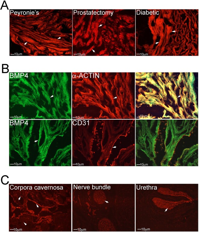
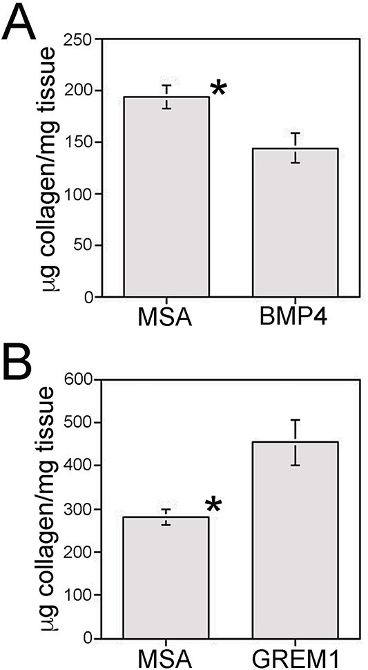
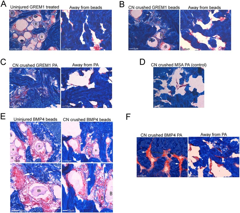
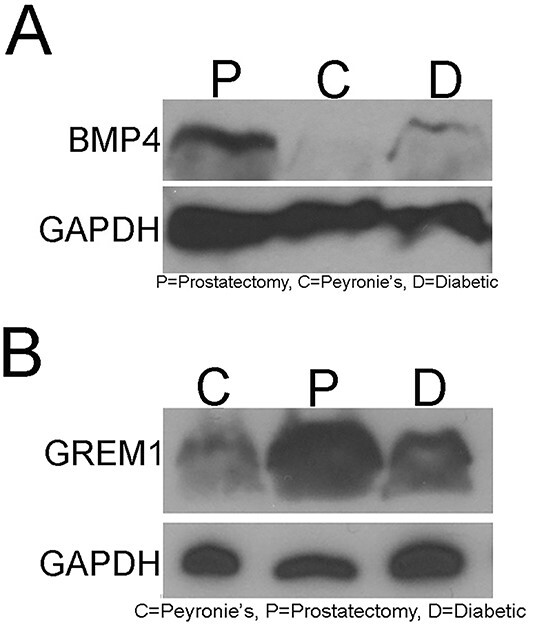
Results: BMP4 and GREM1 proteins were identified in corpora cavernosa smooth muscle of prostatectomy, diabetic, and Peyronie's patients, and in rat smooth muscle, sympathetic nerve fibers, perineurium, blood vessels, and urethra. Collagen decreased 25.4% in rats with CN injury and BMP4 treatment (P = .02) and increased 61.3% with CN injury and GREM1 treatment (P = .005). Trichrome stain showed increased collagen in rats treated with GREM1. Western analysis identified increased BMP4 and GREM1 in corpora cavernosa of prostatectomy and diabetic patients, and after CN injury (1-2 days) in our rat model. Localization of BMP4 and GREM1 changed with SHH inhibition. SHH treatment increased the monomer form of BMP4 and GREM1, altering their range of signaling.

Clinical implications: A better understanding of penile remodeling and how fibrosis occurs with loss of innervation is essential for development of novel ED therapies.

Strengths and limitations: The relationship between SHH, BMP4, GREM1, and collagen is complex in the penis.

Conclusion: BMP4 and GREM1 are downstream targets of SHH that impact collagen and may be useful in collaboration with SHH to prevent penile remodeling and ED.

Keywords: BMP4; GREM1; Peyronie’s; Sonic hedgehog; cavernous nerve injury; collagen; diabetes; erectile dysfunction.


Figures

Figure 1
Figure 1
Immunohistochemistry (IHC) of patient and rat penis tissue assayed for BMP4, ACTA2, and CD31. (A) BMP4 protein is localized in corpora cavernosa of Peyronie’s, prostatectomy and diabetic patients (red stain). (B) Dual IHC analysis for BMP4/ACTA2 and BMP4/CD31 in corpora cavernosa of Peyronie’s patients localized BMP4 to smooth muscle (yellow stain) but not in the endothelium (no overlap of staining). (C) IHC of Sprague Dawley rat penis localized BMP4 protein in corpora cavernosa smooth muscle, fibroblasts, sympathetic nerve fibers, perineurium and blood vessels of the dorsal nerve bundle, and epithelium of the urethra. Arrows indicate staining. Magnification was ×250.
Figure 2
Figure 2
Immunohistochemistry (IHC) of patient and rat penis tissue were assayed for GREM1, ACTA2, and CD31. (A) GREM1 protein is abundant in corpora cavernosa of Peyronie’s, prostatectomy, and diabetic patients (red stain). (B) Dual IHC analysis for GREM1/ACTA2 and GREM1/CD31 in corpora cavernosa of Peyronie’s patients localized GREM1 to smooth muscle (yellow stain) but not in the endothelium (no overlap of staining). (C) IHC of Sprague Dawley rat penis localized GREM1 protein in corpora cavernosa smooth muscle, fibroblasts, sympathetic nerve fibers, perineurium, and blood vessels of the dorsal nerve bundle, and epithelium of the urethra. Arrows indicate staining. Magnification was ×250.
Figure 3
Figure 3
(A) Hydroxyproline quantification of collagen in corpora cavernosa from rats that underwent cavernous nerve (CN) injury and treatment with BMP4 (n = 6) protein by peptide amphiphile (PA) for 14 days showed 25.4% decreased collagen (P = .02) in comparison with mouse serum albumin (MSA) control samples (n = 4). (B) Hydroxyproline quantification of collagen in corpora cavernosa from rats that underwent CN injury and treatment with GREM1 (n = 7) protein by PA for 14 days showed 61.3% increased collagen (P = .005) in comparison with MSA control samples (n = 7). Asterisk indicates significant differences.
Figure 4
Figure 4
Trichrome stain was performed on uninjured and cavernous nerve (CN)–injured rat penis that was treated with GREM1 and BMP4 by Affi-Gel beads and peptide amphiphile (PA) for 14 days. (A, B) collagen (blue stain) increased near the bead vehicle in uninjured (n = 4) and CN-injured (n = 4) rat corpora cavernosa treated with GREM1 in comparison with the untreated control regions away from the Affi-Gel bead vehicle. (C, D) Trichrome stain of CN-crushed and GREM1-treated (n = 10) rat penis delivered by PA for 14 days showed increased collagen compared with the untreated region away from the PA vehicle–treated and mouse serum albumin (MSA)–treated (n = 9) controls. E) BMP4 protein treatment via Affi-Gel beads of uninjured (n = 4) and CN-crushed (n = 4) rat penis for 14 days showed unchanged collagen; however, smooth muscle (red stain) increased near the bead vehicle. Enlarged regions near the Affi-Gel bead vehicle clearly depict smooth muscle cells and fibers. (F) Trichrome stain of CN-injured and BMP4-treated (n = 6) rat penis by PA for 14 days showed increased smooth muscle in comparison with the untreated region away from the PA delivery vehicle and to MSA control samples (D, n = 9). Arrows indicate increased collagen or smooth muscle. B = Affi-Gel bead delivery vehicle. Magnification was ×250 to ×400.
Figure 5
Figure 5
Western analysis of Sprague Dawley rat sham (S) and cavernous nerve (CN)–crushed (1-7 days) corpora cavernosa (n = 4 for each group) showed increased BMP4 (A) and GREM1 (B) at 1 to 2 days after CN injury in comparison with sham control samples, and by 4 to 7 days, BMP4 and GREM1 returned to baseline. A total of 100 μg protein was loaded into each well, and GAPDH was used as a control for protein loading and was unchanged.
Figure 6
Figure 6
Western analysis identified low-intensity protein bands for BMP4 (A) and GREM1 (B) in corpora cavernosa tissue from control Peyronie’s patients (C). In comparison with Peyronie’s control samples (n = 11), increased BMP4 (A) and GREM1 (B) protein band intensity was observed in corpora cavernosa tissue from prostatectomy (P) (n = 9) and diabetic (D) (n = 9) patients. A total of 100 μg protein was loaded into each well, and GAPDH was used as a control for protein loading.
Figure 7
Figure 7
Western analysis identified monomer and dimer forms of BMP4 and GREM1 proteins in corpora cavernosa tissue from cavernous nerve (CN)–injured Sprague Dawley rats treated with mouse serum albumin/bovine serum albumin (control, n = 7) by peptide amphiphile (PA) for 9 days. Western analysis performed on corpora cavernosa of CN-injured rats treated with Sonic hedgehog (S) (n = 8) protein by PA for 9 days showed increased GREM1 and BMP4 monomer, and decreased GREM1 dimer at 9 days after injury in comparison with mouse serum albumin (MSA)/bovine serum albumin (BSA) control samples (C) (n = 7). A total of 100 μg protein was loaded into each well, and β-actin was used as a control for protein loading.
Figure 8
Figure 8
Immunohistochemistry (IHC) analysis for BMP4 protein was performed on penis of Sprague Dawley rats treated in vivo with 5E1 Sonic hedgehog (SHH) inhibitor (n = 6), IgG (n = 6), or phosphate-buffered saline (PBS) (n = 3) control samples. (A) IHC for BMP4 protein in untreated tissue away from the Affi-Gel bead vehicle showed normal BMP4 staining in corpora cavernosa smooth muscle. With 2 days of SHH inhibition, IHC showed altered localization of BMP4 to cells near the Affi-Gel bead delivery vehicle. (B) IgG- and PBS-treated control samples showed normal BMP4 staining around the Affi-Gel beads. (C) IHC with the primary omitted did not show a secondary effect. (D) By day 4, SHH inhibition caused extensive remodeling of the corpora cavernosa, and BMP4 was present in many more cells around the bead vehicle and in the remodeled tissue. (E) The change in localization of BMP4 from smooth muscle to fibroblasts was confirmed by positive P4HB (fibroblast marker) staining and negative ACTA2 (smooth muscle). Arrows indicate staining. B = Affi-Gel bead vehicle. Magnification was ×250.
Figure 9
Figure 9
Immunohistochemistry (IHC) analysis for GREM1 protein was performed on penis of Sprague Dawley rats treated in vivo with 5E1 Sonic hedgehog (SHH) inhibitor (n = 6) or IgG (control, n = 6). (A) IHC for GREM1 protein in untreated tissue away from the Affi-Gel bead vehicle showed normal GREM1 staining in corpora cavernosa smooth muscle. With 2 days of SHH inhibition, IHC showed altered localization of GREM1 to cells near the Affi-Gel bead delivery vehicle. (B) IgG-treated control samples showed normal GREM1 staining around the Affi-Gel beads. (C) IHC with the primary omitted did not show secondary effect. (D) By day 4, SHH inhibition caused extensive remodeling of the corpora cavernosa, and GREM1 was present in many more cells around the bead vehicle and in fibroblasts of the extensively SHH-inhibited region. Arrows indicate staining. B = Affi-Gel bead vehicle. Magnification was ×250.

Similar articles

Cited by

References

    1. Feldman HA, Goldstein I, Hatzichristou DG, Krane RJ, McKinlay JB. Impotence and its medical and psychosocial correlates: results of the Massachusetts male aging study. J Urol. 1994;151(1):54–61. 10.1016/S0022-5347(17)34871-1 - DOI - PubMed
    1. Heruti R, Shochat T, Tekes-Manova D, Ashkenazi I, Justo D. Prevalence of erectile dysfunction among young adults: results of a largescale survey. J Sex Med. 2004;1(3):284–291. 10.1111/j.1743-6109.04041.x - DOI - PubMed
    1. Nguyen HMT, Gabrielson AT, Hellstrom WJG. Erectile dysfunction in young men—a review of the prevalence and risk factors. Sex Med Rev. 2017;5(4):508–520. 10.1016/j.sxmr.2017.05.004 - DOI - PubMed
    1. Kendirci M, Hellstrom WJ. Current concepts in the management of erectile dysfunction in men with prostate cancer. Clin Prostate Cancer. 2004;3(2):87–92. 10.3816/CGC.2004.n.017 - DOI - PubMed
    1. Johansson E, Steineck G, Holmberg L, et al. SPCG-4 investigators. Long-term quality-of-life outcomes after radical prostatectomy or watchful waiting: the Scandinavian prostate cancer Group-4 randomized trial. Lancet Oncol. 2011;12(9):891–899. 10.1016/S1470-2045(11)70162-0 - DOI - PubMed

Publication types