. 2024 Apr 30;21(5):379-390.
doi: 10.1093/jsxmed/qdae016.

SHH regulates penile morphology and smooth muscle through a mechanism involving BMP4 and GREM1

Affiliations

SHH regulates penile morphology and smooth muscle through a mechanism involving BMP4 and GREM1

Jiangping Deng et al. J Sex Med. .

Abstract

Background: The cavernous nerve (CN) is frequently damaged in prostatectomy and diabetic patients with erectile dysfunction (ED), initiating changes in penile morphology including an acute and intense phase of apoptosis in penile smooth muscle and increased collagen, which alter penile architecture and make corpora cavernosa smooth muscle less able to relax in response to neurotransmitters, resulting in ED.

Aim: Sonic hedgehog (SHH) is a critical regulator of penile smooth muscle, and SHH treatment suppresses penile remodeling after CN injury through an unknown mechanism; we examine if part of the mechanism of how SHH preserves smooth muscle after CN injury involves bone morphogenetic protein 4 (BMP4) and gremlin1 (GREM1).

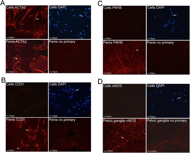
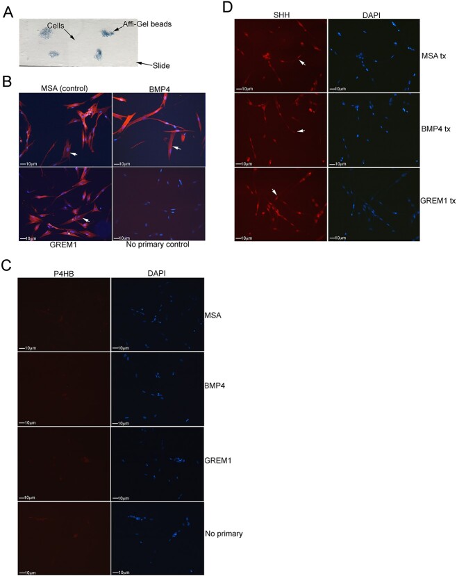
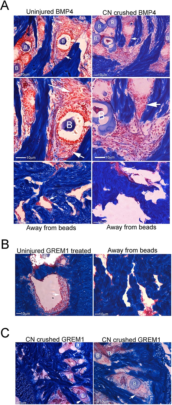
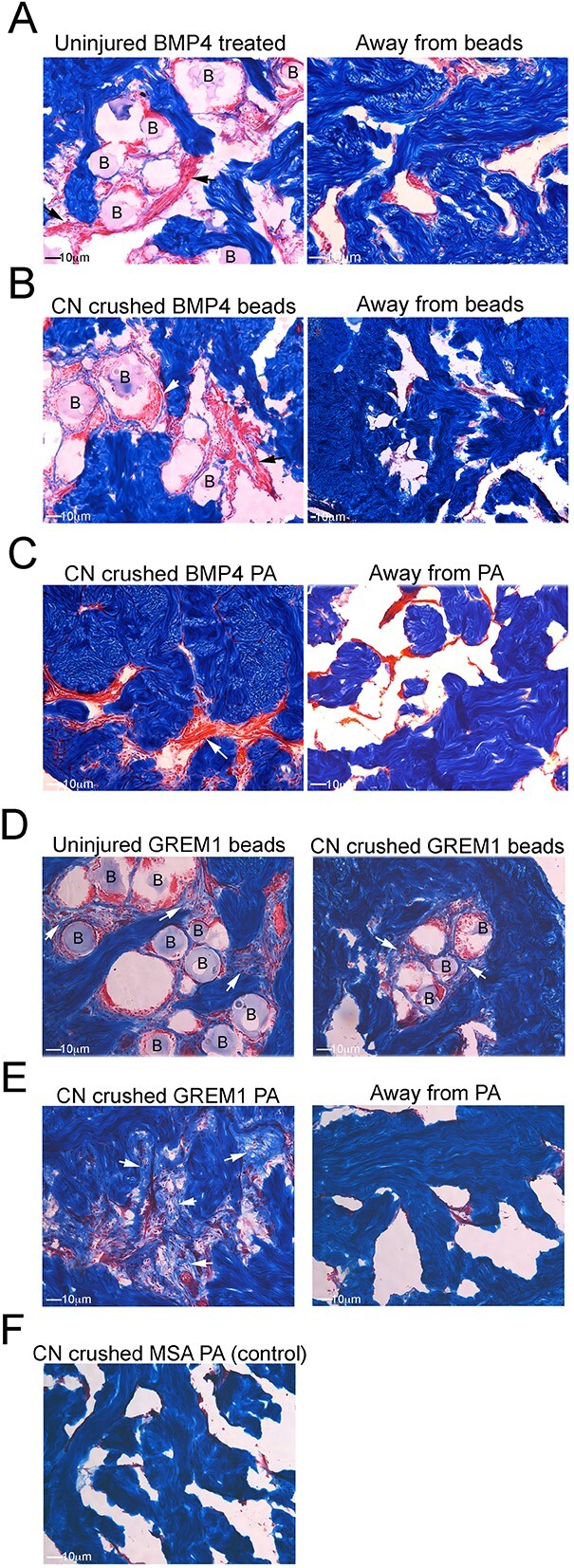
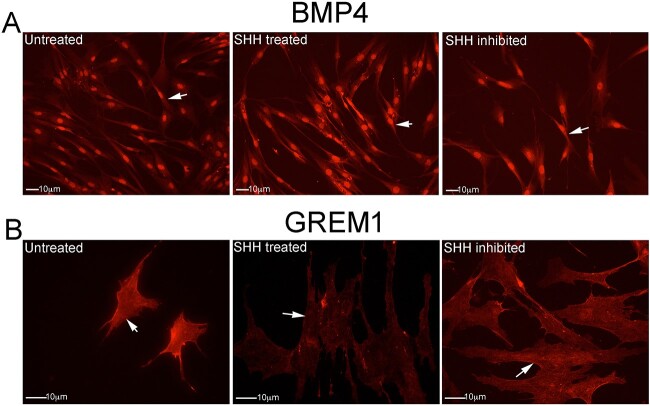
Methods: Primary cultures of smooth muscle cells were established from prostatectomy, diabetic, hypertension and Peyronie's (control) (N = 18) patients. Cultures were characterized by ACTA2, CD31, P4HB, and nNOS immunohistochemical analysis. Patient smooth muscle cell growth was quantified in response to BMP4 and GREM1 treatment. Adult Sprague Dawley rats underwent 1 of 3 surgeries: (1) uninjured or CN-injured rats were treated with BMP4, GREM1, or mouse serum albumin (control) proteins via Affi-Gel beads (N = 16) or peptide amphiphile (PA) (N = 26) for 3 and 14 days, and trichrome stain was performed; (2) rats underwent sham (N = 3), CN injury (N = 9), or CN injury and SHH PA treatment for 1, 2, and 4 days (N = 9).

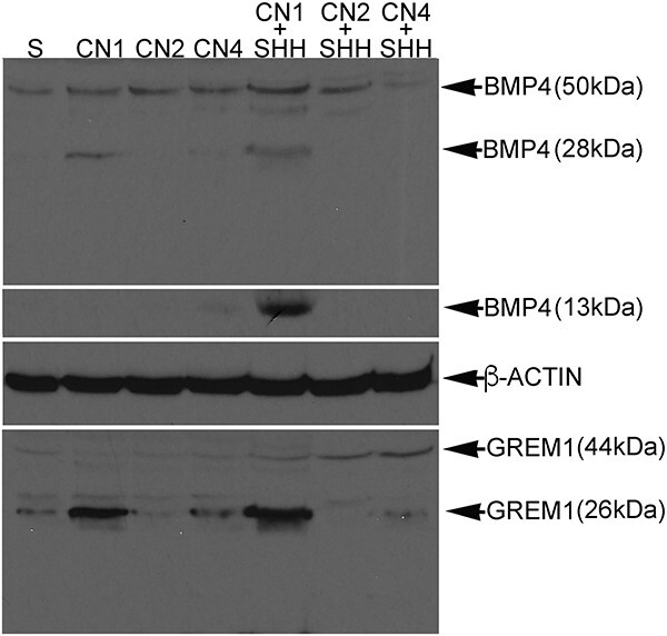
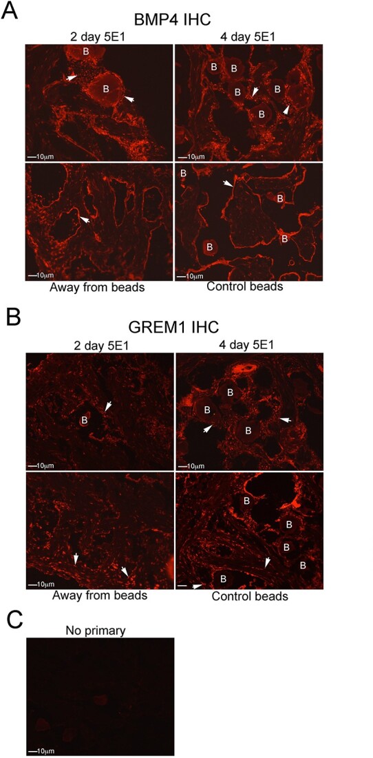
Outcomes: Western analysis for BMP4 and GREM1 was performed; (3) rats were treated with 5E1 SHH inhibitor (N = 6) or IgG (control; N = 6) for 2 and 4 days, and BMP4 and GREM1 localization was examined. Statistics were performed by analysis of variance with Scheffé's post hoc test.

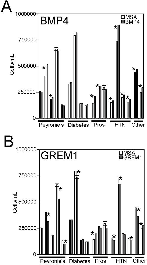
Results: BMP4 increased patient smooth muscle cell growth, and GREM1 decreased growth. In rats, BMP4 treatment via Affi-Gel beads and PA increased smooth muscle at 3 and 14 days of treatment. GREM1 treatment caused increased collagen and smooth muscle at 3 days, which switched to primarily collagen at 14 days. CN injury increased BMP4 and GREM1, while SHH PA altered Western band size, suggesting alternative cleavage and range of BMP4 and GREM1 signaling. SHH inhibition in rats increased BMP4 and GREM1 in fibroblasts.

Clinical implications: Understanding how SHH PA preserves and regenerates penile morphology after CN injury will aid development of ED therapies.

Strengths and limitations: SHH treatment alters BMP4 and GREM1 localization and range of signaling, which can affect penile morphology.

Conclusion: Part of the mechanism of how SHH regulates corpora cavernosa smooth muscle involves BMP4 and GREM1.

Keywords: BMP4; GREM1; Peyronie’s; Sonic hedgehog; cell culture; diabetes; erectile dysfunction; prostatectomy; smooth muscle.


Figures

Figure 1
Figure 1
Patient primary corpora cavernosa cell cultures were characterized by immunohistochemistry (IHC) analysis for ACTA2 (smooth muscle), CD31 (endothelium), P4HB (fibroblast), and nNOS (neuronal marker). (A) ACTA2 staining was present in all cells, by comparison with DAPI, indicating that they were smooth muscle. Penis tissue from Sprague Dawley rats, which was used as a positive control, stained abundantly for ACTA2, and negative control samples with the primary antibody omitted did not show staining, indicating the absence of secondary artifact. IHC showed the absence of endothelial cells (CD31) (B), fibroblasts (P4HB) (C), and neuronal cells (nNOS) (D). Penis control tissue showed abundant staining for CD31 (B) and P4HB (C). Rat PG was used as a positive control for nNOS and had positive staining in neurons that innervate the penis (D). Arrows indicate staining. Magnification was ×250.
Figure 2
Figure 2
(A) BMP4, GREM1, or mouse serum albumin (MSA) (control) proteins were delivered by Affi-Gel beads to patient smooth muscle cells that were grown on slides to examine their morphology. (B) ACTA2 stained strongly in all cells of the MSA-treated control samples, indicating that they were smooth muscle. DAPI costaining was performed to indicate all cells. There was no change in cell morphology or smooth muscle staining with BMP4 and GREM1 treatment. Control samples with the primary antibody omitted showed only DAPI staining to indicate the cells. (C) Immunohistochemistry (IHC) for P4HB, a fibroblast marker, did not show staining in MSA-, BMP4-, or GREM1-treated cells. (D) IHC for Sonic hedgehog (SHH) showed some staining in the cytoplasm, but the primary staining was observed in the nucleus. There was no change in staining with BMP4 and GREM1 treatment. Arrows indicate staining. Magnification was ×250.
Figure 3
Figure 3
Growth of smooth muscle cells from Peyronie’s (n = 5), diabetic (n = 4) prostatectomy (n = 3), hypertension (n = 4), and other (n = 2) patients were quantified after treatment with either BMP4 (A) or GREM1 (B) proteins in comparison with mouse serum albumin (MSA)–treated control patients. BMP4 treatment significantly increased growth in the majority of patients (A). GREM1 treatment significantly decreased growth in the majority of patients (B). Asterisks denote significant differences in cell growth. Where error bars do not appear, it is because the error is too small to appear graphically.
Figure 4
Figure 4
Trichrome stain was performed on uninjured and cavernous nerve (CN)–injured adult Sprague Dawley rat penis that was treated with Affi-Gel beads soaked in BMP4 or GREM1 proteins for 3 days. (A) With BMP4 treatment, uninjured and CN-injured corpora cavernosa tissue show increased smooth muscle (red stain) in the region of the beads in comparison with the untreated region away from the bead vehicle. Enlarged region near the bead vehicle showed individual smooth muscle cells and developing smooth muscle fibers. GREM1 treatment of uninjured (B) and CN-injured (C) corpora cavernosa tissue showed increased collagen interspersed with smooth muscle in comparison with the region of untreated corpora cavernosa away from the bead vehicle. B: Affi-Gel bead vehicle. Arrows indicate staining. Magnification was ×250.
Figure 5
Figure 5
Trichrome stain was performed on uninjured and cavernous nerve (CN)–injured adult Sprague Dawley rats that were treated with BMP4 or GREM1 proteins by Affi-Gel beads or peptide amphiphile (PA) for 14 days. BMP4 treatment of uninjured (A) and CN-injured (B) corpora cavernosa tissue by Affi-Gel beads showed increased smooth muscle near the bead vehicle in comparison with control samples away from the beads (B). (C) Smooth muscle was similarly increased with BMP4 delivery by PA. GREM1 protein increased collagen near the Affi-Gel beads (D) and with PA delivery (E). Mouse serum albumin (MSA)–treated CN-injured control samples did not show changes in morphology (F). B: Affi-Gel bead vehicle. Arrows indicate staining. Magnification was ×250.
Figure 6
Figure 6
Western analysis for BMP4 and GREM1 were performed on proteins isolated from adult Sprague Dawley rat corpora cavernosa 1 to 4 days after cavernous nerve (CN) injury, 1 to 4 days after CN injury with Sonic hedgehog (SHH) peptide amphiphile (PA) treatment, and sham control samples. Western for BMP4 showed a band at ~50 kDa in sham control samples. After CN injury, an additional band was observed at ~28 kDa (alternative cleavage). With SHH PA treatment of CN-injured rats, the 50-kDa band increased at day 1 and declined by day 4, and an additional 13-kDa band was observed at day 1. Western for GREM1 showed a band at 26 kDa on day 1 after CN injury. A 44-kDa GREM1 band was barely detectable in the sham and CN-injured rats; however, with SHH treatment the 44-kDa band increased from 1 to 4 days. ACTA2 was constant in all samples, indicating even protein loading.
Figure 7
Figure 7
The impact of Sonic hedgehog (SHH) inhibitor on BMP4 and GREM1 localization was examined in adult Sprague Dawley rats that were treated with SHH inhibitor (n = 6) or IgG/phosphate-buffered saline (PBS) control (n = 6) via Affi-Gel beads for 2 and 4 days. Immunohistochemistry (IHC) for BMP4 (A) showed increased staining in fibroblast cells near the bead vehicle at 2 and 4 days of SHH inhibition in comparison with IgG/PBS treated control samples, which only showed staining in smooth muscle. Untreated tissue away from the Affi-Gel bead vehicle showed normal BMP4 staining. (B) IHC for GREM1 showed increased staining in fibroblast cells near the bead vehicle at 2 and 4 days of SHH inhibition. Untreated tissue away from the Affi-Gel bead vehicle showed normal GREM1 staining in smooth muscle. (C) Control samples with the primary antibody omitted showed the absence of secondary artifact. B: Affi-Gel bead vehicle. Arrows indicate staining. Magnification was ×250.
Figure 8
Figure 8
Immunohistochemistry (IHC) analysis for BMP4 and GREM1 proteins was performed on patient smooth muscle cells that were treated with Sonic hedgehog (SHH) protein, SHH inhibitor, or no treatment (control). (A) BMP4 protein was abundantly expressed, with no apparent change in cellular localization with SHH treatment or SHH inhibition. (B) GREM1 protein decreased with SHH treatment in comparison with untreated control samples. Arrows indicate staining. Magnification was ×250.

Similar articles

Cited by

References

    1. Feldman HA, Goldstein I, Hatzichristou DG, Krane RJ, McKinlay JB. Impotence and its medical and psychosocial correlates: results of the Massachusetts male aging study. J Urol. 1994;151(1):54–61. 10.1016/S0022-5347(17)34871-1 - DOI - PubMed
    1. Hakim LS, Goldstein I. Diabetic sexual dysfunction. Endocrinol Metab Clin N Am. 1996;25(2):379–400. 10.1016/S0889-8529(05)70329-7 - DOI - PubMed
    1. Kouidrat Y, Pizzol D, Costo T, et al. High prevalence of erectile dysfunction in diabetes; a systematic review and meta-analysis of 145 studies. Diabet Med. 2017;34(9):1185–1192. 10.1111/dme.13403 - DOI - PubMed
    1. Kendirci M, Hellstrom WJ. Current concepts in the management of erectile dysfunction in men with prostate cancer. Clin Prostate Cancer. 2004;3(2):87–92. 10.3816/CGC.2004.n.017 - DOI - PubMed
    1. Johansson E, Steineck G, Holmberg L, et al. SPCG-4 investigators. Long-term quality-of-life outcomes after radical prostatectomy or watchful waiting: the Scandinavian prostate cancer Group-4 randomized trial. Lancet Oncol. 2011;12(9):891–899. 10.1016/S1470-2045(11)70162-0 - DOI - PubMed