Comment
. 2024 Apr 1;142(4):310-319.
doi: 10.1001/jamaophthalmol.2024.0002.

Polarization-Sensitive OCT Imaging of Scleral Abnormalities in Eyes With High Myopia and Dome-Shaped Macula

Affiliations
Comment

Polarization-Sensitive OCT Imaging of Scleral Abnormalities in Eyes With High Myopia and Dome-Shaped Macula

Kyoko Ohno-Matsui et al. JAMA Ophthalmol. .

Abstract

Importance: The relevance of visualizing scleral fiber orientation may offer insights into the pathogenesis of pathologic myopia, including dome-shaped maculopathy (DSM).

Objective: To investigate the orientation and density of scleral collagen fibers in highly myopic eyes with and without DSM by polarization-sensitive optical coherence tomography (PS-OCT).

Design, setting, and participants: This case series included patients with highly myopic eyes (defined as a refractive error ≥6 diopters or an axial length ≥26.5 mm) with and without a DSM examined at a single site in May and June 2019. Analysis was performed from September 2019 to October 2023.

Exposures: The PS-OCT was used to study the birefringence and optic axis of the scleral collagen fibers.

Main outcomes and measures: The orientation and optic axis of scleral fibers in inner and outer layers of highly myopic eyes were assessed, and the results were compared between eyes with and without a DSM.

Results: A total of 72 patients (51 [70.8%] female; mean [SD] age, 61.5 [12.8] years) were included, and 89 highly myopic eyes were examined (mean [SD] axial length, 30.4 [1.7] mm); 52 (58.4%) did not have a DSM and 37 (41.6%) had a DSM (10 bidirectional [27.0%] and 27 horizontal [73.0%]). Among the 52 eyes without DSM, the 13 eyes with simple high myopia had primarily inner sclera visible, displaying radially oriented fibers in optic axis images. In contrast, the entire thickness of the sclera was visible in 39 eyes with pathologic myopia. In these eyes, the optic axis images showed vertically oriented fibers within the outer sclera. Eyes presenting with both horizontal and bidirectional DSMs had clusters of fibers with low birefringence at the site of the DSM. In the optic axis images, horizontally or obliquely oriented scleral fibers were aggregated in the inner layer at the DSM. The vertical fibers located posterior to the inner fiber aggregation were not thickened and appeared thin compared with the surrounding areas.

Conclusions and relevance: This study using PS-OCT revealed inner scleral fiber aggregation without outer scleral thickening at the site of the DSM in highly myopic eyes. Given the common occurrence of scleral pathologies, such as DSM, and staphylomas in eyes with pathologic myopia, recognizing these fiber patterns could be important. These insights may be relevant to developing targeted therapies to address scleral abnormalities early and, thus, mitigate potential damage to the overlying neural tissue.


Conflict of interest statement

Conflict of Interest Disclosures: Dr Ohno-Matsui reported receiving a research grant from Tomey Corporation and being a consultant for Santen and CooperVision outside the submitted work. Dr Banerjee reported having a patent for JP 2023097533 pending. Dr Yamanari reported having patents for JP 6463051 B2, US 9593936 B2, EP 2995245 B1, JP 6542178 B2, and JP 7332131 B2 issued to Tomey Corporation. No other disclosures were reported.

Figures

Figure 1.
Figure 1.. Polarization-Sensitive Optical Coherence Tomography (PS-OCT) Images of a Highly Myopic Eye Without a Dome-Shaped Macula or Pathologic Myopia
Images of the right eye of a 30-year-old man with a refractive error of −14.9 diopters and an axial length of 28.9 mm showing a tessellated fundus without myopic atrophic lesions. A and B, Birefringence images obtained by PS-OCT. Images of the horizontal (A) and vertical (B) sections across the fovea showing a mixture of low-density (green) and high-density (yellow) birefringent fibers. The color map is intentionally saturated for better views of low birefringence structures. C and D, PS-OCT optic axis images. Yellow, blue, green, and pink indicate the vertical, horizontal, and the 2 oblique scan directions, respectively. C, In the horizontal scan, a horizontal fiber (blue) can be seen to run from the optic disc toward the macula along the inner surface of the sclera. Obliquely running fibers (green) can be seen especially in the macular area posterior to the horizontal fibers. Vertically running fibers (yellow) are seen only temporal to the optic nerve. D, In a vertical scan image, the horizontal fibers (blue) are seen in the macular region and oblique fibers (pink and green) are seen superior and inferior to the macula. The outer sclera is not clearly seen. deg indicates degrees.
Figure 2.
Figure 2.. Polarization-Sensitive Optical Coherence Tomography Images of an Eye With Pathologic Myopia and Without a Dome-Shaped Macula
Images of the right eye of a 46-year-old woman with a refractive error of −23.5 diopters and an axial length of 32.0 mm showing diffuse choroidal atrophy. A, Horizontal section of birefringence image across the fovea shows low birefringence (green) in general, but high birefringence (yellow) is seen in the outer scleral layer in the macular area and in the area temporal to the optic nerve. The blood vessel emissary (arrowhead) penetrates the sclera slightly temporal to the fovea and is discontinuous with the birefringence. B, Vertical section across the fovea showing that the fibers have low birefringence (green) in the inner layer of the sclera in the macular region. Fibers with high birefringence (yellow) are seen in the outer scleral layer in the macular area and in the outer sclera superior and inferior to the macula. C, Horizontally scanned optic axis image shows horizontal fibers (blue) running from the optic nerve toward the macula along the inner surface of the sclera. Vertically running fibers (yellow) are seen posterior to the horizontal fibers in a wide area of the posterior sclera. D, Vertically scanned image showing horizontal fibers (blue) in the inner layer of the sclera in the macular region. The sclera consists mainly of vertical (yellow) and oblique (green) fibers. deg indicates degrees.
Figure 3.
Figure 3.. Polarization-Sensitive Optical Coherence Tomography Images of a Highly Myopic Right Eye With a Bidirectional Dome-Shaped Macula (DSM)
Images of the right eye of a 76-year-old woman with an implanted intraocular lens and an axial length of 30.1 mm showing diffuse choroidal atrophy. A, The image of a horizontal section across the fovea shows low birefringence fibers (green, indicated by white arrowhead) in general, while high birefringence fibers (yellow, indicated by magenta arrowhead) are seen only temporal to the optic nerve. B, The vertical section across the fovea shows low birefringence fibers (green, indicated by white arrowhead) at the DSM. In contrast, the fibers with high birefringence (yellow, indicated by blue arrowheads) are seen superior and inferior to the DSM. C, In the horizontal scan image of the optic axis, the DSM shows a mixture of horizontal (blue) and oblique (pink) fibers. The horizontal fibers are seen extensively nasal and temporal to the DSM along the inner sclera. The vertical fibers (yellow) can be seen in the outer sclera, especially temporal to the optic nerve. D, In a vertically scanned image, a mixture of horizontal (blue) and oblique (green and pink) fibers is seen at the DSM. The outer layer of the sclera superior and inferior to the DSM consists of vertical fibers (yellow). deg indicates degrees.
Figure 4.
Figure 4.. Polarization-Sensitive Optical Coherence Tomography Images of a Highly Myopic Right Eye With a Horizontal Dome-Shaped Macula (DSM)
Images of the right eye of a 48-year-old woman with an implanted intraocular lens and an axial length of 30.8 mm showing diffuse choroidal atrophy. A, The horizontal section of the birefringence image across the fovea shows mainly low birefringence fibers (green) in the macula. Fibers with high birefringence are seen between the optic nerve and the macula. B, The vertical section across the fovea shows an aggregation and thickening of fibers with low birefringence (green) at the DSM. The sclera superior and inferior to the DSM has high birefringence (yellow). C, In the horizontal scan image of the optic axis, the relatively thick horizontal fibers (blue) are seen in a wide area. A thin layer of vertical fibers (yellow) is seen in the outer sclera of the macula. Vertical fibers are seen in the outer sclera between the optic nerve and the macula. D, In a vertically scanned image, the DSM appears as an aggregation and thickening of horizontal (blue) and oblique (pink) fibers. Posterior to these fibers, vertical fibers (yellow) are seen in the outer sclera. The outer sclera consists of vertical fibers that are not thickened but appear compressed. The outer sclera superior and inferior to the DSM consists mainly of vertical fibers (yellow). deg indicates degrees.
Figure 5.
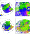
Figure 5.. Streamline Renderings of the Optic Axis for the Eyes Shown in Figures 1 and 2
A and B, Streamlined images recorded with an oblique angle of the camera (A) and from the interior of the eye (B) rendered by ParaView for the participant shown in Figure 1. B, The course of the inner scleral fibers is seen clearly. A concentric peripapillary ring can be seen around the optic nerve head. The inner scleral fibers spread radially from this ring toward the periphery. C and D, Streamlined images recorded with an oblique angle of the camera (C) and from the exterior of the eye (D) rendered by ParaView for the participant shown in Figure 2. The course of the outer scleral fibers can be clearly seen to run almost vertically in parallel with the concentric peripapillary circles.

Comment on

References

    1. Hogan MJ, ed. The sclera. In: Histology of the Human Eye. WB Saunders; 1971.
    1. de la Maza MS, Tauber J, Foster CS. Structural considerations of the sclera. In: de la Maza MS, Tauber J, Foster CS, eds. The Sclera. Springer; 2012:1-27.
    1. Gaucher D, Erginay A, Lecleire-Collet A, et al. . Dome-shaped macula in eyes with myopic posterior staphyloma. Am J Ophthalmol. 2008;145(5):909-914. doi:10.1016/j.ajo.2008.01.012 - DOI - PubMed
    1. Soudier G, Gaudric A, Gualino V, et al. . Long-term evolution of dome-shaped macula: increased macular bulge is associated with extended macular atrophy. Retina. 2016;36(5):944-952. doi:10.1097/IAE.0000000000000806 - DOI - PubMed
    1. Imamura Y, Iida T, Maruko I, Zweifel SA, Spaide RF. Enhanced depth imaging optical coherence tomography of the sclera in dome-shaped macula. Am J Ophthalmol. 2011;151(2):297-302. doi:10.1016/j.ajo.2010.08.014 - DOI - PubMed