

Effect of different palatal expanders with miniscrews in surgically assisted rapid palatal expansion: A non-linear finite element analysis
- PMID: 38451569
- PMCID: PMC10914319
- DOI: 10.1590/2177-6709.29.1.e2423195.oar
Effect of different palatal expanders with miniscrews in surgically assisted rapid palatal expansion: A non-linear finite element analysis
Abstract
Introduction: Surgically assisted rapid palatal expansion (SARPE) has been the treatment of choice in subjects presenting skeletally mature sutures.
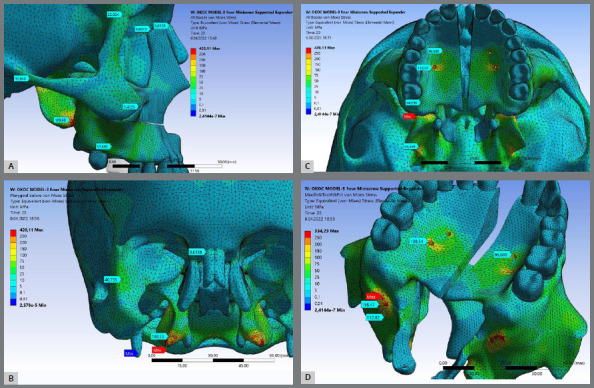
Objective: The purpose of this study was to analyze stress distribution and displacement of the craniofacial and dentoalveolar structures resulting from three types of palatal expanders with surgical assistance using a non-linear finite element analysis.
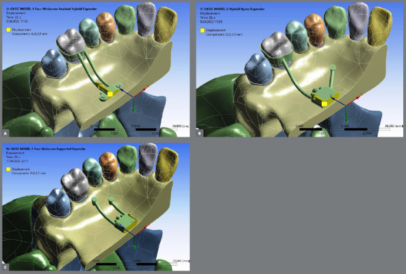
Material and methods: Three different palatal expanders were designed: Model-I (tooth-bone-borne type containing four miniscrews), Model-II (tooth-bone-borne type containing two miniscrews), and Model-III (bone-borne type containing four miniscrews). A Le Fort I osteotomy was performed, and a total of 5.0 mm palatal expansion was simulated. Nonlinear analysis (three theory) method (geometric nonlinear theory, nonlinear contact theory, and nonlinear material methods) was used to evaluate stress and displacement of several craniofacial and dentoalveolar structures.
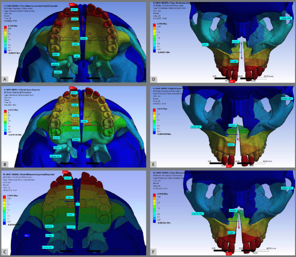
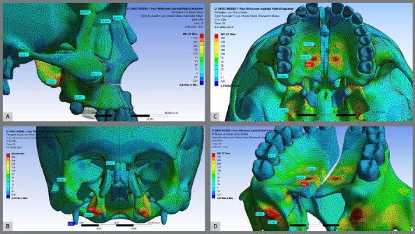
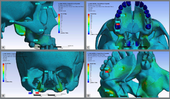
Results: Regardless of the maxillary expander device type, surgically assisted rapid palatal expansion produces greater anterior maxillary expansion than posterior (ANS ranged from 2.675 mm to 3.444 mm, and PNS ranged from 0.522 mm to 1.721 mm); Model-I showed more parallel midpalatal suture opening pattern - PNS/ANS equal to 54%. In regards to ANS, Model-II (1.159 mm) and Model-III (1.000 mm) presented larger downward displacement than Model-I (0.343 mm). PNS displaced anteriorly more than ANS for all devices; Model-III presented the largest amount of forward displacement for PNS (1.147 mm) and ANS (1.064 mm). All three type of expanders showed similar dental displacement, and minimal craniofacial sutures separation. As expected, different maxillary expander designs produce different primary areas and levels of stresses (the bone-borne expander presented minimal stress at the teeth and the tooth-bone-borne expander with two miniscrews presented the highest).
Conclusions: Based on this finite element method/finite element analysis, the results showed that different maxillary expander designs produce different primary areas and levels of stresses, minimal displacement of the craniofacial sutures, and different skeletal V-shape expansion.
Introdução:: A expansão rápida da maxila assistida cirurgicamente (ERMAC) tem sido o tratamento de escolha em indivíduos que apresentam suturas esqueleticamente maduras.
Objetivo:: O objetivo deste estudo foi avaliar, utilizando uma análise não linear com elementos finitos, a distribuição de tensões e os deslocamentos das estruturas craniofaciais e dentoalveolares gerados por três tipos de expansores palatinos usados na ERMAC.
Material e Métodos:: Três tipos de expansores palatinos foram projetados: Modelo I (dento-osseossuportado com quatro mini-implantes), Modelo II (dento-osseossuportado com dois mini-implantes) e Modelo III (osseossuportado com quatro mini-implantes). Uma osteotomia Le Fort I foi realizada e foi simulada uma expansão palatina total de 5,0 mm. Um método de análise não linear (três teorias - teoria da não-linearidade geométrica, teoria do contato não linear e métodos para materiais não lineares) foi utilizado para avaliar a tensão e o deslocamento de diversas estruturas craniofaciais e dentoalveolares.
Resultados:: Independentemente do tipo de aparelho expansor palatino, a ERMAC produziu maior expansão anterior da maxila do que posterior (ENA variou de 2,675 mm a 3,444 mm e ENP variou de 0,522 mm a 1,721 mm); o Modelo I apresentou padrão de abertura mais paralela da sutura palatina mediana, com ENP/ENA igual a 54%. Com relação à ENA, o Modelo II (1,159 mm) e o Modelo III (1,000 mm) apresentaram maior deslocamento para baixo do que o Modelo I (0,343 mm). A ENP deslocou-se mais para anterior do que a ENA com todos os aparelhos; o Modelo III apresentou o maior deslocamento para anterior da ENP (1,147 mm) e da ENA (1,064 mm). Os três tipos de expansores apresentaram deslocamento dentário semelhante e separação mínima das suturas craniofaciais. Como esperado, diferentes designs de expansores palatinos produzem diferentes áreas primárias e níveis de tensões (o expansor osseossuportado apresentou tensão mínima nos dentes, e o expansor dento-osseossuportado com dois mini-implantes apresentou o maior).
Conclusões:: Com base nesse estudo de elementos finitos, os resultados mostraram que diferentes designs de expansores palatinos produzem diferentes áreas primárias e níveis de tensão, com deslocamento mínimo das suturas craniofaciais e diferentes expansões esqueléticas em forma de V.
Conflict of interest statement
The authors report no commercial, proprietary or financial interest in the products or companies described in this article.
Figures

Similar articles
-
Effect of bone-borne rapid maxillary expanders with and without surgical assistance on the craniofacial structures using finite element analysis.Am J Orthod Dentofacial Orthop. 2014 May;145(5):638-48. doi: 10.1016/j.ajodo.2013.12.029. Am J Orthod Dentofacial Orthop. 2014. PMID: 24785928
-
The comparison of biomechanical effects of the conventional and bone-borne palatal expanders on late adolescence with unilateral cleft palate: a 3-dimensional finite element analysis.BMC Oral Health. 2022 Dec 13;22(1):600. doi: 10.1186/s12903-022-02640-1. BMC Oral Health. 2022. PMID: 36514035 Free PMC article.
-
Comparison of 3 different bone-borne type expansion appliances used in surgically-assisted rapid palatal expansion: A finite element analysis.Am J Orthod Dentofacial Orthop. 2023 Mar;163(3):e23-e33. doi: 10.1016/j.ajodo.2022.12.001. Epub 2022 Dec 24. Am J Orthod Dentofacial Orthop. 2023. PMID: 36572581
-
Maxillary transverse deficiency, with closed intermaxillary suture, does bone-anchored appliance during SARPE cause predictable, and stable maxillary expansion compared to the tooth-borne appliance during SARPE - Systematic review.J Stomatol Oral Maxillofac Surg. 2023 Feb;124(1S):101344. doi: 10.1016/j.jormas.2022.11.020. Epub 2022 Nov 24. J Stomatol Oral Maxillofac Surg. 2023. PMID: 36435445
-
Comparison of Different Types of Palatal Expanders: Scoping Review.Children (Basel). 2023 Jul 21;10(7):1258. doi: 10.3390/children10071258. Children (Basel). 2023. PMID: 37508755 Free PMC article.
Cited by
-
Non-Linear Biomechanical Evaluation and Comparison in the Assessment of Three Different Piece Dental Implant Systems for the Molar Region: A Finite Element Study.J Funct Biomater. 2025 Jan 9;16(1):17. doi: 10.3390/jfb16010017. J Funct Biomater. 2025. PMID: 39852573 Free PMC article.
-
Long-term structural and functional nasomaxillary evolution of children with mouth-breathing after rapid maxillary expansion: An 8-year follow-up study.Korean J Orthod. 2025 Mar 25;55(2):95-104. doi: 10.4041/kjod24.102. Epub 2025 Feb 10. Korean J Orthod. 2025. PMID: 39924972 Free PMC article.
References
-
- Proffit WR, Fields HW, Jr, Moray LJ. Prevalence of malocclusion and orthodontic treatment need in the United States estimates from the NHANES III survey. Int J Adult Orthodon Orthognath Surg. 1998;13(2):97–106. - PubMed
-
- Melsen B. A histological study of the influence of sutural morphology and skeletal maturation on rapid palatal expansion in children. Trans Eur Orthod Soc. 1972:499–507. - PubMed
-
- Brin I, Hirshfeld Z, Shanfeld JL, Davidovitch Z. Rapid palatal expansion in cats effect of age on sutural cyclic nucleotides. Am J Orthod. 1981;79(2):162–175. - PubMed
-
- Storey E. Tissue response to the movement of bones. Am J Orthod. 1973;64(3):229–247. - PubMed
-
- Ten Cate AR, Freeman E, Dickinson JB. Sutural development structure and its response to rapid expansion. Am J Orthod. 1977;71(6):622–636. - PubMed
MeSH terms
LinkOut - more resources
Full Text Sources
Research Materials
