. 2024 Mar 7;134(9):e173371.
doi: 10.1172/JCI173371.

An in vivo screening platform identifies senolytic compounds that target p16INK4a+ fibroblasts in lung fibrosis

Affiliations

An in vivo screening platform identifies senolytic compounds that target p16INK4a+ fibroblasts in lung fibrosis

Jin Young Lee et al. J Clin Invest. .

Abstract

The appearance of senescent cells in age-related diseases has spurred the search for compounds that can target senescent cells in tissues, termed senolytics. However, a major caveat with current senolytic screens is the use of cell lines as targets where senescence is induced in vitro, which does not necessarily reflect the identity and function of pathogenic senescent cells in vivo. Here, we developed a new pipeline leveraging a fluorescent murine reporter that allows for isolation and quantification of p16Ink4a+ cells in diseased tissues. By high-throughput screening in vitro, precision-cut lung slice (PCLS) screening ex vivo, and phenotypic screening in vivo, we identified a HSP90 inhibitor, XL888, as a potent senolytic in tissue fibrosis. XL888 treatment eliminated pathogenic p16Ink4a+ fibroblasts in a murine model of lung fibrosis and reduced fibrotic burden. Finally, XL888 preferentially targeted p16INK4a-hi human lung fibroblasts isolated from patients with idiopathic pulmonary fibrosis (IPF), and reduced p16INK4a+ fibroblasts from IPF PCLS ex vivo. This study provides proof of concept for a platform where p16INK4a+ cells are directly isolated from diseased tissues to identify compounds with in vivo and ex vivo efficacy in mice and humans, respectively, and provides a senolytic screening platform for other age-related diseases.

Keywords: Aging; Cellular senescence; Drug screens; Fibrosis; Pulmonology.


Conflict of interest statement

Conflict of interest: The authors have declared that no conflict of interest exists.

Figures

Figure 1
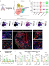
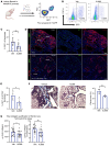
Figure 1. p16Ink4a+ fibroblasts contribute to pathologic fibroblasts in mouse model of lung fibrosis.
(A) Experimental scheme for scRNA-Seq of p16Ink4a+ (GFP+) fibroblasts from the INKBRITE lung after bleomycin-induced fibrosis (14 dpi). (B) Violin plot showing profibrotic gene expressions in the different p16Ink4a+ fibroblast subsets in vivo. (C) Visualization of Acta2, Cthrc1, Col1a1, and Tagln expression pattern within the fibroblast subsets. (D) Representative images showing Cthrc1 (RNAscope in situ), ACTA2, TAGLN, and COL1A1 (immunostaining) in lung sections of bleomycin-injured INKBRITE mice (14 dpi) colocalized with GFP (arrows, p16Ink4a+ fibroblasts). Scale bars: 100 μm. (E) qPCR analysis of purified GFP+ and GFP fibroblasts from bleomycin-treated INKBRITE lungs (n = 5–6 biological replicates, experiment repeated twice). (F) qPCR analysis of cultured GFP+ and GFP fibroblasts isolated from fibrotic INKBRITE lungs after treatment of recombinant TGF-β1 or vehicle (n = 3 technical replicates, experiment repeated twice). Data are represented as mean ± SD. *P < 0.05; **P < 0.01; ***P < 0.001; 2-tailed Student’s t test (E) or 1-way ANOVA (F).
Figure 2
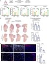
Figure 2. p16INK4a expression primes lung fibroblasts to augment the fibrotic response.
(A) Transcript analysis of cultured primary human lung fibroblasts isolated from control cadaveric donors transduced with 2 lentiviral vectors to overexpress (OE) human p16INK4a with doxycycline induction followed by addition of TGF-β1. (n = 3 technical replicates, experiment repeated twice). (B) Representative H&E sections of Dermo1Cre/+;p16fl/fl and control (p16fl/fl) animals injured with bleomycin to induce lung fibrosis. (C) Hydroxyproline assay to quantify collagen in the left lung of Dermo1Cre/+;p16fl/fl and control animals 14 days following bleomycin injury (n = 5 control, 9 mutant biological replicates). (D) Representative IHC showing ACTA2 and COL1 immunostaining in lung sections of bleomycin-injured Dermo1Cre/+;p16fl/fl and control (p16fl/fl) animals (14 dpi). (E) IHC quantification of ACTA2+ and COL1+ fibroblasts from D (n = 5 control, 9 mutant biological replicates. Scale bars: 200 μm. Data are represented as mean ± SD. *P < 0.05; **P < 0.01; ***P < 0.001; ****P < 0.0001; 2-tailed Student’s t test (A, C, and E).
Figure 3
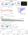
Figure 3. HTS targeting p16Ink4a+ fibroblasts isolated from fibrotic INKBRITE lungs.
(A) Schematic outline of the HTS to identify compounds targeting p16Ink4a+ (GFP+) fibroblasts from the fibrotic INKBRITE lungs. (B) Scatter plot showing hit results from each well containing compound (pink) or vehicle (green). Y-axis indicates %GFP+ cells in each well after compound exposure. Compounds exceeding 3 σ for lowest %GFP were selected for validation. (C) Cell count GFP+ and GFP fibroblasts of the top senolytic candidates. (D) Biologic pathways targeted by the top senolytic candidates. (E) Schematic outline of dose-response analysis of the top senolytic candidate from the primary screen. (F) Top candidates emerging from the secondary validation using dose-response with lowest IC50 values, including trichostatin A, XL888, and ganetespib.
Figure 4
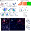
Figure 4. Validation of candidate senolytic compounds using mouse PCLS derived from fibrotic INKBRITE lungs.
(A) Experimental scheme for ex vivo culture of mouse PCLS derived from fibrotic INKBRITE mouse to test senolytic candidates. (B) Bright field and GFP images of cultured PCLS. Scale bars: 2,000 μm. (C) Gating strategy to analyze GFP+ fibroblasts from mouse PCLS by flow cytometry. (D) Quantification of GFP+ fibroblasts in the PCLS cultured with vehicle or XL888 (1 μM) for 5 days (n = 10 technical replicates, experiment repeated twice). (E and F) Immunofluorescence analysis (E) and quantification (F) of ACTA2, COL1A1, and GFP in mouse PCLS treated with vehicle or XL888 (1 μM). (n = 9 technical replicates, experiment repeated twice). Scale bars: 50 μm. Data are represented as mean ± SD. *P < 0.05; **P < 0.01; ***P < 0.001; 2-tailed Student’s t test (D, F).
Figure 5
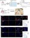
Figure 5. XL888 deletes p16Ink4a+ fibroblasts and attenuates fibrotic remodeling in vivo.
(A) Schematic outline of animal experiments to validate in vivo efficacy of candidate senolytics. (B and C) Flow cytometry analysis of GFP+ fibroblasts (% of fibroblasts that are GFP+) in bleomycin-injured lungs of vehicle or XL888 delivered INKBRITE animals (n = 11–12 biological replicates, experiment repeated twice). (D and E) Immunofluorescence analysis (D) and quantification (E) of GFP+ cells among ACTA2+ fibroblasts in the lungs of vehicle or XL888-treated INKBRITE mice (n = 4 biological replicates, experiment repeated twice). Scale bars: 100 μm. (F) Representative images (left) and quantification of Masson’s trichrome staining of lung sections from indicated group of mice after bleomycin injury (n = 4 biological replicates). Scale bars: 1,000 μm. (G) Quantitative analysis of collagen in lung homogenates from vehicle or XL888 treated animals injured with bleomycin (n = 19–20 biological replicates, experiment repeated twice). Data are represented as mean ± SD. *P < 0.05; **P < 0.01; 2-tailed Student’s t test (C); or 1-tailed Student’s t test (EG).
Figure 6
Figure 6. Human p16INK4a+ fibroblasts contribute to pathologic fibroblasts in IPF.
(A) UMAP plot of fibroblast subsets seen in normal human and IPF lungs. (B) Violin plot showing CDKN2A expression in the pathologic fibroblast cluster of IPF fibroblasts. (C) Violin plots showing the profibrotic genes in the different fibroblast subsets. (D) Visualization of CDKN2A, CTHRC1, COL1A1, and ACTA2 expression patterns in human lung fibroblasts in IPF and control donor lungs. (E) Representative images showing ACTA2+p16INK4a+ pathologic fibroblasts (arrows) in lung sections of controls and individuals with IPF. Scale bars: 100 μm. (F) qPCR analysis of genes enriched in pathologic fibroblasts in p16INK4a-hi and p16INK4a-lo fibroblasts isolated from lungs of patients with IPF (n = 9 technical replicates, experiments repeated with separate IPF donor fibroblasts at least 3 times). Data are represented as mean ± SD. *P < 0.05; **P < 0.01; ***P < 0.001; 2-tailed Student’s t test (F).
Figure 7
Figure 7. XL888 targets human p16INK4a+ fibroblasts from IPF lungs in vitro and ex vivo.
(A) (Left) Schematic outline of dose-escalation challenge of candidate senolytics on p16INK4a-hi and p16INK4a-lo fibroblasts isolated from IPF lungs (Right) Ratio of p16INK4a-hi and p16INK4a-lo fibroblast cell count after treatment of senolytics and XL888 with dose escalation (n = 3 technical replicates, experiments repeated with separate IPF donor fibroblasts at least 3 times). (B) Schematic diagram depicting ex vivo culture of IPF lung with XL888 treatment and bright field images of cultured human PCLS. Scale bars: 2,000 μm. (C) Immunofluorescence analysis and quantification of ACTA2+ p16INK4a+ cells in vehicle or XL888-treated hPCLS (n = 12 slices per condition, sampled from 4 IPF donors independently, each color represents a different donor). Scale bars: 100 μm. (D) Immunofluorescence analysis and quantification of CTHRC1+ p16INK4a+ cells in vehicle or XL888-treated hPCLS (n = 12 slices per condition, sampled from 4 IPF donors independently, each color represents a different donor). Scale bars: 100 μm. Data are represented as mean ± SD. *P < 0.05; **P < 0.01; ***P < 0.001; 2-tailed Student’s t test (A, C, and D).

Comment in

  • A pipeline for senolytics

References

    1. Hayflick L, Moorhead PS. The serial cultivation of human diploid cell strains. Exp Cell Res. 1961;25:585–621. doi: 10.1016/0014-4827(61)90192-6. - DOI - PubMed
    1. Gorgoulis V, et al. Cellular senescence: defining a path forward. Cell. 2019;179(4):813–827. doi: 10.1016/j.cell.2019.10.005. - DOI - PubMed
    1. Burd CE, et al. Monitoring tumorigenesis and senescence in vivo with a p16(INK4a)-luciferase model. Cell. 2013;152(1–2):340–351. doi: 10.1016/j.cell.2012.12.010. - DOI - PMC - PubMed
    1. Baker DJ, et al. Clearance of p16Ink4a-positive senescent cells delays ageing-associated disorders. Nature. 2011;479(7372):232–236. doi: 10.1038/nature10600. - DOI - PMC - PubMed
    1. Demaria M, et al. An essential role for senescent cells in optimal wound healing through secretion of PDGF-AA. Dev Cell. 2014;31(6):722–733. doi: 10.1016/j.devcel.2014.11.012. - DOI - PMC - PubMed

Publication types

MeSH terms