. 2024 Mar 7;134(10):e172716.
doi: 10.1172/JCI172716.

DNA topoisomerase II inhibition potentiates osimertinib's therapeutic efficacy in EGFR-mutant non-small cell lung cancer models

Affiliations

DNA topoisomerase II inhibition potentiates osimertinib's therapeutic efficacy in EGFR-mutant non-small cell lung cancer models

Zhen Chen et al. J Clin Invest. .

Abstract

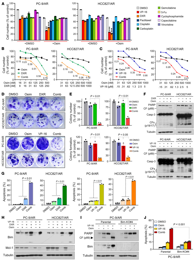
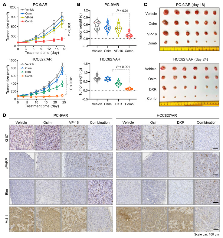
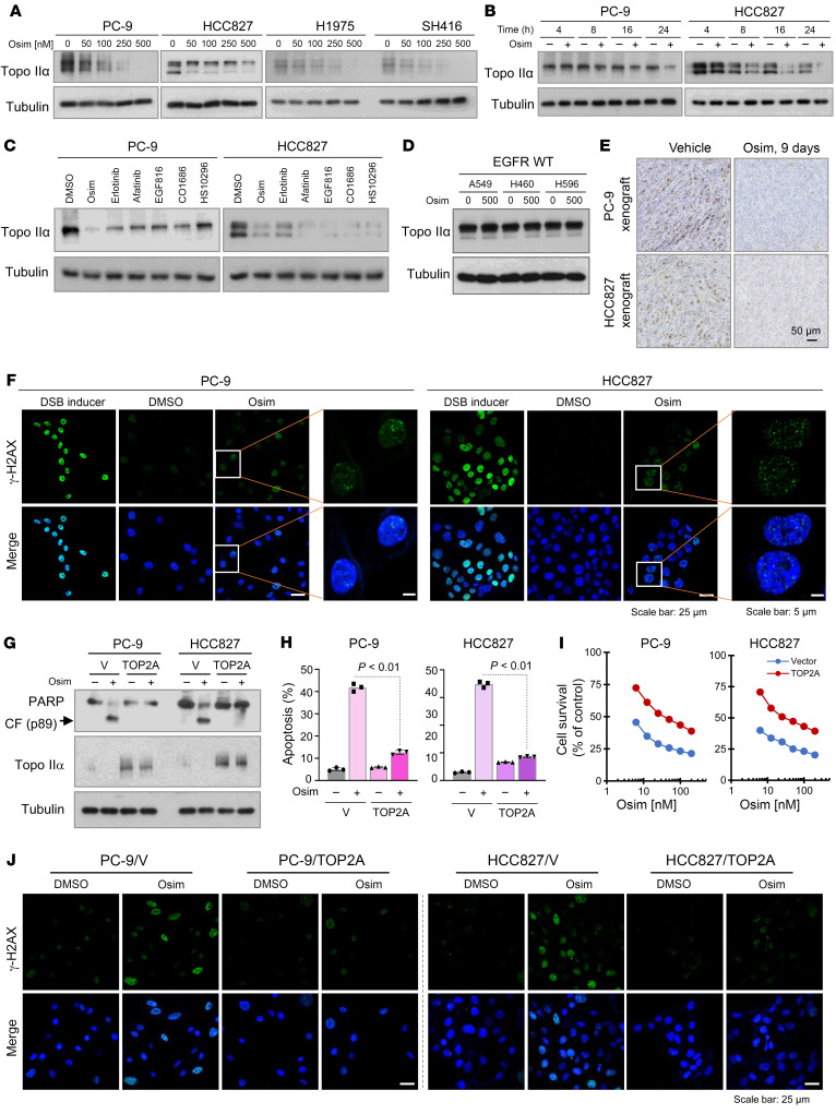
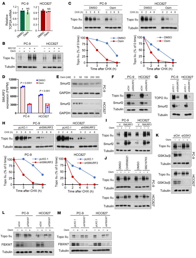
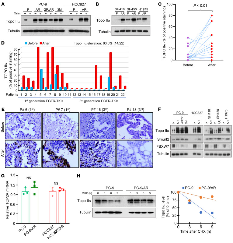
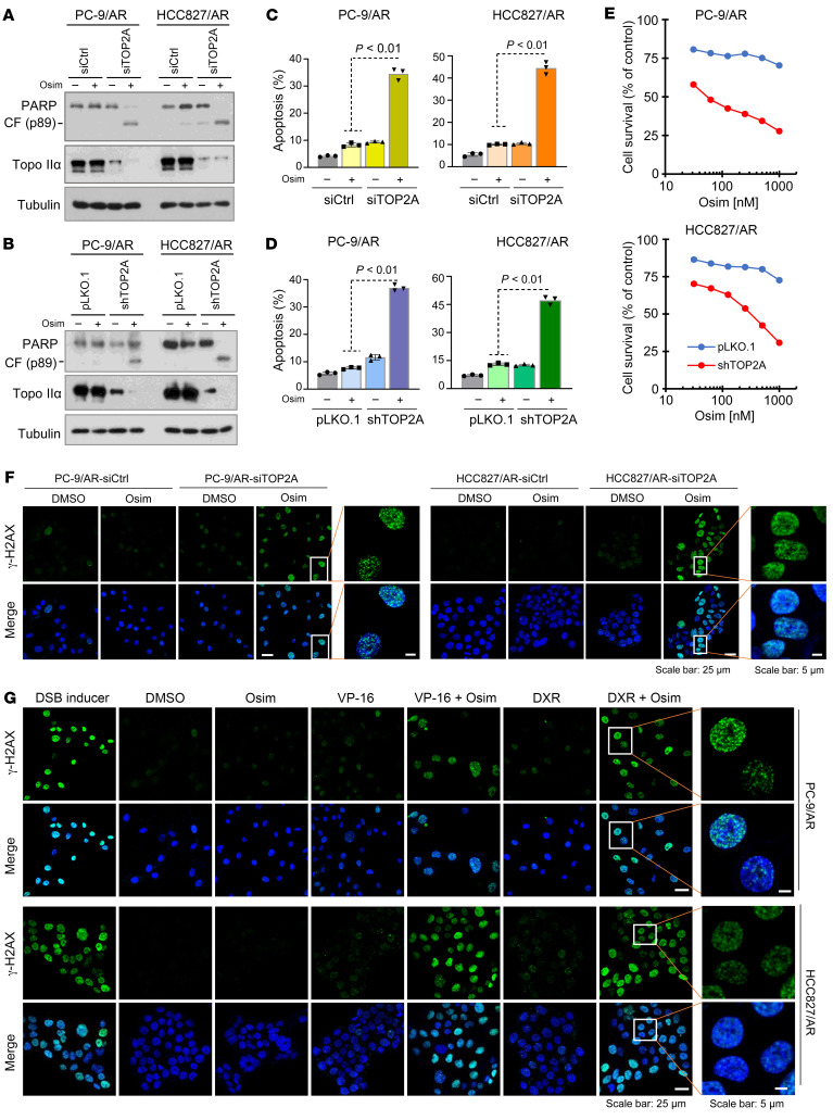
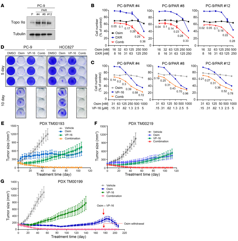
Development of effective strategies to manage the inevitable acquired resistance to osimertinib, a third-generation EGFR inhibitor for the treatment of EGFR-mutant (EGFRm) non-small cell lung cancer (NSCLC), is urgently needed. This study reports that DNA topoisomerase II (Topo II) inhibitors, doxorubicin and etoposide, synergistically decreased cell survival, with enhanced induction of DNA damage and apoptosis in osimertinib-resistant cells; suppressed the growth of osimertinib-resistant tumors; and delayed the emergence of osimertinib-acquired resistance. Mechanistically, osimertinib decreased Topo IIα levels in EGFRm NSCLC cells by facilitating FBXW7-mediated proteasomal degradation, resulting in induction of DNA damage; these effects were lost in osimertinib-resistant cell lines that possess elevated levels of Topo IIα. Increased Topo IIα levels were also detected in the majority of tissue samples from patients with NSCLC after relapse from EGFR tyrosine kinase inhibitor treatment. Enforced expression of an ectopic TOP2A gene in sensitive EGFRm NSCLC cells conferred resistance to osimertinib, whereas knockdown of TOP2A in osimertinib-resistant cell lines restored their susceptibility to osimertinib-induced DNA damage and apoptosis. Together, these results reveal an essential role of Topo IIα inhibition in mediating the therapeutic efficacy of osimertinib against EGFRm NSCLC, providing scientific rationale for targeting Topo II to manage acquired resistance to osimertinib.

Keywords: Apoptosis; Lung cancer; Oncology; Telomeres; Therapeutics.


Conflict of interest statement

Conflict of interest: TL is a consultant or on advisory boards for Eisai, Merck, Roche/Genentech, Jazz Pharmaceuticals, Catalyst, OncoC4, Amgen, AstraZeneca, Pfizer, Regeneron, Janssen, Novartis, Novocure, and Sanofi. SSR is a consultant or on advisory boards for AstraZeneca, BMS, Merck, Roche, Tesaro, and Amgen.

Figures

Figure 1
Figure 1. Topo II inhibitors in combination with osimertinib synergistically decrease cell survival, inhibit colony formation and growth, and induce Bim-dependent apoptosis in osimertinib-resistant EGFRm NSCLC cell lines.
(AC) The given cell lines were treated with 250 nM osimertinib (Osim), 1.25 μM VP-16, 125 nM DXR, 5 nM paclitaxel, 10 μM cisplatin, 25 μM carboplatin, 25 nM gemcitabine, 20 nM 5-FU, 25 μM cyclophosphamide, 25 μM capecitabine, or 10 nM vincristine alone or in combination (A) or with varied concentrations of the tested agents either alone or in combination (B and C) for 3 days. Cell numbers were then measured using the SRB assay. Data represent mean ± SD of 4 replicate determinations. **P < 0.01; ***P < 0.001 compared with each agent alone. The fixed suboptimal concentrations of the tested agents used in A were chosen based on their concentration-dependent survival curves. (D and E) The tested cell lines seeded in 12-well plates were treated with 50 nM osimertinib, 10 nM (PC-9/AR) or 50 nM (HCC827/AR) DXR, 150 nM VP-16, or the indicated combinations, which were repeated with fresh medium every 3 days. After 10 days, the cells were fixed, stained with crystal violet dye, imaged (D) and counted (E). Columns are mean ± SD of triplicate determinations. (FJ) The tested cell lines were exposed to 200 nM osimertinib, 100 nM (PC-9/AR) or 250 nM (HCC827/AR) DXR, 1 μM VP-16, or the indicated combinations for 48 hours (F, G, I, and J) or 16 hours (H). The proteins of interest were detected with Western blotting (F, H, and I), and apoptotic cells were detected with annexin V staining/flow cytometry (E and J). Each column represents mean ± SD of triplicate treatments. Statistical differences were conducted with 2-sided unpaired Student’s t test for 2 groups (J) or 1-way ANOVA test (A, E, and G) for multiple groups.
Figure 2
Figure 2. The combination of osimertinib with DXR or VP-16 effectively inhibits the growth of osimertinib-resistant EGFRm NSCLC xenografts in vivo, with modulation of several critical protein biomarkers in tumor tissues.
PC-9/AR or HCC27/AR cells grown in nu/nu mice as xenograft tumors (n = 6/group) were treated with vehicle, osimertinib (Osim) alone (5 mg/kg, daily, oral gavage), DXR alone (1 mg/kg/d, daily, i.p.), VP-16 alone (1 mg/kg/d, daily, i.p.), or the indicated combinations. Tumor sizes were measured at the indicated time points (A). At the end of treatment, tumors in each group were also weighed (B) and photographed (C). The data in each group represent mean ± SEM of 6 tumors from 6 mice. The proteins of interest as indicated were stained with IHC. (D) Statistical differences among multiple groups were conducted with 1-way ANOVA test. Scale bar: 100 μm.
Figure 3
Figure 3. Osimertinib, as well as other EGFR-TKIs, decreases the levels of Topo IIα and induces γ-H2AX foci formation in EGFRm NSCLC cells and tissues, and overexpression of ectopic TOP2A attenuates the effects of osimertinib on induction of apoptosis, decreasing cell survival, and increasing γ-H2AX foci formation.
(AD) The given cell lines were exposed to varied concentrations of osimertinib (Osim) for 24 hours (A), 200 nM osimertinib for different times (B), 200 nM different EGFR-TKIs for 24 hours (C), or 500 nM osimertinib for 24 hours (D). Proteins of interest were detected with Western blotting. (E) Topo IIα in tissues was detected with IHC. Scale bar: 50 μm. (F and J) The indicated cell lines were exposed to 250 nM osimertinib for 16 hours and then stained with anti–γ-H2AX antibody and DAPI. DSB inducer here served as a positive control and was used at 100 μM for 1 hour of treatment. Scale bar: 25 μm (F and J); 5 μm (F, high-magnification images). (GI) The indicated cell lines were exposed to DMSO or 200 nM osimertinib for 18 hours (G) or 24 hours (H) or treated with different concentrations of osimertinib for 3 days (I). The proteins of interest were detected with Western blotting (G), and apoptotic cells were detected with annexin V staining/flow cytometry (H). Each bar in H represents mean ± SD of triplicate treatments. Cell numbers were measured by the SRB assay and are expressed as mean ± SD of 4 replicate determinations (I). Statistical differences between 2 groups were conducted with 2-sided unpaired Student’s t test.
Figure 4
Figure 4. Osimertinib does not affect TOP2A transcription but promotes GSK3-dependent and FBXW7-mediated Topo IIα protein degradation associated with suppression of SMURF2 expression.
(A) The tested cell lines were exposed to 200 nM osimertinib (Osim) for 16 hours. TOP2A mRNA was detected with quantitative reverse transcription PCR. NS, not significant with 2-sided unpaired Student’s t test. (B) The tested cell lines were pretreated with 10 μM MG132 for 30 minutes and then cotreated with DMSO or 200 nM osimertinib for another 6 hours. (C) Both PC-9 and HCC827 cells were treated with 200 nM osimertinib for 16 hours followed by the addition of 10 μg/mL CHX and then harvested at the indicated times. (D) RNA-Seq detection of SMURF2 mRNA expression in the given cell lines exposed to 100 nM osimertinib for 14 hours. (E) The tested cell lines were exposed to varied concentrations of osimertinib as indicated for 24 hours. (F and G) The tested cell lines were transfected with the given siRNAs or infected with lentiviruses carrying the given shRNA for 48 hours. (H) The tested cell lines were exposed to 10 μg/mL CHX and then harvested at different times as indicated. (I) The tested cell lines were treated with 200 nM osimertinib for 24 hours. (J) Both PC-9 and HCC827 were pretreated with 10 μM CHIR99021 or SB216763 for 30 minutes and then cotreated with 200 nM osimertinib for an additional 16 hours. (K and L) The tested cell lines were transfected with scrambled GSK3 (K) or FBXW7 (L) siRNA for 48 hours followed by treatment with 200 nM osimertinib for another 24 hours. (M) The indicated cell lines expressing pLKO.1 or shFBXW7 were exposed to 200 nM osimertinib for 24 hours. The proteins with the aforementioned treatments were detected with Western blotting. Band intensities were quantified with ImageJ (NIH) software and plotted as percentage of 0 time (C and H).
Figure 5
Figure 5. Topo IIα levels are elevated in EGFRm NSCLC cell lines with acquired resistance to osimertinib and tissue samples from patients with EGFRm NSCLC relapsed from EGFR-TKI treatment, which are resistant to osimertinib modulation and have elevated Smurf2 and decreased FBXW7, unchanged mRNA expression, and increased stability of Topo IIα.
(A, B, and F) Whole-cell protein lysates were prepared from the given osimertinib-resistant cell lines exposed to 1,000 nM osimertinib (Osim) for 24 hours (A) or from untreated given cell lines with similar densities (B and F). The indicated proteins were detected with Western blotting. (CE) Topo IIα in human EGFRm NSCLC tissues before and after relapse from treatment using EGFR-TKIs, including osimertinib, was stained with IHC. Statistical analysis was conducted with 2-sided paired Student’s t test. Original magnification, ×20. (G) TOP2A mRNA expression in the indicated cell lines were detected with quantitative reverse transcription PCR. NS, not significant with 2-sided unpaired Student’s t test. (H) The tested cell lines were exposed to 10 μg/mL CHX and then harvested at different times as indicated for subsequent Western blotting. Band intensities were quantified with ImageJ (NIH) software and plotted as percentage of 0 time.
Figure 6
Figure 6. Genetic knockdown of TOP2A expression in osimertinib-resistant cells restores their response to osimertinib in inducing apoptosis, decreasing cell survival, and increasing DNA damage, similar to the effect of combined osimertinib and Topo II inhibitor on enhancing induction of DNA damage in these resistant cell lines.
(AE) PC-9/AR and HCC827/AR cells transfected with scrambled control or TOP2A siRNA for 48 hours (A and C) or expressing pLKO.1 or shTOP2A (B, D, and E) were exposed to 200 nM osimertinib for 24 hours (A and B), 48 hours (C and D), or 72 hours (E). Topo IIα and PARP cleavage were detected with Western blotting (A and B). Annexin V–positive cells were determined with flow cytometry (C and D). Cell numbers were estimated with the SRB assay (E). The data represent mean ± SD of triplicate (C and D) or 4 replicate (E) determinations. Statistical analysis was conducted with 2-sided unpaired Student’s t test. CF, cleaved form. (F) The indicated cell lines were transfected with scrambled control or TOP2A siRNA for 48 hours and then exposed to 250 nM osimertinib for an additional 24 hours. The cells were then subjected to detection of γ-H2AX foci using IF staining with anti–γ-H2AX antibody. (G) The indicated cell lines were treated with DMSO, 250 nM osimertinib, 100 nM (PC-9/AR) or 250 nM (HCC827/AR) DXR, 1 μM VP-16, or their respective combinations as indicated for 24 hours and then subjected to detection of γ-H2AX foci using IF staining with anti–γ-H2AX antibody. DSB (100 μM for 1 hour) here was used as a positive control. Scale bar: 25 μm (F and G); 5 μm (F and G, high-magnification images).
Figure 7
Figure 7. Osimertinib combined with a Topo II inhibitor synergistically decreases the survival of EGFRm NSCLC cell lines with primary resistance to osimertinib expressing elevated levels of Topo IIα, eliminates DTCs, and regresses different EGFRm PDX tumors in vivo with long-term remissions.
(A) Detection of basal levels of Topo IIα in the indicated cell lines with Western blotting. (B and C) The given cell lines were exposed to varied concentrations of osimertinib (Osim), DXR, or VP-16 alone as indicated and the combination of osimertinib with DXR or VP-16. After 3 days, cell numbers were determined with the SRB assay. The data represent mean ± SD of 4 replicate determinations. (D) The indicated cell lines seeded in 12-well plates were treated with 50 nM osimertinib, 150 nM VP-16, or a combination; these treatments were repeated with fresh medium every 2 days. After 5 or 10 days, the cells were fixed, stained with crystal violet dye, and images were taken. (EG) The indicated PDXs in nude mice (6 tumors/group) were treated with vehicle, 5 mg/kg osimertinib (daily, oral gavage), 1 mg/kg VP-16 (daily, i.p.), or the combination of osimertinib and VP-16. The data represent mean ± SEM of 6 tumors.

References

    1. Siegel RL, et al. Cancer statistics, 2022. CA Cancer J Clin. 2022;72(1):7–33. doi: 10.3322/caac.21708. - DOI - PubMed
    1. Ramalingam SS, et al. Overall survival with osimertinib in untreated, EGFR-mutated advanced NSCLC. N Engl J Med. 2020;382(1):41–50. doi: 10.1056/NEJMoa1913662. - DOI - PubMed
    1. Piper-Vallillo AJ, Sequist LV. Emerging treatment paradigms for EGFR-mutant lung cancers progressing on osimertinib: a review. J Clin Oncol. 2020;38(25):JCO1903123. doi: 10.1200/JCO.19.03123. - DOI - PubMed
    1. Blaquier JB, Ortiz-Cuaran S. Tackling osimertinib resistance in EGFR-mutant non-small cell lung cancer. Clin Cancer Res. 2023;29(18):3579–3591. doi: 10.1158/1078-0432.CCR-22-1912. - DOI - PubMed
    1. Gomatou G, et al. Osimertinib resistance: molecular mechanisms and emerging treatment options. Cancers (Basel) 2023;15(3):841. doi: 10.3390/cancers15030841. - DOI - PMC - PubMed

MeSH terms