. 2024 Mar 26;43(3):113875.
doi: 10.1016/j.celrep.2024.113875. Epub 2024 Mar 6.

Hepatic Snai1 and Snai2 promote liver regeneration and suppress liver fibrosis in mice

Affiliations

Hepatic Snai1 and Snai2 promote liver regeneration and suppress liver fibrosis in mice

Pingping Wang et al. Cell Rep. .

Abstract

Liver injury stimulates hepatocyte replication and hepatic stellate cell (HSC) activation, thereby driving liver regeneration. Aberrant HSC activation induces liver fibrosis. However, mechanisms underlying liver regeneration and fibrosis remain poorly understood. Here, we identify hepatic Snai1 and Snai2 as important transcriptional regulators for liver regeneration and fibrosis. Partial hepatectomy or CCl4 treatment increases occupancies of Snai1 and Snai2 on cyclin A2 and D1 promoters in the liver. Snai1 and Snai2 in turn increase promoter H3K27 acetylation and cyclin A2/D1 expressions. Hepatocyte-specific deletion of both Snai1 and Snai2, but not one alone, suppresses liver cyclin A2/D1 expression and regenerative hepatocyte proliferation after hepatectomy or CCl4 treatments but augments CCl4-stimulated HSC activation and liver fibrosis. Conversely, Snai2 overexpression in the liver enhances hepatocyte replication and suppresses liver fibrosis after CCl4 treatment. These results suggest that hepatic Snai1 and Snai2 directly promote, via histone modifications, reparative hepatocyte replication and indirectly inhibit liver fibrosis.

Keywords: CCl4; CP: Cell biology; CP: Developmental biology; Snai1; Snai2; hepatectomy; hepatocyte replication; histone acetylation; histone modifications; liver fibrosis; liver regeneration.


Conflict of interest statement

Declaration of interests The authors declare no competing interests.

Figures
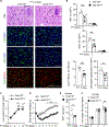
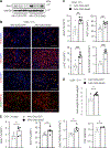
Figure 1.
Figure 1.. Hepatocyte-specific ablation of both Snai1 and Snai2 impairs PH-stimulated liver regeneration
PH was performed on Snai1/2f/f and Snai1/2Δhep male mice at 8 weeks of age. BrdU was injected 10 h before euthanization. (A and B) Liver sections were stained with antibodies to Ki67, BrdU, or HNF4α (2 days after PH). Positive cells were normalized to total (DAPI+) or hepatocytes (HNF4α+). Arrows: binucleate hepatocytes. Scale bar: 200 mm. Day 2: Snai1/2f/f: n = 8, Snai1/2Δhep: n = 6; day 7: n = 8 per group. (C) Liver weight post-PH (normalized to body weight). Day 0: Snai1/2f/f:n = 6, Snai1/2Δhep: n = 7; day 2: Snai1/2f/f:n = 8, Snai1/2Δhep: n = 6; days 7 and 14: n = 8 per group. (D) Body weight changes after PH. Snai1/2f/f: n = 8, Snai1/2Δhep: n = 8. (E and F) PH was performed on Snai1/2f/f and Snai1/2Δhep female mice at 8 weeks of age. (E) Liver sections were stained with anti-Ki67 antibody (2 days after PH). Ki67+ cell number was normalized to total cells (DAPI+). Snai1/2f/f:n = 5, Snai1/2Δhep: n = 5. (F) Liver weight after PH. Day 0: Snai1/2f/f: n = 6, Snai1/2Δhep: n = 5; day 2: n = 5 per group; day 7: Snai1/2f/f: n = 8, Snai1/2Δhep: n = 10. Data are presented as mean ± SEM. *p < 0.05, **p < 0.01, and ***p < 0.001, two-tailed unpaired Student’s t test (B and E) or two-way ANOVA (C, D, and F).
Figure 2.
Figure 2.. Ablation of hepatic Snai1 and Snai2 attenuates hepatocyte proliferation in mice with CCl4-induced acute liver injury
Snai1/2f/f and Snai1/2Δhep male mice (8 weeks) were injected with CCl4 (1 μL/g body weight). BrdU was administrated 10 h before euthanization. Liver sections were stained with the indicated reagents. (A) Representative liver sections 72 h after CCl4 treatment. Scale bar: 200 μm. (B) Ki67+, BrdU+, and pH3+ cells were normalized to total liver cells (DAPI+). Proliferating hepatocytes (BrdU+ HNF4α+) were normalized to total hepatocytes (HNF4α+). Binucleate hepatocyte number was normalized to total liver cells (H&E staining). CCl4 for 24 h: n = 4 per group; 48 h: Snai1/2f/f: n = 6, Snai1/2Δhep: n = 8; 72 h: n = 8 per group; 96 h: n = 6 per group. Data are presented as mean ± SEM. *p < 0.05, **p < 0.01, and ***p < 0.001, two-tailed unpaired Student’s t test.
Figure 3.
Figure 3.. Hepatocyte-specific ablation of Snai1 and Snai2 suppresses liver regeneration in mice with CCl4-induced chronic liver injury
(A and B) C57BL/6J male mice (8 weeks) were treated with CCl4 (0.6 μL/g body weight, twice a week) or vehicle for 24 days. Liver nuclear extracts were immunoblotted with the indicated antibodies. Snai1 and Snai2 levels were normalized to Lamin A/C levels. a.u., arbitrary units. Basal: n = 6; CCl4: n = 7. (C–F) Snai1/2f/f and Snai1/2Δhep male mice (8 weeks) were treated with CCl4 for 24 days. (C and D) Liver sections were stained with the indicated antibodies (CCl4 for 24 days). Positive cells were counted. Binucleate hepatocyte number was normalized to total liver cells (H&E staining). Scale bar: 200 mμm. Snai1/2f/f: n = 9, Snai1/2Δhep: n = 7. (E) Representative livers and liver weight (CCl4 for 24 days). Snai1/2f/f: n = 9, Snai1/2Δhep: n = 7. (F) Body weight after CCl4 treatment. Snai1/2f/f: n = 9, Snai1/2Δhep: n = 7. Data are presented as mean ± SEM. *p < 0.05, **p < 0.01, and ***p < 0.001, two-tailed unpaired Student’s t test (B, D, and E) or two-way ANOVA (F).
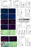
Figure 4.
Figure 4.. Snai2 overexpression increases liver growth
C57BL/6J male mice (8 weeks old) were transduced with AAV8-CAG-Snai2 or AAV8-CAG-GFP vectors for 2 weeks and then treated with a single dose of CCl4 for 72 h (1 μL/g body weight) (A–D) or with CCl4 for 24 days (0.6 μL/g body weight, twice a week) (E). BrdU was injected 10 h before euthanization (A–D). (A) Liver extracts were immunoblotted with antibodies to Snai2 and GAPDH. (B and C) Liver sections were stained with the indicated antibodies. Ki67+, BrdU+, and pH3+ cells were normalized to total liver cells (DAPI+). Proliferating hepatocytes (BrdU+ HNF4α+) were normalized to total hepatocytes (HNF4α+). AAV-CAG-GFP: n = 6, AAV-CAG-Snai2: n = 7. (D) Liver weight 72 h post-CCl4 treatment. AAV-CAG-GFP: n = 6, AAV-CAG-Snai2: n = 7. (E) Liver cell proliferation and liver weight after CCl4 treatment for 24 days. AAV-CAG-GFP: n = 6, AAV-CAG-Snai2: n = 5. Data are presented as mean ± SEM. *p < 0.05, **p < 0.01, and ***p < 0.001, two-tailed unpaired Student’s t test.
Figure 5.
Figure 5.. Snai1 and Snai2 directly stimulate expression of cyclins A2 and D1 in hepatocytes
(A–C) PH was performed on littermates, and the liver was harvested 2–14 days later. Liver cyclins A2 and D1 mRNA levels were measured by qPCR (normalized to 36B4 expression). Liver extracts were prepared 2 days after PH and immunoblotted with antibodies to cyclin A2, cyclin D1, cyclin E1, and GAPDH. Cyclin levels were normalized to GAPDH levels. (A) Day 0: Snai1/2f/f: n = 6, Snai1/2Δhep: n = 7; day 2: Snai1/2f/f: n = 8, Snai1/2Δhep: n = 6; days 7 and 14: n = 8 per group. (B) n = 4 mice per group. (C) mRNA levels: n = 5 mice per group; protein levels: n = 4 mice per group. (D) Snai1/2f/f and Snai1/2Δhep males (8 weeks) were treated with CCl4 for 48 h (1 μL/g body weight). Liver extracts were immunoblotted with antibodies to cyclin A2, cyclin D1, cyclin E1, and GAPDH. Cyclin levels were normalized to GAPDH levels (n = 4 mice per group). (E) Snai1/2f/f and Snai1/2Δhep males (8 weeks) were treated with CCl4 for 24 days (0.6 μL/g body weight, twice a week). Liver cyclin A2, cyclin D1, and cyclin E1 mRNA (normalized to 36B4, Snai1/2f/f n = 9, Snai1/2Δhep: n = 7) and protein (normalized to GAPDH, Snai1/2f/f: n = 7, Snai1/2Δhep: n = 5) levels were measured by qPCR and immunoblotting, respectively. (F and G) Primary hepatocytes were transduced with the indicated adenoviral vectors for 48 h. (F) Cyclin A2, cyclin D1, and cyclin E1 mRNA levels (normalized to 36B4 levels, n = 3 repeats per group). (G) Cell extracts were immunoblotted with the indicated antibodies. (H) C57BL/6J males (8 weeks) were transduced with AAV-CAG-Snai2 or AAV-CAG-GFP vectors for 2 weeks and then treated with CCl4 for 72 h (1 μL/g body weight). Liver cyclin A2, cyclin D1, and cyclin E1 mRNA (normalized to 36B4, n = 6 mice per group) and protein (normalized to GAPDH, n = 3 mice per group) levels were measured by qPCR and immunoblotting, respectively. Data are presented as mean ± SEM. *p < 0.05, **p < 0.01, and ***p < 0.001, two-tailed unpaired Student’s t test.
Figure 6.
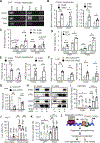
Figure 6.. Snai1 and Snai2 activate Ccna2 and Ccnd1 promoters via histone modifications
(A and B) Primary hepatocytes were transduced with Snai1 (n = 4), Snai2 (n = 4), or β-galactosidase (β-gal) (n = 3) adenoviral vectors for 2 days. (A) ChIP-qPCR images. (B) Occupancies of Snai1 and Snai2 on the Ccna2 and Ccnd1 promoters. (C and D) Wild-type male mice were treated with PH or CCl4 for 48 h. Occupancies of Snai1 and Snai2 on the Ccna2 and Ccnd1 promoters in the liver were measured using ChIP-qPCR. PH: n = 6 per group, control (Con): n = 6, CCl4: n = 5. (E) H3K27ac levels in Ccna2 and Ccnd1 promoters (n = 4 per group). (F) C57BL/6J males (8 weeks) were transduced with AAV8-CAG-Snai2 (n = 6) or AAV8-CAG-GFP (n = 6) vectors for 3 weeks. Snai2 occupancies and H3K27ac levels on Ccna2 and Ccnd1 promoters were measured in the liver by ChIP-qPCR. (G) Livers were harvested from Snai1/2f/f (n = 4) and Snai1/2Δhep (n = 4) males (8 weeks old) and subjected to ChIP to measure H3K27ac levels in Ccna2 and Ccnd1 promoters. (H) Snai2 was coexpressed with p300 in HEK293T cells, immunoprecipitated with anti-Snai2 (or anti-300) antibody, and immunoblotted with anti-p300 (or anti-Snai2) antibody. (I) CBP, p300, or β-gal plasmids were cotransfected with or without Snai2 plasmids in HEK293T cells (with pGL3-Ccna2 cotransfection). CCNA2 promoter H3K27ac levels were measured by ChIP-qPCR (n = 4 per group). (J) Ccna2 luciferase reporter plasmids (pGL3-Ccna2) or pGL3-basic plasmids (Con) were cotransfected with GFP, Snai1, Snai2, or ΔN30 into Huh 7 cells. Luciferase activity was measured 48 h later (n = 3 repeats per group). (K) Ccna2 luciferase reporter plasmids were cotransfected into Huh 7 cells with GFP, CBP, or p300 plasmids in the presence of GFP or Snai2 plasmids. Luciferase activity was measured 48 h later (n = 3 per group). (L) Snai1 and Snai2 recruit CBP and p300 that catalyze H3K27ac (active epigenetic mark) in cyclin A2 and D1 promoters, thereby enhancing hepatocyte proliferation and liver regeneration. Snai1/2-induced hepatocyte renewal suppresses liver fibrosis. Data are presented as mean ± SEM. *p < 0.05, **p < 0.01, and ***p < 0.001, two-tailed unpaired Student’s t test (B–G) and one-way ANOVA (I–K).
Figure 7.
Figure 7.. Hepatocyte-specific ablation of Snai1 and Snai2 enhances CCl4-induced liver fibrosis
(A–F) Snai1/2f/f and Snai1/2Δhep male mice (8 weeks) were treated with CCl4 (0.6 μL/g body weight, twice a week) for 24 days. (A) Plasma ALT levels. Snai1/2f/f: n = 9, Snai1/2Δhep: n = 7. (B and C) Liver sections were stained with the indicated reagents, and positive cells were counted (CCl4 for 24 days). Scale bar: 200 μm. Snai1/2f/f:n = 9, Snai1/2Δhep: n = 7. (D) Liver extracts were immunoblotted with the indicated antibodies. (E) Liver mRNA abundance (normalized to 36B4 expression (CCl4 for 24 days). Snai1/2f/f: n = 9, Snai1/2Δhep: n = 7. (F) Liver hydroxyproline levels (normalized to liver weight). Snai1/2f/f: n = 9, Snai1/2Δhep: n = 7. (G and H) C57BL/6J male mice were transduced with AAV8-CAG-Snai2 or AAV8-CAG-GFP vectors for 2 weeks and then treated with CCl4 for 21 days (0.6 μL/g body weight, twice a week). Liver sections were stained with anti-αSMA antibody or Sirius red. Signal intensity was normalized to view areas. Scale bar: 500 μm. AAV-CAG-Snai2: n = 6, AAV-CAG-GFP: n = 8. Data are presented as mean ± SEM. *p < 0.05, **p < 0.01, and ***p < 0.001, two-tailed unpaired Student’s t test.

Similar articles

Cited by

References

    1. Rui L (2014). Energy metabolism in the liver. Compr. Physiol. 4, 177–197. 10.1002/cphy.c130024. - DOI - PMC - PubMed
    1. Michalopoulos GK (2017). Hepatostat: Liver regeneration and normal liver tissue maintenance. Hepatology 65, 1384–1392. 10.1002/hep.28988. - DOI - PubMed
    1. Trivedi P, Wang S, and Friedman SL (2021). The Power of Plasticity-Metabolic Regulation of Hepatic Stellate Cells. Cell Metabol. 33, 242–257. 10.1016/j.cmet.2020.10.026. - DOI - PMC - PubMed
    1. Bataller R, and Brenner DA (2005). Liver fibrosis. J. Clin. Invest. 115, 209–218. 10.1172/JCI24282. - DOI - PMC - PubMed
    1. Rui L, and Lin JD (2022). Reprogramming of Hepatic Metabolism and Microenvironment in Nonalcoholic Steatohepatitis. Annu. Rev. Nutr. 42, 91–113. 10.1146/annurev-nutr-062220-105200. - DOI - PMC - PubMed

Publication types