. 2024 Apr 17;167(5):e240008.
doi: 10.1530/REP-24-0008. Print 2024 May 1.

Oviduct and endometrial epithelium improve in vitro produced bovine embryo developmental kinetics

Affiliations

Oviduct and endometrial epithelium improve in vitro produced bovine embryo developmental kinetics

L Kirsten Senn et al. Reproduction. .

Abstract

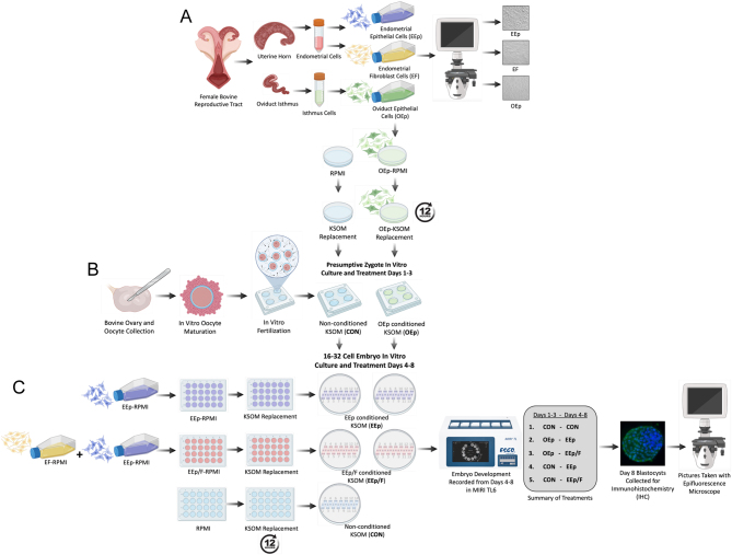
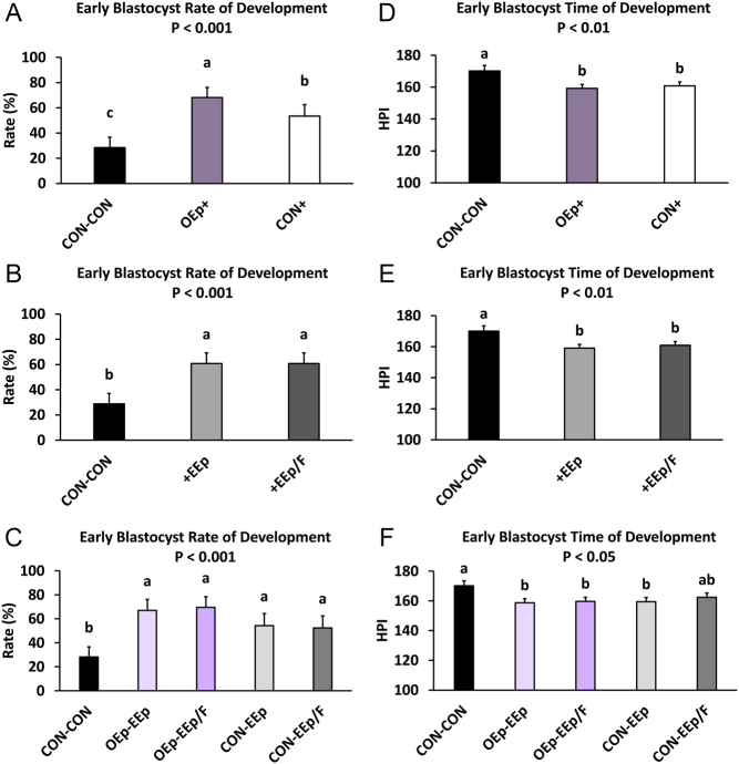
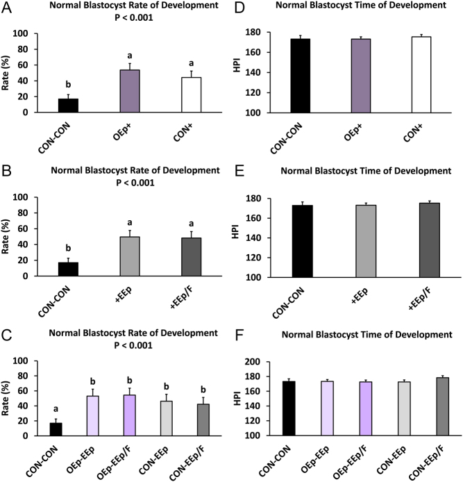
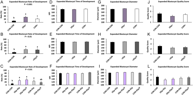
In brief: Standard in vitro produced (IVP) bovine embryo culture media limit embryonic development. Culturing IVP bovine embryos in standard IVP bovine embryo culture media conditioned with oviduct and/or endometrial cells improves blastocyst formation and reduces the time to formation.

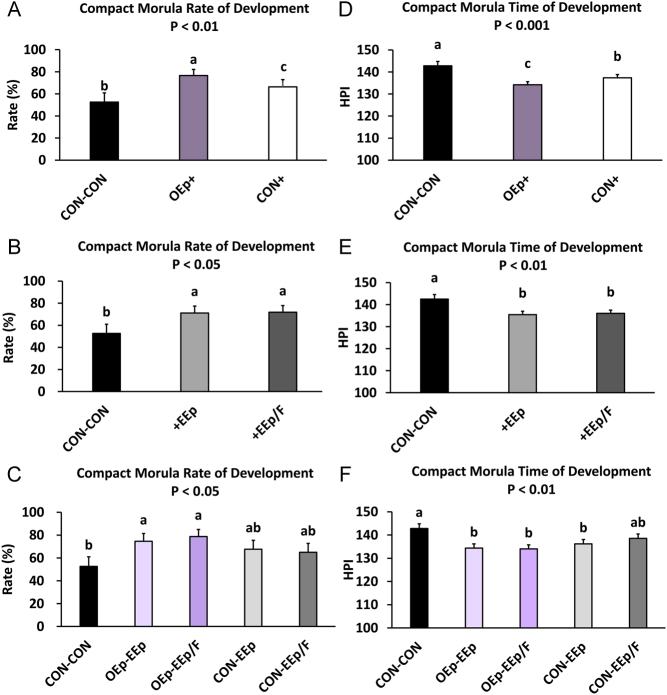
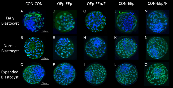
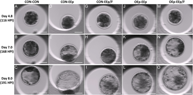
Abstract: In vitro embryo production in cattle greatly impacts blastomere biochemistry, embryo rate of development and pre- and post-transfer survival. In vivo, the bovine embryo migrates through the oviduct isthmus before entering the uterus on approximately day 4 of development where it remains unattached within the uterine lumen until day 20 of gestation. During this time, the embryo is sequentially exposed to oviduct followed by endometrial secretions that support embryonic development. Considering this, we tested the effect of culturing in vitro produced (IVP) bovine embryos sequentially in oviduct epithelial- (OEp; days 1-3) followed by endometrial epithelial- (EEp) or EEp and fibroblast cell (EEp/F; days 4-8)-conditioned media on embryonic development using a time-lapse monitoring system. Compared to control, culturing IVP embryos in EEp- or EEp/F-conditioned media without prior culture in OEp-conditioned media increased blastocyst formation (P < 0.05) and reduced the time to blastocyst formation (P < 0.05). Culturing IVP bovine embryos in OEp-conditioned media followed by EEp- or EEp/F-conditioned media, however, had the greatest impact on embryo developmental kinetics and increased morula and blastocyst formation (P < 0.05) and reduced time to formation (P < 0.05). Day 8 blastocyst cell numbers, diameter and quality were not significantly different, although, blastocyst quality scores were less (indicative of better quality) for all cell-conditioned media compared to control. In conclusion, IVP bovine embryo development may be improved using a sequential embryo culture system involving bovine oviduct followed by endometrial cell-conditioned media.


Conflict of interest statement

The authors declare that there is no conflict of interest that could be perceived as prejudicing the impartiality of the study reported.

Figures

Figure 1
Figure 1
Graphic illustrating the study experimental design. (A) For each female bovine reproductive tract (n = 6), the uterine horn and oviduct isthmus ipsilateral to the corpus luteum were dissected and used to isolate endometrial epithelial (EEp) and fibroblast cells (EF) and oviduct epithelial cells (OEp), respectively. (B) Approximately 2 days before bovine presumptive zygotes (PZ) were to be cultured in conditioned medium, OEp were plated into a petri dish. A second petri dish received culture medium (Roswell Park Memorial Institute; RPMI) without cells. Once OEp reached confluency, the culture medium was removed from both dishes and replaced with fresh potassium simplex optimized medium (KSOM). The KSOM was allowed to incubate in both dishes for 12 h, producing OEp-conditioned medium and non-cell-conditioned (CON) medium. Bovine PZ, produced using standard in vitro embryo production methods, were then cultured (group culture; 20–35) with the CON (n = 807) or OEp (n = 549) medium in four-well plates from days 1 to 3 of development. On day 3, PZ rate of cleavage and formation of 8–16 cells were measured manually. (C) Approximately 2 days before 16–32-cell embryos were to be transferred into a second conditioned medium, EEp or EEp and EF were plated into 24-well plates, the latter of which resulted in EEp/F cocultures. A third 24-well plate received RPMI without cells. Once the cells reached confluency, medium was removed from all dishes and replaced with fresh KSOM. The KSOM was allowed to incubate in the 24-well plates for 12 h producing CON, EEp, and EEp/F-conditioned media. On day 4 of development, 16–32-cell embryos from the previous CON medium were equally transferred into the new CON, EEp, EEp/F medium in MIRI culture coins (7–14/coin; n = 77). Similarly, 16–32-cell embryos from the previous OEp medium were equally transferred into the EEp or EEp/F medium in MIRI culture coins (7–14/coin; n = 77). The embryos were then monitored continuously from days 4 to 8 of development in the MIRI time lapse 6 (TL6) embryo incubator. On day 8, blastocysts were removed from the MIRI and stained for trophoblast marker caudal-type homeobox 2, and cell nuclei with 4′,6-diamidino-2-phenylindole in preparation of TE and ICM cell counts. MIRI time lapse videos of developing embryos were used to measure embryo structure formation (compact morula and early, normal, and expanded blastocyst) and time to structure formation as well as day 8 expanded blastocyst diameter and quality. Graphic was created using illustrations from BioRender.com.
Figure 2
Figure 2
Images of confluent female reproductive tract cells used to condition potassium simplex optimized medium (KSOM). D, Image of cocultured endometrial fibroblast (EF) and epithelial cells (EEp) used to prepare the EEp/F-conditioned KSOM. A, oviduct epithelial cells (OEp); B, endometrial epithelial cells (EEp), C, endometrial fibroblast cells (EF). Bar = 500 µm.
Figure 3
Figure 3
Percentage of compact morula development and time of development during sequential embryo culture in bovine reproductive cell-conditioned potassium simplex optimized medium (KSOM). (A and D; statistical analysis 1; SA1) Percentage of compact morula development and time of development (hours post-insemination; (HPI)) between days 4 and 8 for embryos sequentially cultured in non-cell-conditioned KSOM from days 1 to 3 followed by days 4 to 8 (CON-CON) and embryos cultured in oviduct epithelial cell-conditioned KSOM or CON from days 1 to 3 (OEp+ or CON+, respectively) followed by culture in endometrial epithelial (EEp) and endometrial epithelial and fibroblast cell (EEp/F; coculture) conditioned KSOM (EEp and EEp/F data combined) from days 4 to 8. (B and E; SA2) Percentage of compact morula development and time of development (HPI) between days 4 and 8 for embryos sequentially cultured in CON-CON and embryos cultured in EEp or EEp/F cell-conditioned KSOM from days 4 to 8 (+EEp or +EEp/F, respectively) after prior culture in oviduct epithelial cell-conditioned KSOM (OEp) and CON (data combined) from days 1 to 3. (C and F; SA3) Percentage of compact morula development and time of development between days 4 and 8 for embryos cultured sequentially from days 1 to 3 followed by days 4 to 8 in the different combinations of cell-conditioned and non-cell-conditioned media. Data are presented as least squares means (LSM) ± s.e. of the LSM (s.e.m .). Number (n) of observations for each treatment are presented in Supplementary Table 2.
Figure 4
Figure 4
Percentage of early blastocyst development and time of development during sequential embryo culture in bovine reproductive cell-conditioned potassium simplex optimized medium (KSOM). (A and D; statistical analysis 1; SA1) Percentage of early blastocyst development and time of development (hours post insemination; (HPI)) between days 4 and 8 for embryos sequentially cultured in non-cell-conditioned KSOM from days 1 to 3 followed by days 4 to 8 (CON-CON) and embryos cultured in oviduct epithelial cell- conditioned KSOM or CON from days 1 to 3 (OEp+ or CON+, respectively) followed by culture in endometrial epithelial (EEp) and endometrial epithelial and fibroblast cell (EEp/F; coculture) conditioned KSOM (EEp and EEp/F data combined) from days 4 to 8. (B and E; SA2) Percentage of early blastocyst development and time of development between days 4 and 8 for embryos sequentially cultured in CON-CON and embryos cultured in EEp or EEp/F cell-conditioned KSOM from days 4 to 8 (+EEp or +EEp/F, respectively) after initial culture in oviduct epithelial cell-conditioned KSOM (OEp) and CON (data combined) from days 1 to 3. (C and F; SA3) Percentage of early blastocyst development and time of development (HPI) between days 4 and 8 for embryos cultured sequentially from days 1 to 3 followed by days 4–8 in the different combinations of cell conditioned and non-cell-conditioned media. Data are presented as least squares means (LSM) ± s.e. of the LSM (s.e.m.). Number (n) of observations for each treatment are presented in Supplementary Table 2.
Figure 5
Figure 5
Percentage of normal blastocyst development and time of development during sequential embryo culture in bovine reproductive cell-conditioned potassium simplex optimized medium (KSOM). (A and D; statistical analysis 1; SA1) Percentage of normal blastocyst development and time of development (hours post insemination (HPI)) between days 4 and 8 for embryos sequentially cultured in non-cell-conditioned KSOM from days 1 to 3 followed by days 4–8 (CON-CON) and embryos cultured in oviduct epithelial cell-conditioned KSOM or CON from days 1 to 3 (OEp+ or CON+, respectively) followed by culture in endometrial epithelial (EEp) and endometrial epithelial and fibroblast cell (EEp/F; coculture) conditioned KSOM (EEp and EEp/F data combined) from days 4 to 8. (B and E; SA2) Percentage of normal blastocyst development and time of development (HPI) between days 4 and 8 for embryos sequentially cultured in CON-CON and embryos cultured in EEp or EEp/F cell-conditioned KSOM from days 4 to 8 (+EEp or +EEp/F, respectively) after initial culture in oviduct epithelial cell-conditioned KSOM (OEp) and CON (data combined) from days 1 to 3. (C and F; SA3) Percentage of normal blastocyst development and time of development (HPI) between days 4 and 8 for embryos cultured sequentially from days 1 to 3 followed by days 4–8 in the different combinations of cell-conditioned and nonconditioned media. Data are presented as least squares means (LSM) ± s.e. of the LSM (s.e.m.). Number (n) of observations for each treatment are presented in Supplementary Table 2.
Figure 6
Figure 6
Percentage of expanded blastocyst development and time of development during sequential embryo culture in bovine reproductive cell-conditioned potassium simplex optimized medium (KSOM) as well as day 8 expanded blastocyst diameter and quality score (1–4, excellent/good to degenerating/dead, respectively). (A, D, G, and J; statistical analysis 1; SA1) Percentage of expanded blastocyst development and time of development (hours post insemination; (HPI)) between days 4 and 8 as well as day 8 expanded blastocyst diameter and quality score for embryos sequentially cultured in non-cell-conditioned KSOM from days 1–3 followed by days 4–8 (CON-CON) and embryos cultured in oviduct epithelial cell-conditioned KSOM or CON from days 1–3 (OEp+ or CON+, respectively) followed by culture in endometrial epithelial (EEp) and endometrial epithelial and fibroblast cell (EEp/F; coculture) conditioned KSOM (EEp and EEp/F data combined) from days 4 to 8. (B, E, H, and K; SA2) Percentage of expanded blastocyst development and time of development (HPI) between days 4 and 8 as well as day 8 expanded blastocyst diameter and quality score for embryos sequentially cultured in CON-CON and embryos cultured in EEp or EEp/F cell-conditioned KSOM from days 4 to 8 (+EEp or +EEp/F, respectively) after initial culture in oviduct epithelial cell-conditioned KSOM (OEp) and CON (data combined) from days 1 to 3. (C, F, I, and L; SA3) Percentage of expanded blastocyst development and time of development (HPI) between Days 4 and 8 as well as day 8 expanded blastocyst diameter and quality score for embryos cultured sequentially from days 1 to 3 followed by days 4 to 8 in different combinations of cell conditioned and non-cell-conditioned media. Data are presented as least squares means (LSM) ± s.e. of the LSM (s.e.m.). Number (n) of observations for each treatment are presented in Supplementary Table 2.
Figure 7
Figure 7
Immunohistochemistry (IHC) images of early (A–M), normal (B–N), and expanded (C–O) day 8 bovine blastocysts after sequential culture in non-cell-conditioned (CON) potassium simplex optimized medium (KSOM) or oviduct epithelial cell (OEp)-conditioned KSOM followed by either CON or endometrial epithelial cell (EEp) or endometrial epithelial and fibroblast cell (EEp/F) conditioned KSOM. During IHC, blastocyst TE caudal-type homeobox 2 (CDX-2) was stained using an indirect immunostaining technique involving a green, fluorescent labeled antibody. Blastocysts were then mounted in Fluoromount G containing 4’,6-diamidino-2-phenylindole (DAPI; blue) to stain all blastomere nuclei. Images of the blastocysts were taken using a camera and microscope equipped for fluorescent green and blue detection and the number of green labeled TE cells were counted and subtracted from the total number of cells (blue) to determine the number of ICM cells. The panel above presents merged fluorescent images of select blastocysts. Overall, there was no effect of treatment on the number or percentage of cells or ratio of TE to ICM cells. Bar = 50 µm. Early, normal, and expanded blastocyst cell number data is presented in Supplementary Tables 4, 5, and 6.
Figure 8
Figure 8
MIRI TL6 images of IVP bovine embryos on days 4.8 (116 post insemination (HPI)), 7 (168 HPI) and 8 (191 HPI) of development after culture in bovine reproductive cell or non-cell-conditioned potassium simplex optimized media (KSOM). (A–C) An embryo cultured from days 1 to 3 (CON) followed by days 4–8 (CON) in non-cell-conditioned KSOM (CON-CON). (D–F) An embryo cultured in non-cell-conditioned KSOM from days 1–3 followed by endometrial epithelial cell (EEp) conditioned KSOM from days 4 to 8 of development (CON-EEp). (G–I) An embryo cultured in non-cell-conditioned KSOM from days 1 to 3 followed by endometrial epithelial and fibroblast cell (EEp/F) conditioned KSOM from days 4 to 8 of development (CON-EEp/F). (J–L) An embryo cultured in oviduct epithelial cell (OEp)-conditioned KSOM from days 1 to 3 followed by EEp-conditioned KSOM from days 4 to 8 of development (OEp-EEp). (M-O) An embryo cultured in OEp-conditioned KSOM from days 1 to 3 followed by EEp/F-conditioned KSOM from days 4 to 8 of development. Bar = 50 µm.

Similar articles

Cited by

References

    1. Avilés M Gutiérrez-Adán A & Coy P. 2010Oviductal secretions: will they be key factors for the future ARTs? Molecular Human Reproduction 16896–906. (10.1093/molehr/gaq056) - DOI - PubMed
    1. Biase FH, Rabel C, Guillomot M, Hue I, Andropolis K, Olmstead CA, Oliveira R, Wallace R, Le Bourhis D, Richard C, et al.2016Massive dysregulation of genes involved in cell signaling and placental development in cloned cattle conceptus and maternal endometrium. PNAS 11314492–14501. (10.1073/pnas.1520945114) - DOI - PMC - PubMed
    1. Biggers JD McGinnis LK & Raffin M. 2000Amino acids and preimplantation development of the mouse in protein-free potassium simplex optimized medium. Biology of Reproduction 63281–293. (10.1095/biolreprod63.1.281) - DOI - PubMed
    1. Bó GA & Mapletoft RJ. 2013Evaluation and classification of bovine embryos. Animal Reproduction 10344–348.
    1. Brooks K Burns GW Moraes JGN & Spencer TE. 2016Analysis of the uterine epithelial and conceptus transcriptome and luminal fluid proteome during the peri-implantation period of pregnancy in Sheep1. Biology of Reproduction 9588–88. (10.1095/biolreprod.116.141945) - DOI - PubMed

Substances

LinkOut - more resources