. 2024 Mar 8;383(6687):1111-1117.
doi: 10.1126/science.adk9469. Epub 2024 Mar 7.

Prophage proteins alter long noncoding RNA and DNA of developing sperm to induce a paternal-effect lethality

Affiliations

Prophage proteins alter long noncoding RNA and DNA of developing sperm to induce a paternal-effect lethality

Rupinder Kaur et al. Science. .

Abstract

The extent to which prophage proteins interact with eukaryotic macromolecules is largely unknown. In this work, we show that cytoplasmic incompatibility factor A (CifA) and B (CifB) proteins, encoded by prophage WO of the endosymbiont Wolbachia, alter long noncoding RNA (lncRNA) and DNA during Drosophila sperm development to establish a paternal-effect embryonic lethality known as cytoplasmic incompatibility (CI). CifA is a ribonuclease (RNase) that depletes a spermatocyte lncRNA important for the histone-to-protamine transition of spermiogenesis. Both CifA and CifB are deoxyribonucleases (DNases) that elevate DNA damage in late spermiogenesis. lncRNA knockdown enhances CI, and mutagenesis links lncRNA depletion and subsequent sperm chromatin integrity changes to embryonic DNA damage and CI. Hence, prophage proteins interact with eukaryotic macromolecules during gametogenesis to create a symbiosis that is fundamental to insect evolution and vector control.


Figures

Fig. 1.
Fig. 1.. CifA is an in vitro DNase and RNase, and CifB is a DNase.
(A) Schematic representation showing the Cif recombinant proteins used in the study. Domain annotations are based on structural homology-based analyses (18). The full histidine (His)— tagged CifA protein was generated with (CifAQY) and without (CifA) substitutions in the QxxxY motif. Because the full length CifB protein was too large to be recombinantly expressed in E. coli, purified His-glutathione S-transferase-tagged CifB variants were generated with the amino terminus, N-terminal nuclease domain (NTND), and C-terminal nuclease domain (CTND), refered to CifB▵D, along with engineered substitutions in the QxxxY motif (CifBQY). (B) Nuclease activities of the Cif proteins against single-stranded (ss) DNA, double-stranded (ds) DNA, and ssRNA substrates. CifB▵D cleaves both ss- and dsDNA but not RNA. QxxxY motif substitutions ablate CifB DNase activity CifA is both a DNase and RNase. The CifAQY mutant ablates ssDNase activity, whereas dsDNase and ssRNase activities remain intact. Commercial DNase and RNase enzymes were used as positive controls, and no CifA or CifB proteins were added to the reaction mixtures for negative controls. EDTA was added to a 20-fold molar excess over Mg2+ to inhibit the reactions. Samples were run in a 10% polyacrylamide-tris-borate EDTA gel. Single-letter abbreviations for the amino acid residues referenced throughout are as follows: Q, Glu; Y, Tyr; A, Ala; G, Gly.
Fig. 2.
Fig. 2.. CifA in situ RNase activity depletes AAGAG lncRNA in association with CI.
Fluorescent in situ hybridization (FISH) assay was performed on testes (n = 15) squashes from <8-hours-old males to visualize and quantify the AAGAG repeat lncRNA abundance in primary spermatocytes (25). (A) Compared with the wMel-negative and transgenic non-CI control line CifA▵bnlsB, AAGAG lncRNA (red) is less abundant in primary spermatocytes, marked by 4′,6-diamidino-2-phenylindole (DAPI) staining (blue), of wMel+ and dual-transgenic CifAB lines, respectively. CifA expression depletes lncRNA, whereas CifB does not. (B) lncRNA signal intensity per spermatocyte was quantified in ImageJ, as described in fig. S3, and graphed.The y axis corresponds to the genotype labels shown on the left side of (A). (C) Hatch rate assay confirmed that AAGAG_KD(+) symbiotic males with depleted lncRNA result in stronger CI than negative control Scramble_KD(+) males. Each dot represents the percentage of embryos that hatched from a single male and female pair. (D) Representative lncRNA-FISH and quantification data are shown for AAGAG_KD(+) and Scramble_KD(+) sibling males to those used in the panel 2C hatch rate assay. Vertical bars in (B), (C), and (D) represent the median. *P < 0.05; **P < 0.01; ****P < 0.0001; calculated by Mann-Whitney pairwise comparison test. The skull icon represents CI, and the fly icon represents no CI.
Fig. 3.
Fig. 3.. CifAB in situ DNase activity enhances DNA damage in elongating spermatids.
TUNEL staining on testes squashes from <8-hours-old males was performed to visualize and quantify sperm bundles with DNA breaks. (A) Representative images of spermatid bundles with DNA damage from each treatment group are shown. Images were taken using Keyence All-in-One confocal microscope at 100× magnification. Compared with the wMel– control (empty arrowheads), DNA break signals marked by TUNEL (green) were highly intense in the wild-type wMel+ and dual transgenic CifAB lines at the canoe stage of spermiogenesis (solid arrowheads). DAPI staining (blue) labels the spermatid nuclei. Upon individual expression, CifB induced higher DNA damage than CifA. (B) TUNEL signal intensity from spermatid bundles was quantified in ImageJ and graphed. Vertical bars represent the median. (C) Total sperm bundles and those with fragmented DNA were manually counted. The numbers of testes investigated are shown in parentheses next to the genotype. Uncropped images of testes from CI versus non-CI males with total damaged bundles with high versus low TUNEL intensity are shown in fig. S5. **P < 0.01; ****P < 0.0001; calculated by Mann-Whitney pairwise comparison test.
Fig. 4.
Fig. 4.. Late-stage CI embryos suffer from DNA damage.
(A to C) (Top) Representative images of 2- to 3-hours-old CI embryos (wMel+ males × wMel– females) during cellular blastoderm formation (nuclear cycles 12 to 14) exhibit intense pH2Av signals (red) indicative of DNA damage compared with that of rescue embryos (wMel+ males × wMel+ females). pH2Av is a histone H2Av variant that phospohorylates in response to DNA damage (42, 43). (Bottom) Magnified images of boxed regions in (A), (B), and (C) top panels, respectively. (A) CI embryonic nuclei on polar ends undergo shrinkage and are distantly spaced with condensed chromatin (white arrows), likely becoming apoptotic leading to embryonic death. DAPI (gray) staining labels the embryonic nuclei. (B) In CI embryos, a CI-defining chromatin bridging is marked by an asterisk colocalized with DNA damage signal (red). In the same embryo, nuclei in the middle (empty arrowhead) developed normally with no pH2Av signals. A white solid arrowhead indicates a distinct missegregating chromosomal feature in the region between the middle and polar end of the embryo that contains condensed chromatin with DNA damage (white arrows). (C) A rescue embryo develops normally with evenly spaced embryonic nuclei lacking DNA damage. Full embryo confocal images were acquired with a Zeiss LSM880 confocal microscope at 63× magnification by using the tile-scan feature and automated stitching. (D) Data show the quantification of pH2Av signal intensity per embryo derived from CI and non-CI–rescue crosses. Values in parentheses correspond to the number of embryos scored. Vertical bars represent the median. A horizontal dotted line separates wild-type and transgenic cross types. ****p value < 0.0001; ns, not significant; calculated by Mann-Whitney pairwise comparison test.
Fig. 5.
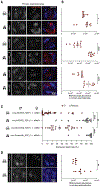
Fig. 5.. CI induction and ablation directly link with AAGAG lncRNA depletion, impairment of histone-to-protamine transition, and DNA damage in embryos.
(A) RNA-FISH on testes (n = 15) from <8-hour-old males of CI-inducing CifA4B and non-CI CifA3B mutant males (Fig. 2) showed that the CifA4B mutant depletes AAGAG lncRNA, whereas CifA3B does not. (B) CI embryos (nos;cifA4B males × wMel– females) exhibited intense pH2Av signals (red) at the polar end (white arrow), which was indicative of DNA damage, compared with non-CI embryos (nos;cifA3B males × wMel– females). Dotted periphery is drawn around the embryo shape. (C) CI and non-CI testes were immunostained to quantify histone-retaining spermatid bundles (purple) during late canoe stage of spermiogenesis (13). Non-CI lines (CifA3B and CifA▵bnlsB) showed significantly less spermatid bundles with retained histones compared with that of CI groups (CifA4B and CifAB). (D) Mature sperm from seminal vesicles were stained with fluorescent Chromomycin A3 (CMA3) stain (green) to detect protamine deficiency (13). CI sperm (CifA4B and CifAB) lack protamines compared with non-CI sperm (CifA3B and nos;wMel–). (E) pH2Av signal intensity quantification per embryo derived from CI and non-CI crosses. Vertical bars represent mean, and error bars represent standard deviation. **P < 0.01; ****P < 0.0001; ns, not significant; calculated by Mann-Whitney pairwise comparison test.

References

    1. Barr JJ, mSystems 4, e00105–19 (2019). - PubMed
    1. Weinert LA, Araujo-Jnr EV, Ahmed MZ, Welch JJ, Proc. Biol. Sci 282, 20150249 (2015). - PMC - PubMed
    1. Hoffmann AA, Turelli M, Harshman LG, Genetics 126, 933–948 (1990). - PMC - PubMed
    1. Brucker RM, Bordenstein SR, Trends Ecol. Evol 27, 443–451 (2012). - PubMed
    1. Caputo B et al., Pest Manag. Sci 76, 1324–1332 (2020). - PubMed

Publication types

LinkOut - more resources