. 2024 Mar 8;383(6687):eadi7342.
doi: 10.1126/science.adi7342. Epub 2024 Mar 8.

Vitamin A resolves lineage plasticity to orchestrate stem cell lineage choices

Affiliations

Vitamin A resolves lineage plasticity to orchestrate stem cell lineage choices

Matthew T Tierney et al. Science. .

Abstract

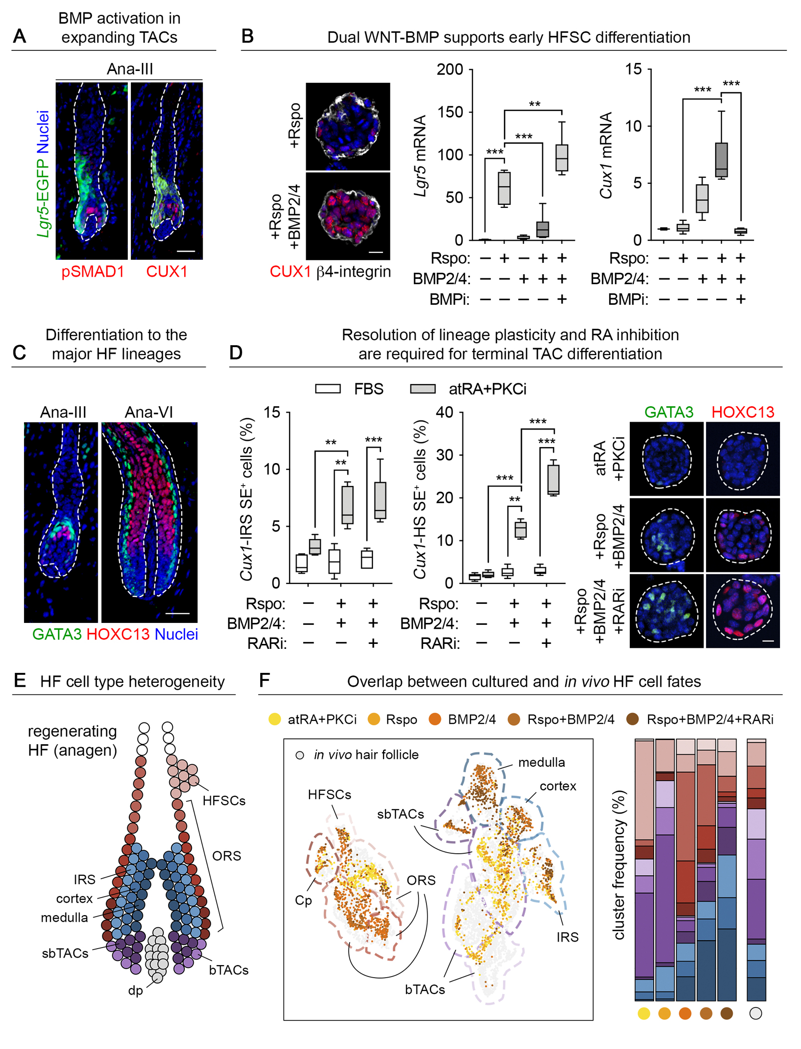
Lineage plasticity-a state of dual fate expression-is required to release stem cells from their niche constraints and redirect them to tissue compartments where they are most needed. In this work, we found that without resolving lineage plasticity, skin stem cells cannot effectively generate each lineage in vitro nor regrow hair and repair wounded epidermis in vivo. A small-molecule screen unearthed retinoic acid as a critical regulator. Combining high-throughput approaches, cell culture, and in vivo mouse genetics, we dissected its roles in tissue regeneration. We found that retinoic acid is made locally in hair follicle stem cell niches, where its levels determine identity and usage. Our findings have therapeutic implications for hair growth as well as chronic wounds and cancers, where lineage plasticity is unresolved.


Conflict of interest statement

Competing interests: MTT and EF are listed as inventors on a US provisional patent application filed by The Rockefeller University encompassing aspects of this work. No other authors have competing interests to declare.

Figures

Fig. 1:
Fig. 1:. A screen to resolve lineage plasticity in tissue stem cells ex vivo.
(A) Cultured HFSCs were stably transduced with a lentiviral Klf5-EGFP reporter (left). Flow cytometry quantification (center) showed that serum replacement reduced Klf5 reporter expression in 3D cultures. Colony diameter plateaued over time in serum replacement media, in contrast to unrestrained outgrowth in serum-replete media (right) (n=5 technical replicates across three purified HFSC lines). (B) Quantification of Klf5-EGFP+ cells after screening a library of small molecules implicated in wound repair identified several small molecule ‘hits’ (n=5). The dashed line indicates baseline EGFP levels in B-27 media with vehicle-only controls. The significant hits that repressed Klf5-EGFP were: atRA, the PKCa/b inhibitor Gö6976, and FR180204, a competitive inhibitor of ERK1/2. (C and D) Immunofluorescence (C) and immunoblot (D) analyses show that serum replacement and treatment with atRA and PKCi (n=6) resolves lineage plasticity, as judged by elevated HFSC master regulator SOX9 and silenced EpSC transcriptional regulator KLF5. Keratin profiles reflect the loss of epidermal differentiation and wound-induced suprabasal markers (KRT10, KRT6), uniform expression of pan-skin progenitor marker KRT14, and gain of KRT15, a HFSC marker. The epidermal-dermal interface of sagittal skin section is delineated by a dashed line. epi, epidermis; sg, sweat gland; dp, dermal papilla. Scale bar, 20μm. (E) UMAP representation and unsupervised k-nearest-neighbor-based clustering of single cell transcriptomes from 3,757 cultured HFSCs reveal that RA-PKCi co-treatment groups cells in near-uniform clusters distinct from wound-like and epidermal cell types (left, center). Annotated cluster assignments made using gene signatures derived from independent bulk sequencing of in vivo HFSCs, EpSCs, and lineage traced HFSCs isolated at intermediate stages of wound repair confirmed that atRA+PKCi-treated cells had a strong HFSC signature and reduced EpSC and wound signatures (right).
Fig. 2:
Fig. 2:. Retinoic acid acts at the chromatin level to resolve lineage plasticity.
(A) atRA-RAR/RXR activity is detected in the HFSC niche in vivo as judged by a retinoic acid response element reporter (RARE-RFP; H2B-GFP transduction control) in adult skin epithelium. ALDH1A2 immunolabeling confirms the co-presence of this rate-limiting atRA production enzyme. Both are transiently lost after an abrasion wound (left). The epidermal-dermal interface is delineated by a dashed line. Scale bar, 50μm. The kinetics of RARE-RFP activity during wound repair was measured by flow cytometry (n=5). Lineage plasticity (percentage of SOX9+KLF5+ cells in the HF) is overlaid (right; n=10–12 HFs per mouse, n=8 mice). Uninj., uninjured. (B) Conditional Rxra ablation in Sox9-CreER+ HFSCs during telogen results in ectopic KLF5 expression. The epidermal-dermal interface is delineated by dashed lines. Arrows denote double immunolabeled SOX9+KLF5+ cells, reflective of lineage plasticity and quantified at right (n=5). Scale bar, 50μm. Ctrl., control. (C) Line plots and heatmap of ATAC-seq signal intensity in atRA-stimulated chromatin peaks from wild-type and Rxra cKO HFSCs cultured in FBS or atRA+PKCi media (left; n=2). HOMER motif analysis of Rxra-dependent chromatin domains differentially opened in atRA+PKCi media (top right). Note that atRA-sensitive peaks are highly enriched for SOX motifs. Venn diagrams of overlapping (opened or closed) Rxra-dependent chromatin peaks (ATAC-seq) and DEGs (RNA-seq) (bottom right). Selected genes are listed by culture condition, color-coded if previously determined to be a super-enhancer regulated gene in cultured or in vivo HFSCs (16). Notable among these Rxra-dependent, atRA-regulated genes is Sox9, encoding a pioneer factor essential for activating key HFSC genes directly and silencing EpSC genes indirectly (34). (D) Venn diagram showing transcripts and accessible chromatin peaks that are atRA and RXRa sensitive and co-bound by RARγ and RXRa (left). Representative ATAC peaks and CNR binding profiles for RARγ and RXRa at direct atRA target genes (right).
Fig. 3:
Fig. 3:. atRA cooperates in a dose-dependent manner with BMPs and WNTs to influence the balance between HFSC quiescence and activation.
(A) BMP activity in SOX9+ quiescent bulge HFSCs in vivo. The epidermal-dermal interface is denoted by dashed lines; asterisk refers to autofluorescence in the sebaceous gland. Scale bar, 50μm. (B) The percentage of EdU+ cultured HFSCs is reduced by BMP6 (left; n=6). Quiescent HFSC markers Nfatc1 (center) and HFSC-associated Cxcl14-EGFP super-enhancer reporter activity (right) are induced by co-treating with BMP6 and low-dose RA (10 nM). Gates outlined in red denote EGFP+ cells (n=5–6). (C) During telogen, TCF4 and Lgr5-EGFP are expressed in HFSCs. Upon hair cycle induction, Lgr5-EGFP tracks with the ORS while LEF1 is upregulated in TACS and lower ORS (arrows). The epidermal-dermal interface is denoted by dashed lines; asterisks refer to autofluorescence in the sebaceous gland. Scale bar, 50μm. (D) The TCF4:LEF1 switch is accurately modeled in atRA-treated cultured HFSCs by exposure to Rspo, with associated SOX9 downregulation (left). Scale bar, 50μm. High-dose atRA (100 nM) is required for a Rspo-dependent increase in cultured HFSC proliferation, quantified by flow cytometry (right; n=6).
Fig. 4:
Fig. 4:. atRA-mediated directed differentiation to HF lineages via combinatorial niche signaling.
(A) Downstream of WNT-activated HFSCs in the early anagen phase of the hair cycle, BMP signaling (measured by pSMAD1 immunolabeling) distinguishes CUX1+ TACs of the hair ‘bulb’ and is mutually exclusive to Lgr5-EGFP in the ORS. The epidermal-dermal interface is denoted by dashed lines. Scale bar, 50μm. (B) atRA-treated cultured HFSCs respond similarly: BMPs upregulate Cux1 in Rspo-stimulated HFSCs while antagonizing Lgr5-EGFP activity (n=6). Scale bar, 20μm. (C) In full or late anagen, the HOXC13+ HS and GATA3+ channel IRS lineages emerge. The epidermal-dermal interface is denoted by dashed lines. Scale bar, 50μm. (D) In culture, only when lineage plasticity (FBS) is resolved (atRA+PKCi) are HFSCs able to cooperate with WNT and BMP signals and activate these downstream hair lineages. Note that the HS lineage is further promoted by retinoic acid receptor inhibition (RARi). Quantification is by flow cytometry for reporter activity driven by Cux1 enhancer elements active in the IRS and HS, respectively (16) (n=5). GATA3 and HOXC13 immunofluorescence are shown at right. HFSC colonies are outlined by white dashed lines. Scale bar, 20μm. (E) Scheme depicting the HFSC-derived cell fates of the anagen-phase hair follicle. (F) UMAP clustering of cultured HFSCs treated with atRA and PKCi and additionally exposed to Rspo, BMP or RARi as noted. Harmony (57) was used to place these data in the context of a reference dataset of autologous, FACS-purified cells from skin HFs (left; 5,333 cultured cells, 6,007 in vivo HF cells). Shifts in cultured HFSC identity, mapped onto annotated clusters, were consistent with the establishment of traits of HFSC or ORS, Rspo-activated TACs and differentiating IRS or HS lineages after WNT-BMP co-stimulation, respectively (right).
Fig. 5:
Fig. 5:. atRA/RAR/RXR dynamics regulate stem cell fate switching during wound repair in vivo.
(A) Scheme depicting the steps and critical enzymes involved in atRA synthesis, degradation, and atRA-RAR-RXR mediated transcriptional activity. (B) Rxra was conditionally ablated in telogen-phase HFSCs of Sox9-CreER+ mice before abrasion (shallow) wounding. Note markedly altered tissue repair with Rxra cKO 7 days post-injury, including prolonged KRT6 (epidermal hyperproliferation) and blocked hair regeneration (left). The epidermal-dermal interface is denoted by dashed lines. Scale bar, 50μm. Quantification of lineage-traced HFSCs that mobilized to the epidermis at the expense of retention in the HF after Rxra cKO after 30 days post-injury by flow cytometry (n=6 to 8). (C) Transduction of Sox9CreER;Rosa26-LSL-YFP adult skin epithelium with a transgene encoding the atRA-degrading protein CYP26B1 (constitutive H2B-RFP control; left). Tamoxifen treatment activated Cyp26b1 and enabled YFP+ HFSC lineage tracing after wounding. RFP+, i.e. Cyp26b1-overexpressing cells are present in uninjured HFs and epidermis but become overrepresented in the repaired epidermis relative to the HF after wound repair (center left). The epidermal-dermal interface is denoted by dashed lines; arrows refer to YFP+RFP+ cells of HFSC origin that overexpress Cyp26b1. Scale bar, 50μm. Quantification by flow cytometry (center right; n=11). Sustained Cyp26b1 also precluded hair formation in HFSC skin grafts (right; n=5). (D) Mice were fed a normal or vitamin A-deficient diet and wounded in telogen. After repair was complete, YFP+ HFSCs were assessed for their contribution to skin barrier restoration or HF regeneration. Topical atRA applied during the first week post-wound accelerated lineage plasticity resolution (left, n=10 to 12 HFs per mouse, n=5 mice) and skewed HFSC contribution to hair regeneration, as measured by flow cytometry (right, n=10–12 HFs per mouse, n=5 mice), whereas vitamin A loss slowed HF regrowth at intermediate stages of repair (center, n=9–12 HFs, n=5 mice). Hair regeneration and HFSC fate were rescued by topical atRA treatment. Veh., vehicle.

References

    1. Hsu Y-C, Li L, Fuchs E, Emerging interactions between skin stem cells and their niches. Nat Med 20, 847–856 (2014). - PMC - PubMed
    1. Morrison SJ, Spradling AC, Stem cells and niches: mechanisms that promote stem cell maintenance throughout life. Cell 132, 598–611 (2008). - PMC - PubMed
    1. Ge Y, Fuchs E, Stretching the limits: from homeostasis to stem cell plasticity in wound healing and cancer. Nat Rev Genet 19, 311 (2018). - PMC - PubMed
    1. Ge Y, Gomez NC, Adam RC, Nikolova M, Yang H, Verma A, Lu CP-J, Polak L, Yuan S, Elemento O, Fuchs E, Stem Cell Lineage Infidelity Drives Wound Repair and Cancer. Cell 169, 636–650.e14 (2017). - PMC - PubMed
    1. Shook BA, Wasko RR, Mano O, Rutenberg-Schoenberg M, Rudolph MC, Zirak B, Rivera-Gonzalez GC, López-Giráldez F, Zarini S, Rezza A, Clark DA, Rendl M, Rosenblum MD, Gerstein MB, Horsley V, Dermal Adipocyte Lipolysis and Myofibroblast Conversion Are Required for Efficient Skin Repair. Cell Stem Cell 26, 880–895.e6 (2020). - PMC - PubMed

Publication types

MeSH terms