Meta-Analysis
. 2024 Mar 7;22(3):e3002006.
doi: 10.1371/journal.pbio.3002006. eCollection 2024 Mar.

FAM81A is a postsynaptic protein that regulates the condensation of postsynaptic proteins via liquid-liquid phase separation

Affiliations
Meta-Analysis

FAM81A is a postsynaptic protein that regulates the condensation of postsynaptic proteins via liquid-liquid phase separation

Takeshi Kaizuka et al. PLoS Biol. .

Abstract

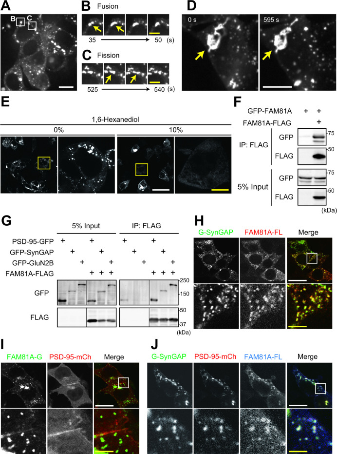
Proteome analyses of the postsynaptic density (PSD), a proteinaceous specialization beneath the postsynaptic membrane of excitatory synapses, have identified several thousands of proteins. While proteins with predictable functions have been well studied, functionally uncharacterized proteins are mostly overlooked. In this study, we conducted a comprehensive meta-analysis of 35 PSD proteome datasets, encompassing a total of 5,869 proteins. Employing a ranking methodology, we identified 97 proteins that remain inadequately characterized. From this selection, we focused our detailed analysis on the highest-ranked protein, FAM81A. FAM81A interacts with PSD proteins, including PSD-95, SynGAP, and NMDA receptors, and promotes liquid-liquid phase separation of those proteins in cultured cells or in vitro. Down-regulation of FAM81A in cultured neurons causes a decrease in the size of PSD-95 puncta and the frequency of neuronal firing. Our findings suggest that FAM81A plays a crucial role in facilitating the interaction and assembly of proteins within the PSD, and its presence is important for maintaining normal synaptic function. Additionally, our methodology underscores the necessity for further characterization of numerous synaptic proteins that still lack comprehensive understanding.


Conflict of interest statement

The authors have declared that no competing interests exist.

Figures

Fig 1
Fig 1. Meta-analysis of the PSD proteome datasets.
(A and B) Overlap of the detected proteins in 35 PSD proteome datasets. “Protein complex” in the top panel indicates 995 proteins included in 15 datasets of the PSD protein complex. (C) Venn diagram describes the overlap of PSD fraction and PSD protein complex. (D) Histogram of the number of detected datasets. (E) The percentage of proteins that belong to indicated GO terms. (F and G) SynGO enrichment analysis of 123 proteins detected in more than 20 proteome datasets. The results of localization (Cellular Component; CC) (F) and function (Biological Process; BP) (G) are shown as -log10 Q-value. PSD, postsynaptic density.
Fig 2
Fig 2. FAM81A, a PSD protein expressed in the higher vertebrate brain.
(A) Hypothetical PSD proteins which are poorly characterized. Proteins identified in at least 8 datasets are listed. The blue and yellow bars indicate the dataset number of unbiased and candidate-based approaches. The name of proteins is described as the official symbol of the mouse gene. (B) Twenty-one PSD proteome datasets in which FAM81A (FAM81A) was identified. (C) Homologs of FAM81A. Red and blue items indicate orthologs of FAM81A and FAM81B, respectively. Green items indicate a common ortholog of FAM81A and 2 in amphibians, fish, and invertebrates. The phylogenetic tree was described with Kalign (https://www.ebi.ac.uk/Tools/msa/kalign/). Hs: Homo sapiens, Mm: Mus musculus, Gg: Gallus gallus, Pp: Paroedura picta, Xl: Xenopus laevis, Dr: Danio rerio. Ci: Ciona intestinalis, Bl: Branchiostoma lanceolatum, Sp: Strongylocentrotus purpuratus, Ac: Aplysia californica. (D) Expression pattern of FAM81A and FAM81 B in human tissues. The data was obtained from NCBI Gene. RPKM: Reads Per Kilobase of exon per Million mapped reads. (E) Sequence conservation of human FAM81A, FAM81B, and major PSD proteins in mouse (Mm) and zebrafish (Dr). The percentage of identical or similar residues was evaluated using Gene2Function. PSD, postsynaptic density.
Fig 3
Fig 3. FAM81A is a PSD protein that partially colocalizes with NMDA receptor.
(A) Enrichment of FAM81A in PSD. S1 fraction and PSD-I fraction were prepared from brain homogenate of adult mice. (B) Interaction of FAM81A with PSD-95 in synapse. Crude synaptosome fraction of adult mouse forebrain was subjected to immunoprecipitation using anti-PSD-95 antibody. (C and D) Distribution of FAM81A in the brain. Immunohistochemistry of FAM81A was performed on the sagittal section of the adult mouse brain. (E) Colocalization of FAM81A and NMDA receptor. Immunohistochemistry of FAM81A and GluN1 was performed on the section of the adult mouse brain. The bottom part of the dentate gyrus is described with high magnification. (F and G) Relative abundance of individual proteins in mouse PSD (F) and synaptosome fractions (G) was estimated using iBAQ intensities calculated from our published proteomic data [19]. Scale bar: 2 mm (C), 10 μm (D), or 5 μm (E). NMDA, N-methyl-D-aspartate; PSD, postsynaptic density.
Fig 4
Fig 4. Formation of FAM81A condensates at PSD and cytoplasm in neuron.
(A) Localization of FAM81A at PSD in the cultured neuron. Primary mouse hippocampal neurons were transfected with FAM81A-GFP, PSD-95-mCherry, and BFP at DIV19. Two days later, the cells were fixed and observed with a confocal microscope. (B–G) Live-imaging of primary cultured mouse hippocampal neurons (DIV16-18) transfected with FAM81A-GFP and DsRed (30 min/frame). (B) Overview of the observed neuron. Regions magnified in panels C, E, F, and G are described. See also S1 Movie. (C and D) Accumulation of FAM81A on the PSD upon spine maturation. The signal intensity of 2 spines shown in panel C is quantified and plotted in panel D. (E) Puncta of FAM81A-GFP on the PSD. (F) Puncta of FAM81A-GFP at soma. (G) Puncta of FAM81A-GFP at dendritic shaft. Scale bars: (A) 50 μm (white) or 10 μm (yellow). (B–G) 10 μm (white) or 5 μm (yellow). DIV, day in vitro; PSD, postsynaptic density.
Fig 5
Fig 5. Domain structure of FAM81A required for condensation.
(A) Condensate formation of FAM81A mutants. HEK293T cells were transfected with indicated FAM81A mutants, and 24 h later, the cells were fixed and observed with a confocal microscope. (B) Primary structure of mouse FAM81A. The coiled-coil domains (CC1 and CC2) and LCR were identified using SMART. Evolutional conservation was analyzed using ConSurf. The probability of disorder is assessed using IUPred2A. Truncate mutants described at the bottom were used in (A). (C–E) Quantitative analysis of FAM81A droplets described in (A). Number of FAM81A condensates per cell (C) and size of FAM81A condensates. (D) Maximum size of FAM81A condensates in individual cells (E) are described. The numbers of analyzed cells expressing full length, Δ1–36, Δ37–74, Δ75–106, Δ107–157, Δ158–187, N-half, or C-half FAM81A are 98, 85, 87, 109, 95, 101, 93, or 83, respectively. Scale bars: 50 μm (white) or 10 μm (yellow). *P < 0.05. LCR, low complexity region; SMART, Simple Modular Architecture Research Tool.
Fig 6
Fig 6. Condensation-mediated localization of FAM81A on PSD enlarges dendritic spines.
(A) Impaired PSD localization of the condensation-deficient FAM81A mutant. Primary cultured mouse hippocampal neurons were transfected with full-length or Δ107–157 FAM81A mutants together with DsRed at DIV19. Two days later, the cells were fixed and observed with a confocal microscope. (B) Enlargement of dendritic spines by accumulation of FAM81A on PSD. The diameter of 1,337 dendritic spines on neurons expressing Δ75–106 FAM81A and 560 spines on neurons expressing full-length FAM81A were quantified. (C) Interaction of FAM81A mutants with PSD-95. HEK293T cells were transfected with PSD-95-GFP and indicated FAM81A mutants tagged with FLAG; 24 h later, cells were lysed, and immunoprecipitation was performed with an anti-FLAG antibody. Then, immunoblotting was performed with anti-GFP or anti-FLAG antibodies. Scale bars: 50 μm (white) or 10 μm (yellow). *P < 0.05. DIV, day in vitro; PSD, postsynaptic density.
Fig 7
Fig 7. Interaction and co-localization of FAM81A with core synaptic molecules.
(A–D) Live-imaging of HEK293T cells transfected with FAM81A-GFP. (A) Overview of the observed cells. Regions magnified in panels B and C are described. See also S2 Movie. (B and C) Representative movie images of punctate structures undergoing fusion (B) and fission (C). (D) Enlarged rigid structure with a stable shape. See also S3 Movie. (E) The effect of 1,6-hexanediol on FAM81A positive structures. HEK293T cells were transfected with FAM81A-GFP, and 24 h later, the medium was changed to fresh medium, including the indicated concentration of 1,6-hexanediol. Ten min later, the cells were fixed. (F and G) Interaction between FAM81A or interaction of FAM81A and PSD-95, SynGAP, or GluN2B. HEK293T cells were transfected with indicated plasmids, and 24 h later, cells were lysed, and immunoprecipitation was performed with an anti-FLAG antibody. Then, immunoblotting was performed with anti-GFP or anti-FLAG antibodies. (H–J) Localization of FAM81A on SynGAP-positive droplets. HEK293T cells were transfected with indicated plasmids, and 24 h later, cells were fixed and observed with confocal microscopy. For panels H and J, cells were subjected to immunocytochemistry using an anti-FLAG antibody before observation. Scale bars: 10 μm (white) or 5 μm (yellow) in (A–D), 50 μm (white) or 10 μm (yellow) in (E), and 20 μm (white) or 4 μm (yellow) in (H–J), respectively. PSD, postsynaptic density.
Fig 8
Fig 8. FAM81A facilitates LLPS of postsynaptic proteins in vitro.
(A) Confocal microscopic images of LLPS of FAM81A with PSD-95, GluN2B carboxyl tail, and SynGAP1. iFluor 488-labeled PSD-95, NirFP-labeled GluN2B, and iFluor 405-labeled SynGAP1 (3 μm each) were mixed with increasing concentrations (0, 1, and 3 μm) of iFluor 568-labeled FAM81A. Scale bars: 5 μm. (B) The histogram of droplet size distribution. **P < 0.01, ***P < 0.001 by one-way analysis of variance (ANOVA) followed by the Tukey–Kramer test. LLPS, liquid–liquid phase separation; PSD, postsynaptic density.
Fig 9
Fig 9. FAM81A modulates PSD size and neuronal activity.
Primary cultured mouse hippocampal neurons were infected with lentivirus-encoding FAM81A shRNAs at DIV14. At DIV21, the neurons were subjected to immunocytochemistry (A and B) or electrophysiological recording using the MEA (C and D). (A and B) Neurons were fixed and labeled with anti-PSD-95 antibody. Representative dendrite images (A) and the mean size of PSD-95 puncta (B) are described. The numbers of analyzed neurons is 51 (mock), 47 (shFAM81A #1), and 49 (shFAM81A #2), respectively. (C and D) Neurons were subjected to extracellular electrophysiological recording using MEA. Representative signals (C) and spike frequency (D) are described. Scale bars: 50 μm (A, white), 10 μm (A, yellow), 10 s (C, x-axis), and 100 μV (C, y-axis). *P < 0.05. DIV, day in vitro; MEA, multielectrode array; PSD, postsynaptic density.

Similar articles

Cited by

References

    1. Dosemeci A, Weinberg RJ, Reese TS, Tao-Cheng JH. The postsynaptic density: There is more than meets the eye. Front Synaptic Neurosci. 2016;8(AUG):1–8. doi: 10.3389/fnsyn.2016.00023 - DOI - PMC - PubMed
    1. Zhu J, Shang Y, Zhang M. Mechanistic basis of MAGUK-organized complexes in synaptic development and signalling. Nat Rev Neurosci [Internet]. 2016;17(4):209–223. doi: 10.1038/nrn.2016.18 - DOI - PubMed
    1. Kaizuka T, Takumi T. Postsynaptic density proteins and their involvement in neurodevelopmental disorders. J Biochem. 2018;163(6):447–455. doi: 10.1093/jb/mvy022 - DOI - PubMed
    1. Carlin RK, Grab DJ, Cohen RS, Siekevitz P. Isolation and characterization of postsynaptic densities from various brain regions: Enrichment of different types of postsynaptic densities. J Cell Biol. 1980;86(3):831–843. doi: 10.1083/jcb.86.3.831 - DOI - PMC - PubMed
    1. Laßek M, Weingarten J, Volknandt W. The synaptic proteome. Cell Tissue Res. 2015;359(1):255–265. doi: 10.1007/s00441-014-1943-4 - DOI - PubMed

Publication types