

Synthesizing Contrast-Enhanced MR Images from Noncontrast MR Images Using Deep Learning
- PMID: 38453408
- PMCID: PMC11286124
- DOI: 10.3174/ajnr.A8107
Synthesizing Contrast-Enhanced MR Images from Noncontrast MR Images Using Deep Learning
Abstract
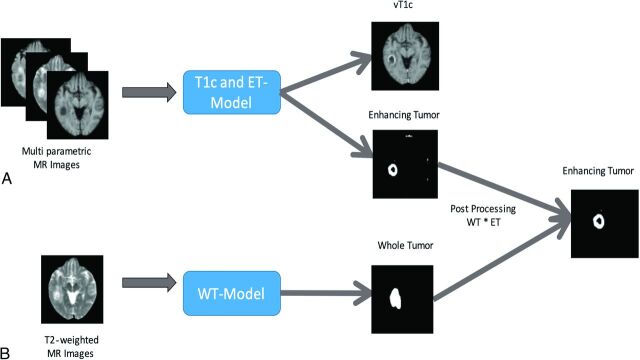
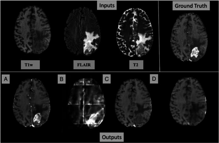
Background and purpose: Recent developments in deep learning methods offer a potential solution to the need for alternative imaging methods due to concerns about the toxicity of gadolinium-based contrast agents. The purpose of the study was to synthesize virtual gadolinium contrast-enhanced T1-weighted MR images from noncontrast multiparametric MR images in patients with primary brain tumors by using deep learning.
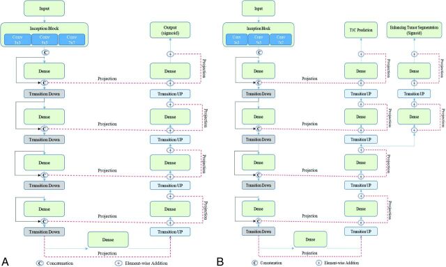
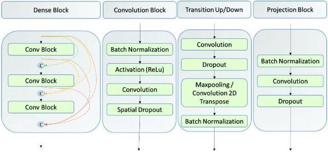
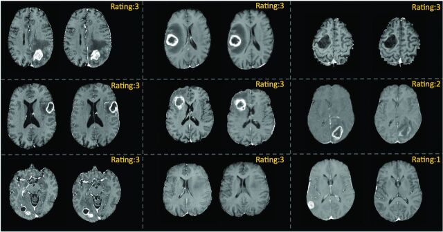
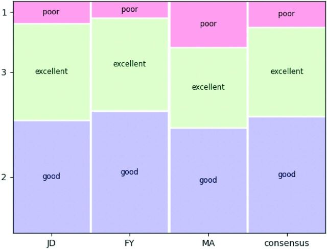
Materials and methods: We trained and validated a deep learning network by using MR images from 335 subjects in the Brain Tumor Segmentation Challenge 2019 training data set. A held out set of 125 subjects from the Brain Tumor Segmentation Challenge 2019 validation data set was used to test the generalization of the model. A residual inception DenseNet network, called T1c-ET, was developed and trained to simultaneously synthesize virtual contrast-enhanced T1-weighted (vT1c) images and segment the enhancing portions of the tumor. Three expert neuroradiologists independently scored the synthesized vT1c images by using a 3-point Likert scale, evaluating image quality and contrast enhancement against ground truth T1c images (1 = poor, 2 = good, 3 = excellent).
Results: The synthesized vT1c images achieved structural similarity index, peak signal-to-noise ratio, and normalized mean square error scores of 0.91, 64.35, and 0.03, respectively. There was moderate interobserver agreement between the 3 raters, regarding the algorithm's performance in predicting contrast enhancement, with a Fleiss kappa value of 0.61. Our model was able to accurately predict contrast enhancement in 88.8% of the cases (scores of 2 to 3 on the 3-point scale).
Conclusions: We developed a novel deep learning architecture to synthesize virtual postcontrast enhancement by using only conventional noncontrast brain MR images. Our results demonstrate the potential of deep learning methods to reduce the need for gadolinium contrast in the evaluation of primary brain tumors.
© 2024 by American Journal of Neuroradiology.
Figures
Similar articles
-
Can Virtual Contrast Enhancement in Brain MRI Replace Gadolinium?: A Feasibility Study.Invest Radiol. 2019 Oct;54(10):653-660. doi: 10.1097/RLI.0000000000000583. Invest Radiol. 2019. PMID: 31261293
-
Deep learning enables reduced gadolinium dose for contrast-enhanced brain MRI.J Magn Reson Imaging. 2018 Aug;48(2):330-340. doi: 10.1002/jmri.25970. Epub 2018 Feb 13. J Magn Reson Imaging. 2018. PMID: 29437269
-
Amplifying the Effects of Contrast Agents on Magnetic Resonance Images Using a Deep Learning Method Trained on Synthetic Data.Invest Radiol. 2023 Dec 1;58(12):853-864. doi: 10.1097/RLI.0000000000000998. Epub 2023 Jul 28. Invest Radiol. 2023. PMID: 37378418 Free PMC article.
-
Contrast-enhanced MRI synthesis using dense-dilated residual convolutions based 3D network toward elimination of gadolinium in neuro-oncology.J Appl Clin Med Phys. 2023 Dec;24(12):e14120. doi: 10.1002/acm2.14120. Epub 2023 Aug 8. J Appl Clin Med Phys. 2023. PMID: 37552487 Free PMC article.
-
Reduction of Gadolinium-Based Contrast Agents in MRI Using Convolutional Neural Networks and Different Input Protocols: Limited Interchangeability of Synthesized Sequences With Original Full-Dose Images Despite Excellent Quantitative Performance.Invest Radiol. 2023 Jun 1;58(6):420-430. doi: 10.1097/RLI.0000000000000955. Epub 2023 Jan 28. Invest Radiol. 2023. PMID: 36735399
Cited by
-
Recommendations on the use of gadolinium-based contrast agents in the diagnosis and monitoring of common adult intracranial tumours.Eur Radiol. 2025 Jun 6. doi: 10.1007/s00330-025-11646-6. Online ahead of print. Eur Radiol. 2025. PMID: 40478344 Review.
References
Publication types
MeSH terms
Substances
Grants and funding
LinkOut - more resources
Full Text Sources
Medical