. 2024 Apr 8;45(4):379-385.
doi: 10.3174/ajnr.A8143.

Clinical Evaluation of a 2-Minute Ultrafast Brain MR Protocol for Evaluation of Acute Pathology in the Emergency and Inpatient Settings

Affiliations

Clinical Evaluation of a 2-Minute Ultrafast Brain MR Protocol for Evaluation of Acute Pathology in the Emergency and Inpatient Settings

Min Lang et al. AJNR Am J Neuroradiol. .

Abstract

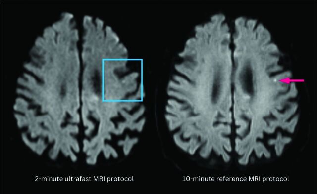
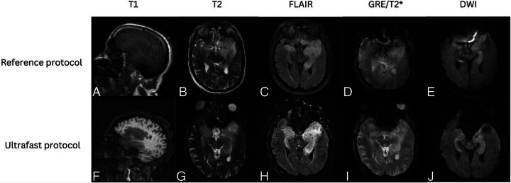
Background and purpose: The use of MR imaging in emergency settings has been limited by availability, long scan times, and sensitivity to motion. This study assessed the diagnostic performance of an ultrafast brain MR imaging protocol for evaluation of acute intracranial pathology in the emergency department and inpatient settings.

Materials and methods: Sixty-six adult patients who underwent brain MR imaging in the emergency department and inpatient settings were included in the study. All patients underwent both the reference and the ultrafast brain MR protocols. Both brain MR imaging protocols consisted of T1-weighted, T2/T2*-weighted, FLAIR, and DWI sequences. The ultrafast MR images were reconstructed by using a machine-learning assisted framework. All images were reviewed by 2 blinded neuroradiologists.

Results: The average acquisition time was 2.1 minutes for the ultrafast brain MR protocol and 10 minutes for the reference brain MR protocol. There was 98.5% agreement on the main clinical diagnosis between the 2 protocols. In head-to-head comparison, the reference protocol was preferred in terms of image noise and geometric distortion (P < .05 for both). The ultrafast ms-EPI protocol was preferred over the reference protocol in terms of reduced motion artifacts (P < .01). Overall diagnostic quality was not significantly different between the 2 protocols (P > .05).

Conclusions: The ultrafast brain MR imaging protocol provides high accuracy for evaluating acute pathology while only requiring a fraction of the scan time. Although there was greater image noise and geometric distortion on the ultrafast brain MR protocol images, there was significant reduction in motion artifacts with similar overall diagnostic quality between the 2 protocols.


Figures

FIG 1.
FIG 1.
A punctate focus of restricted diffusion was less conspicuous on the ultrafast DWI image (left) compared with the reference DWI image (right). The decreased conspicuity may be due to a combination of differences in image quality between the 2 protocols and section positioning leading to partial volume averaging.
FIG 2.
FIG 2.
Bubble plot shows head-to-head comparison between the reference brain MR protocol and the ultrafast ms-EPI protocol. Negative scores indicate preference of the clinical reference protocol; 0 indicates equivalence between the 2 protocols; and positive scores indicate preference of the ultrafast ms-EPI protocol. The reference protocol was preferred by the evaluating neuroradiologists in terms of image noise and geometric distortion (P < .05 for both). The ultrafast protocol was preferred over the reference protocol in terms of motion artifacts (P < .05). There was no significant difference between the 2 protocols in terms of overall diagnostic quality.
FIG 3.
FIG 3.
Patient with a diagnosis of amyloid angiopathy. Reference sagittal T1-weighted (A), axial T2-weighted (B), FLAIR (C), SWI (D), and DWI (E) show scattered foci of susceptibility signal in the left greater than right frontal lobes consistent with chronic microhemorrhages. The findings were more conspicuous on the ultrafast sagittal T1-weighted (F), axial T2-weighted (G), FLAIR (H), SWI (I), and DWI (J) because of motion artifact on the reference MR images.
FIG 4.
FIG 4.
Patient with a diagnosis of herpes simplex virus encephalitis. Reference sagittal T1-weighted (A), axial T2-weighted (B), FLAIR (C), SWI (D), and DWI (E) show prominent T1 hypointensity and T2/FLAIR hyperintensity in the left greater than right mesial temporal lobes with scatter foci of susceptibility signal in the left temporal lobe consistent with microhemorrhages. There was associated restricted diffusion in the left greater than right mesial temporal lobes. These signal abnormalities were all seen with similar conspicuity on the ultrafast sagittal T1-weighted (F), and axial T2-weighted (G), FLAIR (H), SWI (I), and DWI (J).
FIG 5.
FIG 5.
Patient with punctate subacute infarct in the left corona radiata. Reference sagittal T1-weighted (A), axial T2-weighted (B), FLAIR (C), SWI (D), and DWI (E) show a punctate focus of restricted diffusion with associated FLAIR hyperintensity in the left centrum semiovale on a background of white matter T2/FLAIR hyperintensities that likely represent chronic small vessel ischemic disease. The same findings were seen on the ultrafast sagittal T1-weighted (F), axial T2-weighted (G), FLAIR (H), SWI (I), and DWI (J).
FIG 6.
FIG 6.
Patient with a low-grade left thalamic glioma. Reference sagittal T1-weighted (A), and axial T2-weighted (B), FLAIR (C), SWI (D), and DWI (E) show an ill-defined, mildly expansile, T1 hypointense, and T2/FLAIR hyperintense lesion centered in the left thalamus, compatible with diagnosis of low-grade glioma. Similar findings were appreciated on the ultrafast sagittal T1-weighted (F), axial T2-weighted (G), FLAIR (H), SWI (I), and DWI (J).

References

    1. Robinson NB, Gao M, Patel PA, et al. . Secondary review reduced inpatient MRI orders and avoidable hospital days. Clin Imaging 2022;82:156–60 10.1016/j.clinimag.2021.11.014 - DOI - PubMed
    1. Drose JA, Pritchard NL, Honce JM, et al. . Utilizing process improvement methodology to improve inpatient access to MRI. Radiographics 2019;39:2103–10 10.1148/rg.2019190043 - DOI - PubMed
    1. Tokur S, Lederle K, Terris DD, et al. . Process analysis to reduce MRI access time at a German University Hospital. Int J Qual Health Care 2012;24:95–99 10.1093/intqhc/mzr077 - DOI - PubMed
    1. Manwani B, Rath S, Lee NS, et al. . Early magnetic resonance imaging decreases hospital length of stay in patients with ischemic stroke. J Stroke Cerebrovasc Dis 2019;28:425–29 10.1016/j.jstrokecerebrovasdis.2018.10.015 - DOI - PubMed
    1. Andre JB, Bresnahan BW, Mossa-Basha M, et al. . Toward quantifying the prevalence, severity, and cost associated with patient motion during clinical MR examinations. J Am Coll Radiol 2015;12:689–95 10.1016/j.jacr.2015.03.007 - DOI - PubMed

Publication types

LinkOut - more resources